Epidemiological characteristics of sarcoidosis
patients in the city of Rio de Janeiro, Brazil*
Características epidemiológicas de pacientes com sarcoidose na cidade do Rio de Janeiro
Vinicius Lemos-Silva, Paula Barroso Araújo, Christiane Lopes, Rogério Rufino, Cláudia Henrique da Costa
Abstract
Objective: To analyze the epidemiological characteristics of sarcoidosis patients in the city of Rio de Janeiro, Brazil.
Methods: A descriptive, case-control study involving 100 sarcoidosis patients under outpatient treatment between
2008 and 2010 at the Pedro Ernesto University Hospital, located in the city of Rio de Janeiro, Brazil. The diagnosis of sarcoidosis was based on clinical, radiological, biochemical, and histopathological criteria. Results: There was a predominance of females in the 35-40 year age bracket (range, 7-69 years), who accounted for 65% of the sample, although there was a second peak at approximately 55 years of age. The most common symptom was dyspnea (in 47%), and the most common radiological findings were pulmonary and lymph node involvement (stage II; in 43%), followed by stage III (in 20%), stage I (in 19%), stage 0 (in 15%), and stage IV (in 3%). No pleural effusion or digital clubbing was observed at diagnosis. The tuberculin skin test was negative in 94 patients. Spirometric findings at diagnosis were normal in 61 patients; indicative of obstructive lung disease in 21; and indicative of restrictive lung disease in 18. The most common biopsy sites were the lungs (principally by bronchoscopy) and the skin, the diagnosis being confirmed by biopsy in 56% and 29% of the cases, respectively. Treatment with prednisone was initiated in 75% of the patients and maintained for more than 2 years in 19.7%. Conclusions: This study corroborates the findings of previous studies regarding the epidemiological characteristics of sarcoidosis patients.
Keywords: Sarcoidosis/epidemiology; Sarcoidosis/diagnosis; Sarcoidosis/therapy.
Resumo
Objetivo: Analisar as características epidemiológicas de pacientes com sarcoidose na cidade do Rio de Janeiro.
Métodos: Estudo descritivo, caso-controle, envolvendo 100 pacientes com sarcoidose acompanhados no Hospital
Universitário Pedro Ernesto, localizado na cidade do Rio de Janeiro, entre 2008 e 2010. O diagnóstico de sarcoidose foi baseado em critérios clínicos, radiográficos, laboratoriais e histopatológicos. Resultados: A doença predominou em mulheres (65%), na faixa de 35-40 anos (variação: 7-69 anos), embora houvesse um segundo pico na população de aproximadamente 55 anos. A dispneia foi o sintoma mais comum (47%), assim como o achado radiográfico de comprometimento pulmonar e linfonodal (estágio II; 43%), seguido por estágio III (20%), estágio 1(19%), estágio 0 (15%) e estágio IV (3%). Nenhum paciente apresentou derrame pleural ou baqueteamento digital no diagnóstico. O PPD foi não reator em 94 pacientes. Os achados espirométricos no diagnóstico foram normais em 61 pacientes; indicativos de distúrbio ventilatório obstrutivo, em 21; e indicativos de distúrbio ventilatório restritivo, em 18. Os sítios de biópsia mais comuns foram os pulmões (principalmente por broncoscopia) e a pele, que confirmaram o diagnóstico em 56% e 29% dos casos, respectivamente. O tratamento com prednisona foi iniciado em 75% dos pacientes e mantido por mais de 2 anos em 19,7%. Conclusões: Este estudo corrobora vários achados relatados em outros estudos sobre as características epidemiológicas de pacientes com sarcoidose.
Descritores: Sarcoidose/epidemiologia; Sarcoidose/diagnóstico; Sarcoidose/terapia.
* Study carried out at the Pedro Ernesto University Hospital, Faculdade de Ciências Médicas da Universidade do Estado do Rio de Janeiro – FCM-UERJ, Rio de Janeiro State University School of Medical Sciences – Rio de Janeiro, Brazil.
Correspondence to: Cláudia Henrique da Costa. Hospital Universitário Pedro Ernesto, Disciplina de Pneumologia e Tisiologia, Universidade do Estado do Rio de Janeiro, Avenida Vinte e Oito de Setembro, 77, 2º andar, Vila Isabel, CEP 20551-030, Rio de Janeiro, RJ, Brasil.
Tel. 55 21 2587-6348. E-mail: [email protected]
Financial support: Paula Barroso Araújo is a recipient of a Young Investigator Grant from the Universidade do Estado do Rio de Janeiro (UERJ, Rio de Janeiro State University), as well as of a grant from the Fundação de Amparo à Pesquisa do Estado do Rio de Janeiro (FAPERJ, Foundation for the Support of Research in the state of Rio de Janeiro).
Ernesto University Hospital (Protocol no. 2302). The objectives of the study and the methods to be employed were explained in advance to the patients, who were then asked to give written informed consent. The study protocol was in accordance with Brazilian National Health Council Resolution 196/96. Between 2008 and 2010, 111 sarcoidosis patients were regularly treated at the Department of Pulmonology and Tuberculosis outpatient clinic for interstitial diseases, which belongs to the UERJ Faculdade de Ciências Médicas (FCM, School of Medical Sciences) and is located in the city of Rio de Janeiro. Of those 111 patients, 100 agreed to participate in the study. At this writing, all patients were still under outpatient follow-up. The sample size was calculated considering a confidence level (1 − α) of 95%, with a 9.39%
confidence interval. Although the incidence of sarcoidosis in the Brazilian population has yet to be established, it is estimated to be lower than 10 cases/100,000 population,(13) and the number of cases of sarcoidosis in the state of Rio de Janeiro is estimated to be 1,200. Therefore, we considered that a minimum of 63 cases of sarcoidosis would be necessary for the study. Qualitative variables are expressed as percentages and as means or medians and standard errors.
The participants were asked to complete a specific questionnaire and underwent ancillary tests, such as pulmonary function testing, chest X-ray, chest CT, high-resolution electrocardiography, tuberculin skin testing with PPD, determination of calcium levels in serum and urine, determination of hepatic enzyme levels, HIV testing, antineutrophil cytoplasmic antibody testing, rheumatoid factor testing, antinuclear antibody testing, serology for Histoplasma capsulatum and Paracoccidioides brasiliensis, ophthalmologic examination, and dermatological examination. All pulmonary function tests (spirometry and plethysmography) were performed in accordance with the 1991 American Thoracic Society criteria.(14) The tests were performed with a bellows spirometer (Vitatrace; Pró Médico Ltda., Rio de Janeiro, Brazil) coupled to a computer running the SPIROMATIC program (Engelógica Engenharia de Sistemas Ltda, Rio de Janeiro, Brazil), and the measurements were interpreted with the use of the theoretical values devised by
Introduction
Sarcoidosis is a multisystemic inflammatory disease that is characterized histologically by noncaseating granulomas rich in macrophages and T lymphocytes, especially CD4+ T cells.(1,2) Although the disease is of unknown etiology, it is believed that there is an environmental agent, whether infectious or not, that is responsible for triggering the inflammatory response in genetically susceptible hosts.(1-5) The lung is the most commonly affected organ.(3,4,6) Although there is remission in 50% of cases, some patients live with chronic disease for decades.(1,4)
Sarcoidosis affects all ethnic and age groups, being more common before the age of 50 years and being predominant in females.(7) The literature describes epidemiological, radiological, and biochemical differences, as well as different clinical manifestations and different therapeutic responses, in various ethnic groups.(4,6-8) The highest prevalence of sarcoidosis is found in Sweden (121/100,000 population),(9) whereas Spain has the lowest rates in Europe.(10) In the USA, the annual incidence rate ranges from 10.9/100,000 population to 35.5/100,000 population, being higher in African-Americans. (4) Similarly, in South Africa, the prevalence of sarcoidosis is higher in Blacks, being between 10/100,000 population and 20/100,000 population, whereas, in Whites, it is lower than 10/100,000 population.(10) In a recent study, the mean annual incidence of sarcoidosis in African-American women was 71/100,000 population. (11) In Asian countries, sarcoidosis is much less common; the prevalence of the disease in Japan is 1-2 cases/100,000 population.(12) In Latin America, there are few studies of the prevalence of sarcoidosis, and, in Brazil, its incidence is estimated at 10/100,000 population.(13)
The objective of the present study was to evaluate the epidemiological profile of sarcoidosis patients in the city of Rio de Janeiro, Brazil, and to compare their characteristics with those of patients in other countries, for which there are more data about this disease.
Methods
The spirometry results obtained at the first evaluation were within normal parameters in 61 patients. However, they revealed obstructive lung disease in 21 and restrictive lung disease in 18.
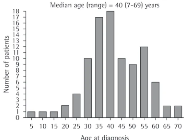
Chest X-rays performed at the time of diagnosis were reviewed by the professional staff working at the outpatient clinic (Figure 2). Nearly half of the patients (43%) were classified as stage II (pulmonary and lymph node involvement) at the first evaluation. The other patients had chest X-rays consistent with stage 0 (15%), stage I (19%), or stage III (20%). Three patients who had had symptoms of sarcoidosis for more than 1 year were classified as stage IV.
Although the lung was the most commonly affected site (in 66%), we found a high percentage of extrapulmonary manifestations (in 80%). The Knudson et al.(15) The diagnosis of sarcoidosis
was confirmed by the finding of noncaseating granulomas (anatomopathological examination) and by the exclusion of other granulomatous infectious diseases (culture for bacteria and fungi). Radiological and biochemical findings consistent with the disease were also taken into consideration.
Results
We analyzed data from 100 patients, all of whom had a confirmed histopathological diagnosis of sarcoidosis and were under follow-up between 2008 and 2010 at the outpatient clinic for interstitial diseases of the FCM/UERJ Department of Pulmonology and Tuberculosis. We excluded patients who were lost to follow-up or for whom not all test results were available. Table 1 summarizes the principal findings in the patients studied.
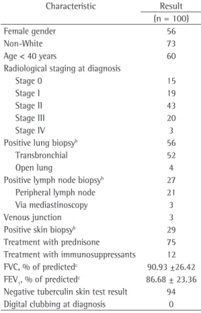
Although the median age at diagnosis was 40 years (range, 7-69 years), 61% of the patients were under 40 (Figure 1). There was a predominance of females (56%). The mean follow-up period was 10.08 ± 7.05 years.
There has been considerable miscegenation in the population the city of Rio de Janeiro, which makes it difficult to define race and ethnicity. Therefore, separating White from non-White (Mulatto or Black) patients, we found that only 27% described themselves as White. No other ethnicities were represented.
With regard to occupation, we found a predominance of homemakers and administrative workers. However, among the 100 patients evaluated, some degree of dust exposure in the work environment was reported by 14 (5 firefighters, 5 construction workers, 1 shoemaker, 1 joiner, 1 floor polisher, and 1 biologist who had been exposed to asbestos). Although 12 patients were smokers and 26 had a history of smoking, the majority (65 patients) had never smoked.
In terms of symptoms, 10 patients were asymptomatic at the time of diagnosis. The most common complaint was dyspnea (in 47 patients), followed by arthralgia (in 23) and dry cough (in 21). Arthritis was uncommon, occurring in only 2 patients who complained of arthralgia. Skin lesions were common (occurring in 29 patients), but, in most cases, they were disregarded by the patients.
Table 1 - Principal characteristics of the study
population.a
Characteristic Result (n = 100)
Female gender 56
Non-White 73
Age < 40 years 60
Radiological staging at diagnosis
Stage 0 15
Stage I 19
Stage II 43
Stage III 20
Stage IV 3
Positive lung biopsyb 56
Transbronchial 52
Open lung 4
Positive lymph node biopsyb 27
Peripheral lymph node 21 Via mediastinoscopy 3
Venous junction 3
Positive skin biopsyb 29
Treatment with prednisone 75 Treatment with immunosuppressants 12 FVC, % of predictedc 90.93 ±26.42
FEV1, % of predictedc 86.68 ± 23.36
Negative tuberculin skin test result 94 Digital clubbing at diagnosis 0
aValues expressed as number of patients, except where
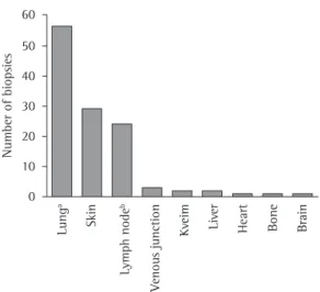
obtained through mediastinoscopy in 3; and of samples obtained through lymph node puncture at the venous junction in 3. The least common biopsy sites were the liver (in 2 patients), heart (in 1), and bones (in 1). Two patients underwent the Kveim-Siltzbach test. Some patients underwent biopsy of more than one site (Figure 3).
In this population, the most common diseases associated with sarcoidosis were systemic arterial hypertension (in 26%) and diabetes mellitus (in 16%). Other diseases identified were nephrolithiasis (in 7 patients); gastroesophageal reflux, cataract, osteoporosis, and coronary artery disease (in 5 patients each); asthma, hypothyroidism, and glaucoma (in 4 patients each); rheumatoid arthritis, cholelithiasis, and symptomatic depression (in 3 patients each); hepatitis B, hepatitis C, dyslipidemia, COPD, and leiomyoma (in 2 patients each); and sickle cell anemia, rectal prolapse, hyperthyroidism, abdominal aortic aneurysm, and epilepsy (in 1 patient each).
As expected, the tuberculin skin test was negative in nearly all (94) of the patients. Two patients with positive tuberculin skin test results had a history of tuberculosis treatment, although there had been no bacteriological confirmation in any of the cases.
Digital clubbing was found in only 1 patient, after years of follow-up and together with the onset of pulmonary arterial hypertension.
Corticosteroid treatment was used in 75 patients, 9 of whom had not completed 2 years of outpatient follow-up. Some patients were treated at the clinic of origin and were already receiving corticosteroids when they were referred to our outpatient clinic. In our outpatient clinic, prednisone treatment is used in patients with target organ involvement, with a maximum initial dose of 40 mg (< 1 mg/kg) and tapering at 4 months. Treatment with corticosteroids is not used, a priori, in patients with sarcoidosis of the skin, lymph nodes, or both. Of the 66 patients under follow-up for more than 2 years, 13 experienced symptom recurrence when the dose was reduced, and it was necessary to maintain a minimal amount of prednisone or use it in combination with immunosuppressants, such as azathioprine or cyclophosphamide (in 12%), chloroquine (in 3%), or thalidomide (in 1%). The choice of treatments depended on disease severity, on the organs affected, on the presence signs and symptoms of the patients at the time
of diagnosis are shown in Table 2. Lymph node sarcoidosis was found in 43 patients, whereas skin sarcoidosis was found in 29.
Most (56%) of the biopsies taken in order to confirm the diagnosis were lung biopsies, and most of those were performed by bronchoscopy, although thoracotomy was required in 4 cases. In 29 patients, the diagnosis was confirmed by biopsy of skin lesions. The diagnosis was confirmed by histopathological examination in 27 patients: of needle-biopsy samples of the peripheral lymph nodes in 21; of samples
Figure 1 - Age of the sarcoidosis patients studied
(n = 100) at the time of diagnosis. Note the bimodal distribution, with a peak of incidence around 40 years of age and a second peak, lower than the first one, at 55 years of age. We found that the diagnosis can be made from the first to the seventh decade of life.
Figure 2 - Radiological staging of the sarcoidosis
The aspect of ethnicity, however, merits attention. The state of Rio de Janeiro has a population that is characterized by considerable miscegenation. According to data from the Instituto Brasileiro de Geografia e Estatística (IBGE, Brazilian Institute of Geography and Statistics),(20) 55.2% of the population consider themselves White, whereas most others consider themselves Black or Mulatto, and other ethnicities, such as indigenous subjects and subjects of Asian descent, are poorly represented. Taking these data into consideration, we used the self-definition method (asking patients to state their ethnicity) in order to define patient ethnicity. Using that method, we found that the percentage of non-White patients was considerable (73%), much higher than the 45% reported by the IBGE for the population of the state. Therefore, we can suggest that sarcoidosis predominantly affects Black and Mulatto patients in this region. However, it should be taken into consideration that the population that seeks treatment at public institutions, as was the case of our study population, belongs to an underprivileged social class, which is characterized by higher proportions of Black and Mulatto individuals. This could constitute a confounding factor in the data analysis. A study of 92 patients conducted in the Brazilian state of Rio Grande do Sul showed that the mean age was similar to that found for the population in the city of Rio de Janeiro (41.8 ± 14.1 years); however, 84% of the patients in that study were White.(16) It is possible that some differences found between the two groups, such as the larger number of patients with constitutional symptoms in the Rio Grande do Sul cohort, can be explained by the intergroup differences in terms of ethnic makeup.
The study designated A Case Control Etiologic Study of Sarcoidosis (ACCESS): Environmental and Occupational Risk Factors was based on data collected from 10 facilities in the United States. In that study, a total of 736 patients with sarcoidosis diagnosed via histopathology were compared with a control group of 706 healthy subjects. The authors reported a negative association between sarcoidosis and smoking. (21) However, other studies(22) failed to confirm
this “protective” effect of smoking. In our study, we found that the proportions of smokers and former smokers were similar to those reported for of hypercalcemia, on previous treatment
complications, and on the impossibility of using corticosteroids. One patient with severe skin lesions showed partial improvement after treatment with thalidomide.
Discussion
Sarcoidosis is a disease with epidemiological characteristics that vary depending on the population studied. In the present study, we found that the patients under follow-up had characteristics similar to those reported in the literature, especially in terms of the most affected age group (61% of the patients were under 40 years of age at the onset of disease) and gender (56% were female).(16) It is interesting that the bimodal distribution of age at diagnosis that we found in our population, with a second peak of incidence at approximately 55 years of age, has been reported by other authors.(17-19)
Table 2 - Signs and symptoms of the patients studied
(n = 100) at the time of diagnosis.a
Signs and symptoms
Patients, n (n = 100)
None 10
Dyspnea 47
Skin lesions 29
Arthralgia 23
Cough 21
Weight loss 8
Asthenia 6
Erythema nodosum 4
Myalgia 3
Wheezing 3
Night sweats 3
Headache 3
Dysesthesia 3
Dysphagia 2
Alopecia 2
Arthritis 2
Fever 2
Eye pain 2
Hemoptysis 2
Hemiparesis 2
Raynaud’s phenomenon 1
Sicca syndrome 1
Chest pain 1
Fainting 1
aSome patients had more than one sign or symptom, and
The ACCESS study(4) reported that approximately half of the patients (51%) had dyspnea at the time of diagnosis. In our cohort, the percentage was similar: 47% had this complaint at the time of diagnosis. Arthralgia was reported by 23 patients, whereas small joint arthritis was found in only 2 cases. Dry cough was part of the clinical profile of 21 patients and, in all cases, it was accompanied by dyspnea. None of the patients had digital clubbing at the time of diagnosis. The most commonly found skin lesions were papules, which were located on the face and trunk, were usually faint, and often went undetected by the patients. However, as in other studies,(26) a variety of lesions were diagnosed, including lesions on scars, hypopigmentation, plaque lesions, and deforming lesions, such as lupus pernio.
Previous studies have shown that the frequency of stage II disease is high among patients with sarcoidosis. González et al. found that 34.7% of the patients had pulmonary and lymph node involvement at the time of diagnosis. (27) In the ACCESS study conducted in the United States, the most common presentation (seen in 39.7%) was lymph node lesions only (stage I), followed by stage II disease (in 36.7%).(4) Our cohort had a higher percentage of stage II chest X-rays at the time of diagnosis (43%), closer to the 48% reported in another study conducted in Brazil.(16) Pooling the patients with pulmonary lesions (stages II, III, and IV), we found that they collectively represented 66% of our sample, compared with 52% for the ACCESS study sample. It is known that pulmonary parenchymal lesions constitute a criterion for the initiation of corticosteroid treatment, as well as being related to more aggressive disease. It is possible that, because our facility is a referral center, we received a larger number of patients with more advanced disease. Among the patients with pulmonary lesions, the most common finding was predominantly central interstitial involvement, with nodular lesions, affecting the peribronchovascular bundle. It is of note that our sample did not comprise any cases of pleural effusion. Although the reported rates of pleural effusion in sarcoidosis range from 1% to 10%, this manifestation has been characterized as rare.(28)
Although nearly half of the patients analyzed (47%) complained of dyspnea, with or without the general population in Brazil (35%),(20) a fact
that questions the negative association between smoking and sarcoidosis in our population.
Occupation and environmental exposure have been reported to be potential triggers of sarcoidosis in genetically susceptible patients. (23,24) After the World Trade Center tragedy, some firefighters who participated in the rescue efforts were diagnosed with sarcoidosis, which suggests that some occupations can be associated with a greater risk for the disease.(25) The multivariate analysis performed in the ACCESS study suggested that the development of sarcoidosis is positively associated with exposure to insecticides and aerosols in the work environment.(21) The same analysis did not confirm that such an association is valid for health professionals.(21) In our cohort, we observed the occurrence of exposure to dust in 14 of the 100 patients evaluated. It is of note that, of those 14 patients, 5 were firefighters and 5 construction workers, which suggests that disease onset resulted from that exposure. However, other comparative studies, with control groups, are needed in order to explore this possibility.
Figure 3 - Biopsy sites for the diagnosis of sarcoidosis. A total of 119 biopsies were performed in 100 patients. Some patients underwent more than one type of biopsy. The patients who underwent lymph node biopsy at the venous junction were considered separately from those who underwent lymph node biopsy, and two different columns represent the former and the latter. aFour open lung biopsies
and 52 transbronchial biopsies. bThree biopsies by
can occur in sarcoidosis.(30) However, in our population, only 1 patient (with fibrosing disease and an unfavorable clinical evolution) had pulmonary arterial hypertension.
The present study presents an evaluation of 100 sarcoidosis patients who were diagnosed by histopathology and by excluding other diseases and who were under follow-up at a university referral center. Our analysis of this cohort of patients in Brazil has corroborated the findings of previous studies regarding the epidemiological characteristics of sarcoidosis patients and has revealed characteristics of our population, potentially furthering understanding of the disease in Brazil.
References
1. Lazar CA, Culver DA. Treatment of sarcoidosis. Semin Respir Crit Care Med. 2010;31(4):501-18.
2. Nunes H, Soler P, Valeyre D. Pulmonary sarcoidosis. Allergy. 2005;60(5):565-82.
3. Moller DR. Potential etiologic agents in sarcoidosis. Proc Am Thorac Soc. 2007;4(5):465-8.
4. Baughman RP, Teirstein AS, Judson MA, Rossman MD, Yeager H Jr, Bresnitz EA, et al. Clinical characteristics of patients in a case control study of sarcoidosis. Am J Respir Crit Care Med. 2001;164(10 Pt 1):1885-9. 5. Rybicki BA, Iannuzzi MC, Frederick MM, Thompson BW,
Rossman MD, Bresnitz EA, et al. Familial aggregation of sarcoidosis. A case-control etiologic study of sarcoidosis (ACCESS). Am J Respir Crit Care Med. 2001;164(11):2085-91.
6. Lazarus A. Sarcoidosis: epidemiology, etiology, pathogenesis, and genetics. Dis Mon. 2009;55(11):649-60.
7. Fernández Fabrellas E. Epidemiology of sarcoidosis [Article in Spanish]. Arch Bronconeumol. 2007;43(2):92-100.
8. Sharma OP. Sarcoidosis around the world. Clin Chest Med. 2008;29(3):357-63, vii.
9. Deubelbeiss U, Gemperli A, Schindler C, Baty F, Brutsche MH. Prevalence of sarcoidosis in Switzerland is associated with environmental factors. Eur Respir J. 2010;35(5):1088-97.
10. Bethlem EP. Sarcoidosis in Brazil. J Bras Pneumol. 2005;31(5):ii-iii.
11. Cozier YC, Berman JS, Palmer JR, Boggs DA, Serlin DM, Rosenberg L. Sarcoidosis in black women in the United States: data from the Black Women’s Health Study. Chest. 2011;139(1):144-50.
12. Morimoto T, Azuma A, Abe S, Usuki J, Kudoh S, Sugisaki K, et al. Epidemiology of sarcoidosis in Japan. Eur Respir J. 2008;31(2):372-9.
13. Bethlem NM. Epidemiology of sarcoidosis in Brazil. Sarcoidosis. 1985;2:162.
14. Lung function testing: selection of reference values and interpretative strategies. American Thoracic Society. Am Rev Respir Dis. 1991;144(5):1202-18.
15. Knudson RJ, Lebowitz MD, Holberg CJ, Burrows B. Changes in the normal maximal expiratory flow-volume
cough, only 39% had plethysmographic abnormalities at the time of diagnosis. It should also be taken into account that 35% of our patients smoked or had a history of smoking, which certainly contributed to the finding of an obstructive pattern in some patients. Other functional tests, such as the measurement of TLC, DLCO, and maximal inspiratory/expiratory pressures, were not performed in all patients at the time of diagnosis. Therefore, these parameters were not analyzed for the present study.
In all of our patients, the diagnosis of sarcoidosis was confirmed by histopathology to detect noncaseating granulomas, which is in accordance with current recommendations. (29) The most common biopsy sites were the lung (in 56%), principally by bronchoscopy, and the skin (in 29%). Therefore, in most cases, the diagnosis of sarcoidosis can be suggested and confirmed by clinicians (pulmonologists and dermatologists). Other cases in our sample required tests such as peripheral lymph node biopsy or lymph node biopsy at the venous junction, which are relatively simple for surgeons to perform. Open lung biopsy by thoracotomy was performed in 4 patients, and lymph node biopsy by mediastinoscopy was performed in 3.
Among granulomatous diseases in Brazil, tuberculosis is much more prevalent than is sarcoidosis. In some cases, diagnostic confusion can arise, especially when the tuberculin skin test result is positive. This occurred in 6 patients. Of those, 2 had a history of tuberculosis treatment, although there was no bacteriological confirmation in either case.
23. Rossman MD, Kreider ME. Lesson learned from ACCESS (A Case Controlled Etiologic Study of Sarcoidosis). Proc Am Thorac Soc. 2007;4(5):453-6.
24. Rossman MD, Thompson B, Frederick M, Iannuzzi MC, Rybicki BA, Pander JP, et al. HLA and environmental interactions in sarcoidosis. Sarcoidosis Vasc Diffuse Lung Dis. 2008;25(2):125-32.
25. Bowers B, Hasni S, Gruber BL. Sarcoidosis in World Trade Center rescue workers presenting with rheumatologic manifestations. J Clin Rheumatol. 2010;16(1):26-7. 26. Abu-Hilal M, Krotva J, Chichierchio L, Obeidat N,
Madanat M. Dermatologic aspects and cutaneous manifestations of sarcoidosis. G Ital Dermatol Venereol. 2010;145(6):733-45.
27. González EL, Vigliano C, Cáneva J. Sarcoidosis. Clinical presentation and prognosis [Article in Spanish]. Medicina (B Aires). 2010;70(6):499-502.
28. Salerno D. Sarcoidosis pleural effusion: a not so common feature of a well known pulmonary disease. Respir Care. 2010;55(4):478-80.
29. Morgenthau AS, Iannuzzi MC. Recent advances in sarcoidosis. Chest. 2011;139(1):174-82.
30. Palmero V, Sulica R. Sarcoidosis-associated pulmonary hypertension: assessment and management. Semin Respir Crit Care Med. 2010;31(4):494-500.
curve with growth and aging. Am Rev Respir Dis. 1983;127(6):725-34.
16. Silva LC, Hertz FT, Cruz DB, Caraver F, Fernandes JC, Fortuna FP, et al. Sarcoidosis in the south of Brazil: a study of 92 patients. J Bras Pneumol. 2005;31(5):398-06. 17. Iannuzzi MC, Rybicki BA, Teirstein AS. Sarcoidosis. N
Engl J Med. 2007;357(21):2153-65.
18. Nunes H, Bouvry D, Soler P, Valeyre D. Sarcoidosis. Orphanet J Rare Dis. 2007;2:46.
19. Milman N, Selroos O. Pulmonary sarcoidosis in the Nordic countries 1950-1982. Epidemiology and clinical picture. Sarcoidosis. 1990;7(1):50-7.
20. Instituto Brasileiro de Geografia e Estatística - IBGE [homepage on the Internet]. Brasília: Ministério do Planejamento, Orçamento. [updated 2010 Nov 29; cited 2011 Feb 02]. Mapa do Site – Indicadores. Available from: http://www.ibge.gov.br/home/mapa_site/mapa_ site.php#populacao
21. Newman LS, Rose CS, Bresnitz EA, Rossman MD, Barnard J, Frederick M, et al. A case control etiologic study of sarcoidosis: environmental and occupational risk factors. Am J Respir Crit Care Med. 2004;170(12):1324-30. 22. Gupta D, Singh AD, Agarwal R, Aggarwal AN, Joshi K,
Jindal SK. Is tobacco smoking protective for sarcoidosis? A case-control study from North India. Sarcoidosis Vasc Diffuse Lung Dis. 2010;27(1):19-26.
About the authors
Vinicius Lemos-Silva
Professor. Department of Clinical Medicine, Faculdade de Ciências Médicas da Universidade do Estado do Rio de Janeiro – FCM-UERJ, Rio de Janeiro State University School of Medical Sciences – Rio de Janeiro, Brazil.
Paula Barroso Araújo
Medical Student. Faculdade de Ciências Médicas da Universidade do Estado do Rio de Janeiro – FCM-UERJ, Rio de Janeiro State University School of Medical Sciences – Rio de Janeiro, Brazil.
Christiane Lopes
3. Medical Student. Faculdade de Ciências Médicas da Universidade do Estado do Rio de Janeiro – FCM-UERJ, Rio de Janeiro State University School of Medical Sciences – Rio de Janeiro, Brazil.
Rogério Rufino
Adjunct Professor. Department of Pulmonology and Tuberculosis. Faculdade de Ciências Médicas da Universidade do Estado do Rio de Janeiro – FCM-UERJ, Rio de Janeiro State University School of Medical Sciences – Rio de Janeiro, Brazil.
Cláudia Henrique da Costa