Original Article
Artigo Original
This is an Open Access article distributed under the terms of the Creative Commons Attribution License, which permits unrestricted use, distribution, and reproduction in any medium, provided the original work is properly cited. ISSN 2317-1782 (Online version)
Comparison between early and delayed
facial nerve decompression in traumatic
facial nerve paralysis - A retrospective study
David Victor Kumar Irugu1Anoop Singh1 Sravan CH2 Achyuth Panuganti3 Anand Acharya4 Hitesh Varma1 Ramya Thota1 Maurizio Falcioni5 Sridhar Reddy3
Keywords
Facial Nerve Fracture Decompression Mastoidectomy Geniculate Ganglion Perforation
Correspondence address: David Victor Kumar Irugu Department of Otorhinolaryngology and Head & Neck Surgery, All India Institute of Medical Sciences – AIIMS
Room No- 4065, ENT Ofice,
4th Floor- Teaching Block, Ansarinagar, New Delhi-110029, India, New Delhi, India.
E-mail: [email protected]
Received: April 11, 2017
Accepted: September 04, 2017
Study carried out at Department of ENT and Head & Neck Surgery, Govt ENT Hospital, Osmania Medical College, Hyderabad, India.
1 Department of Otorhinolaryngology and Head & Neck Surgery, All India Institute of Medical Sciences – AIIMS, New Delhi, India.
2 Department of Surgery, Christian Medical College, Vellore, Tamil Nadu, India.
3 Department of Otorhinolaryngology and Head & Neck Surgery, Osmania Medical College, Hyderabad, India. 4 Department of Otorhinolaryngology and Head & Neck Surgery, Governament Medical College, Nizamabad,
India.
5 Department of Otorhinolaryngology and Head & Neck Surgery, University Hospital, Parma, Italy. Financial support: nothing to declare.
Conlict of interests: nothing to declare. ABSTRACT
INTRODUCTION
Facial nerve paralysis (FNP) is a debilitating and devastating
condition causing disigurement and functional consequences in the form of speech and masticatory dificulties(1-4). Common
causes of FNP are trauma (motor vehicle accidents), iatrogenic (surgery), infections (Bell’s palsy), and tumors(2,3,5-7). Worldwide,
seven to 10% of temporal bone fractures (TBF) are caused
by road trafic accidents, which lead to FNP(1,4,6,7). Temporal bone fractures are classiied into longitudinal, transverse, or
mixed in relation to the petrous pyramid axis(7). Longitudinal
fractures contribute to 80% of TBF, of which 20% cause FNP, whereas transverse fractures constitute 20% of TBF, of which 50% cause FNP(8). Management of FNP with facial
nerve decompression (FND) remains a surgical challenge to otologists and neuro-otologists. In recent times, imaging studies have provided excellent preoperative localization of injury and selection of surgical approach(4,9). Authors who believe
in surgical management have different opinions regarding the timing of decompression(9,10), and the most appropriate time of surgery remains a subject of controversy. The question to
be answered is whether early decompression provides faster improvement of FNP as compared with delayed decompression.
The present study aimed to assess the intraoperative indings
in case of early and delayed microsurgical decompression of FNP and compare their results.
METHODS
Thirty-eight cases of facial nerve paralysis (FNP) were analyzed, and 23 cases of longitudinal temporal bone fractures were included in the study. Ten-year data were collected retrospectively, and the results were analyzed. All patients consented to participate in the study as per the aforementioned Institute protocol.
Fifteen cases of FNP caused by other etiological factors, such as iatrogenic and infections, were excluded from the study. Demography of all 23 cases of FNP is given in Table 1.
Preoperative workup: After complete clinical history and examination, facial weakness was rated according to the House-Brackmann (H-B) grading system. Preoperatively, all patients presented grade V and VI paralysis. Pure tone audiometry was conducted by a trained audiologist to document the hearing status. High-resolution computed tomography (HRCT) scans of 1-mm thick coronal and axial sections were obtained for operative planning (Figure 1). All patients underwent
electro-neuronography (ENoG) to quantify the regenerative
status of the nerve. The decision to decompress the facial nerve was taken based on the following criteria:
1. Fracture line identiied in HRCT of the temporal bone;
2. ENoG >90%.
The early decompression group (EDG) included patients who had undergone surgical decompression within six weeks of FNP onset, whereas the delayed decompression group (DDG)
included patients who had been operated after six weeks of FNP onset.
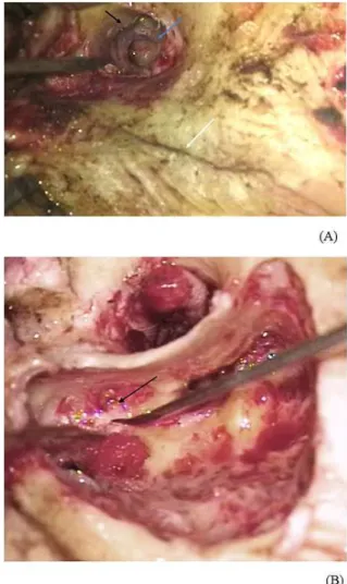
After signing an Informed Consent Form (ICF), all patients were operated under general anesthesia. The retro auricular-transmastoid approach (canal wall up) with posterior tympanotomy and superior
extension technique was used to decompress the facial nerve. While performing the procedure, indings were documented
for further analysis (Table 2; Figure 2A and B).
Table 1. Demography of patient population
P. no. Age/Gender Cause/side/age
of FNP (in days) H-B Grading
1 42yrs/ M RTA/R/20 VI
2 27yrs/M RTA/R/28 V
3 36yrs/M RTA/L/26 VI
4 44yrs/M RTA/L/42 VI
5 52yrs/M RTA/L/150 VI
6 23yrs/M RTA/R/55 VI
7 30yrs/M RTA/L/49 VI
8 28yrs/M RTA/R/90 VI
9 34yrs/M RTA/R/42 VI
10 40yrs/M RTA/L/120 VI
11 20yrs/M RTA/L/30 VI
12 30yrs/M RTA/R/35 VI
13 42yrs/M RTA/L/150 VI
14 55yrs/M AF/L/120 VI
15 26yrs/M AF/L/120 VI
16 28yrs/F AF/R/150 VI
17 35yrs/F AF/L/150 VI
18 40yrs/M RTA/L/23 VI
19 60yrs/M AF/L/90 VI
20 38yrs/M RTA/R/90 with DM V
21 27yrs/M RTA/R/35 VI
22 31yrs/M RTA/L/60 VI
23 51yrs/M AF/L/8 VI
Caption: FNP- Facial Nerve Paralysis; H-B grading - House-Brackmann grading; R - Right; L - Left; M - Male; F - Female; RTA- Road traffic accident; AF - Accidental fall; DM- Diabetes Mellitus
Figure 1. Axial High-resolution Computed Tomography showing
Follow-up varied considerably between individuals, but all the cases were monitored for up to two years. Patients were
monitored weekly during the irst month, then once a month for the next six months, and every six months subsequently.
All patients were subjected to physical rehabilitation in the form of facial tapping and light massage to restore the resting tone on the affected face side. Galvanic stimulation was performed to evoke the muscular contractions. The principle behind this is to reduce muscular atrophy to promote reinnervation of facial muscles.
RESULTS
A total of 23 patients had longitudinal temporal bone fractures causing facial nerve paralysis (FNP). The male: female ratio was 21:2 (91.3%:8.7%), with mean age of 36.5 years. There were 14 (60.9%) cases of left-sided FNP and nine (39.1%) cases of
right-sided FNP. The most common etiology was road trafic
accident (RTA) - 17 cases (73.9%), followed by accidental fall - 6 cases (26.1%).
On clinical examination of the 23 patients, 13 (56.52%) cases presented blood stains/clots in the external auditory canal (EAC) and 12 (52.17%) cases had step deformity of the EAC; 17 (73.91%) cases had intact tympanic membrane, of which 5 (29.41%) cases presented haemotympanum; 12 (70.59%) cases had altered color/dull tympanic membrane; 6 (26.08%) cases presented discharging ears owing to secondary infection. According to the H-B grading system, two patients had grade V (8.7%) and 21 patients had grade VI (91.30%) FNP (Table 2).
Preoperative hearing documented by pure tone audiometry showed conductive hearing loss averaging 40 dB in 21 cases (91.30%) and moderate mixed hearing loss in two cases (8.7%) (Table 2).
All patients underwent high-resolution computed tomography (HRCT) of the temporal bones with 1-mm coronal and axial sections with 3D reconstruction (Figure 1). All the cases
presented visible fracture on the HRCT scans, with other indings
such as soft tissue density in epitympanum, middle ear, and mastoid bone in 12 cases (52.17%); incudo-stapedial (IS) joint dislocation in 15 cases (65.21%); probable nerve injury at the
geniculate ganglion (Figure 1) in 15 cases (65.21%); vertical segment injury in three cases (13.04%). The remaining cases showed suspected injury proximal to the geniculate ganglion, such as impingement of bony spicules. Clinically apparent step
Table 2. Clinical and radiological findings of patients (n=23)
Clinical finding No. of patients radiological No. of patients
Blood stains in EAC 13 Soft tissue density 12
Discharge in EAC 6 IS joint dislocation 15
Step deformity of EAC 12 Nerve injury - site
Geniculate Ganglion 15
Vertical part of facial nerve 3
Tympanic Membrane Step deformity
Intact 17 Anterior Meatal Wall 7
Altered 12 Posterior Meatal Wall 3
Haemotympanum 5
Perforated 6
Preoperative Hearing
Conductive (average of 40dB) 21
Mixed (moderate) 2
Caption: EAC - External auditory canal
deformity was conirmed by HRCT in 10 cases (43.47%), of
which seven cases presented injury on the anterior meatal wall (70%) and three on the posterior meatal wall (30%) (Table 2).
All 23 patients were operated and divided into two groups (early and delayed decompression) depending on the time elapsed between onset of FNP and surgery. This delay ranged from eight to 180 days. Findings on the extra- and intra-fallopian canals in both groups were recorded (Table 3).
1. Early Decompression Group (EDG): It comprised 10 patients (43.48%) who had undergone facial nerve decompression within six weeks of FNP onset.
(A) The extra fallopian canal indings included tympanic
membrane perforation in four cases (40%), hemotympanum in four cases (40%), and IS joint dislocation in eight cases
(80%). Displaced bony segments and other indings such as soft tissue were identiied in three cases (30%); (B) The intra fallopian canal indings included edema in seven
cases (70%), clotted blood in six cases (60%), bony spicules
in eight cases (80%), ibrotic bands in two cases (20%),
and nerve traction injury from fractured bony segments in six cases (60%).
2. Delayed Decompression Group (DDG): It was composed of 13 patients (56.52%) who had undergone facial nerve decompression after six weeks of FNP onset.
(A) The extra fallopian canal indings included tympanic
membrane perforation in two cases (15.38%), hemotympanum in one case (7.69%), and dislocation of IS joint in 10 cases
(76.92%). Displaced bony segments and other indings such as soft tissue were identiied in nine cases (69.23%); (B) The intra fallopian canal indings included edema in two
cases (15.38%), clotted blood in three cases (23.07%),
bony spicules in nine cases (69.23%), ibrotic bands in
10 cases (76.92%), and traction injury from fractured bony
segments in ive cases (38.46%).
Overall, the extra fallopian canal indings in both groups
included tympanic membrane perforation in six cases (26.08%),
hemotympanum in ive cases (21.73%), IS joint dislocation in
18 cases (78.26%), and traction injury of nerve by displaced
bony segments in 12 cases (52.17%); the intra fallopian
canal indings in both groups included edema in nine cases
(39.13%), clotted blood in nine cases (39.13%), bony spicules
in 17 cases (73.91%), ibrous bands in 10 cases (43.47%), and
traction injury of nerve by separated bony fractured segments in 11 cases (47.82%).
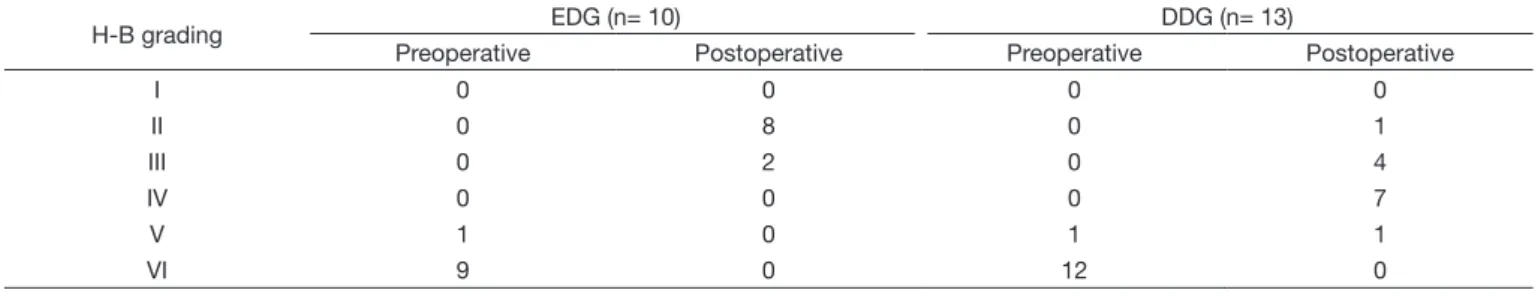
In the EDC, the preoperative status of the facial nerve was grade VI in nine cases (90%) and grade V in one case (10%). Postoperatively, it improved to grade II in eight cases (80%) and grade III in two cases (20%) (Figure 3).
In the DDG, the preoperative status was grade V in one case (7.70%) and grade VI in 12 cases (92.30%). After surgery, it improved to grade II in one case (7.70%), grade III in four cases (30.76%), grade IV in seven cases (53.84%), and grade V in one case (7.70%).
Overall, postoperatively in both groups, nine patients improved to grade II (39.13%), six patients to grade III (26.08%), seven patients to grade IV (30.43%), and one patient to grade
V (4.34%). Pre- and postoperative H-B grading classiication
of patients is shown in Table 4.
Figure 3. Clinical photographs of preoperative H-B grade VI (A to C) and postoperative H-B grade II after six months of follow-up (D to F)
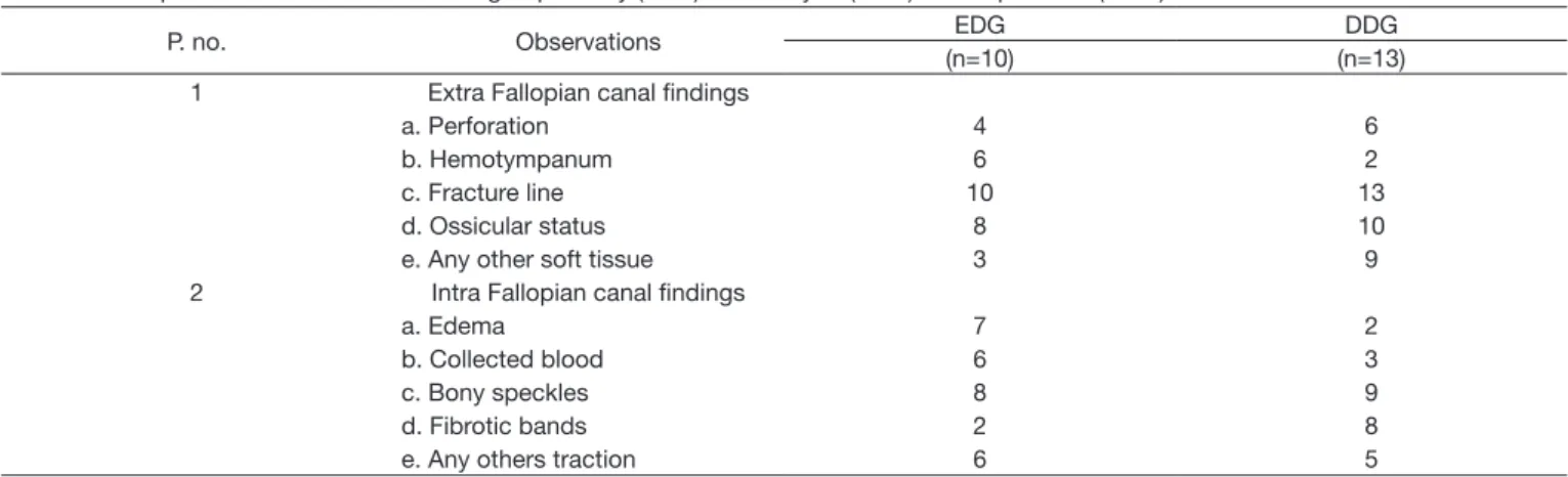
Table 3. Intraoperative observations of both groups: early (EDG) and delayed (DDG) decompression (n=23)
P. no. Observations EDG DDG
(n=10) (n=13)
1 Extra Fallopian canal findings
a. Perforation 4 6
b. Hemotympanum 6 2
c. Fracture line 10 13
d. Ossicular status 8 10
e. Any other soft tissue 3 9
2 Intra Fallopian canal findings
a. Edema 7 2
b. Collected blood 6 3
c. Bony speckles 8 9
d. Fibrotic bands 2 8
DISCUSSION
Facial nerve paralysis (FNP) can be caused by infections,
iatrogenic, road trafic accidents (RTA), and tumors involving
the nerve(2,7). Seven to 10% of the cases of FNP are caused by
temporal bone fractures (TBF)(4,7).
TBF are classiied into longitudinal, transverse, and mixed
depending on the site of fracture in relation to the petrous pyramid axis (4,7,10). Recently, they have been classiied as otic
capsule sparing or otic capsule violating, labyrinthine and extra labyrinthine fractures, respectively(4,5). FNP associated with TBF
can be immediate or delayed in onset(7). According to Ulrich et al.,
the geniculate ganglion is the most common site for FNP in longitudinal TBF(11). In this site, the fracture line usually runs
superior to the external auditory canal (EAC) into the middle ear and along the facial nerve, especially in the perigeniculate area(8,11-13). The mechanism of FNP may be due to traction by
displaced bony segments in the aforementioned area(4,13). Of the
23 participants in this study, 74% (17/23) had been involved in RTA, whereas the remaining 26% (6/23) had accidental fall injuries as the cause of FNP. Most (21/23) patients were male, possibly because of their higher likelihood of involvement in outdoor risk activities.
A thorough evaluation, which is conducted in all cases through clinical, audiological and radiological examinations and electro-diagnostic testing methods, is the key to determine the prognosis of palsy. All these investigations can be used to assess the site of lesion, status of the tympanic membrane and middle ear, hearing level, and selection of surgical approach, offering better counseling to patients regarding prognosis(4-6,10,14).
The House-Brackmann grading system was adopted for preoperative grading of the facial nerve functions because of its reliability and ease of application(3). All patients presented
grade V (8.7%) or grade VI (91.30%) FNP, with conductive hearing loss (average 40dB) caused by hemotympanum, ossicular disruption, and perforation in 21 cases (91.30%) and mixed hearing loss owing to sudden concussion injury to the inner ear in two (8.7%) cases, preoperatively.
High-resolution computed tomography (HRCT) of the temporal bone remains the only important investigation needed for patients affected by TBF, for preoperative surgical planning and better patient counseling. It can detect not only the site of facial nerve involvement, but also other possible severe complications such as meningeal involvement and vascular injury(4-6,8,10,14,15).
All the patients presented fractured line radiologically, which
was conirmed intraoperatively. In contrast, incudo-stapedial
(IS) joint dislocation was suspected in 15 cases (65.21%) on preoperative HRCT, but was detected intraoperatively in 18 patients (78.26%).
Electro-diagnostic tests are based on the principles of electric stimulation of nerve to evoke electromyography response and provide information regarding the degenerative status of the
nerve and, possibility, beneits with early surgical interventions.
Many electro-diagnostic tests are presently available, but electromyography (EMG) and electro-neuronography (ENoG) are more commonly used for FNP. These tests ascertain the degenerative status of the nerve by comparing the side of lesion with the normal side. Fisch advised that ENoG in traumatic FNP causing degeneration >90% within six days of onset of complete FNP needs immediate decompression(6,8,10,12,16,17). In the present
study, all patients presented ENoG scores >90%.
Facial nerve decompression is still a matter of universal debate with respect to the onset of palsy and time of surgery. Many studies have suggested that early decompression provides early expansion of the nerve, relieving edema; it can remove bony spicules impinging on the nerve and drain the blood collected
from the fallopian canal. If decompression is delayed, ibrotic
bands are formed following pathological repair and impinged bone fragments affect nerve conduction. A long delay can compromise blood supply, causing degeneration and shrinkage of the nerve, formation of scar and neuroma, and atrophy of the
peripheral structures. Endoneuronal hematoma and ibrosis are
the main causes of nerve dysfunction(1,7,10,12,14,18).
This study compared early and delayed decompression with respect to onset of FNP and time of surgery, and the results are shown in Table 4. These results encourage performance of early decompression when compared with delayed decompression.
A wide variety of surgical approaches to decompress the nerve in FNP are available. In otic capsule violating fractures, which are associated with sensorineural hearing loss (SNHL) in
a signiicant proportion of cases, the translabyrinthine approach
is preferred for complete nerve decompression; however, in otic capsule sparing fractures, with preserved cochlear and 8th
nerve function, the transmastoid supralabyrinthine and middle cranial fossa approaches provide appropriate exposure without violating the labyrinth, depending on the site of lesion and mastoid pneumatization. In well pneumatized mastoids with nerve involvement limited to the perigeniculate area, transmastoid
Table 4. Comparison of outcome (H-B grading) between the early (EDG) and delayed (DDG) decompression groups
H-B grading EDG (n= 10) DDG (n= 13)
Preoperative Postoperative Preoperative Postoperative
I 0 0 0 0
II 0 8 0 1
III 0 2 0 4
IV 0 0 0 7
V 1 0 1 1
supralabyrinthine access provides enough exposure, whereas a fracture involving the labyrinthine segment of the facial nerve can be better accessed via middle fossa approach. The literature reports that the facial nerve should be decompressed from the labyrinthine segment to the stylomastoid foramen(1,5-8,10,16,17).
In view of this, and of the fact that more than 90% of the patients in this study presented pure conductive deafness, the transmastoid approach was chosen, including incus removal to provide working space in the supra labyrinthine area. Removal of the incus is also helpful in reducing the risk of SNHL, which can occur due to contact of the rotating burr with the intact ossicular chain while working in narrow space(7,10); in addition,
many patients already presented IS joint dislocation. The incus can be repositioned as the last step of surgery. In the population of this study, the most common site of injury was at the geniculate ganglion, involved in 15 cases (65.21%), with injury to the vertical part present in three cases (13.04%), and compression nerve injury of the geniculate ganglion by edema/impingement of bony spicules or dislodged incus present in the remaining cases. Bony spicules, blood collected inside the fallopian canal, nerve edema, and dislodged incus were the predominant
indings in the group explored early on, whereas bony spicules impinging on the nerve and ibrotic bands compressing the facial
nerve were the factors related with FNP in the cases explored in delayed fashion.
Rehabilitation enables reintegration of stomatognathic functions such as suction, chewing, and swallowing, as well
as cosmetic appearance to improve quality of life. The various
modalities were used as described in the literature, including ice/thermal stimulation, massage therapy, and isometric/isotonic facial muscle exercises. All patients were subjected to physical and speech-language pathology rehabilitation and galvanic stimulation postoperatively(19,20).
Although the sample size was not large enough to analyze
statistical signiicance, 80% of the patients undergoing early
decompression achieved grade II in the H-B status and the remaining 20% attained improvement to grade III, whereas most of the patients (85%) undergoing delayed decompression achieved improvement to grade III or IV and only one patient (7.5%) achieved grade II, postoperatively in both groups. The results are presented in Table 4. All patients were monitored for up to two years. The data show a trend towards a better outcome with early decompression; however, they need to be statistically validated using a larger sample size.
CONCLUSION
Despite the limited data available, this study demonstrates that early surgical decompression provides better results in terms of facial nerve function improvement, possibly because it enables early expansion of the nerve by removing impinged bony particles, avoiding compression from dislodged ossicles or bony step deformities, and by reducing the traction injury from displaced bony segments. In delayed decompression cases,
the presence of ibrotic bands may not allow nerve regeneration
because of the existing irreversible changes; in this situation, facial nerve grafting should be considered.
ACKNOWLEDGEMENTS
The authors are grateful to the superintendent of Govt ENT Hospital/Osmania Medical College, Hyderabad, Telangana, India, whose assistance was fundamental to the completion of this study.
REFERENCES
1. McKennan KX, Chole RA. Facial paralysis in temporal bone trauma. Am J Otol. 1992;13(2):167-72.
2. Cha HE, Baek MK, Yoon JH, Yoon BK, Kim MJ, Lee JH. Clinical features and management of facial nerve paralysis in children: analysis of 24 cases. J Laryngol Otol. 2010;124(4):402-6. PMid:20025809. http:// dx.doi.org/10.1017/S0022215109991812.
3. Reitzen SD, Babb JS, Lalwani AK. Significance and reliability of the House-Brackmann grading system for regional facial nerve function. Otolaryngol Head Neck Surg. 2009;140(2):154-8. PMid:19201280. http://dx.doi.org/10.1016/j.otohns.2008.11.021.
4. Rajati M, Rad MP, Irani S, Khorsandi MT, Zarandy MM. Accuracy of high-resolution computed tomography in locating facial nerve injury sites in temporal bone trauma. Eur Arch Otorhinolaryngol. 2014;271(8):2185-9. PMid:24081792. http://dx.doi.org/10.1007/s00405-013-2709-4. 5. Darrouzet V, Duclos JY, Liguoro D, Truilhe Y, De Bonfils C, Bebear JP.
Management of facial paralysis resulting from temporal bone fractures: our experience in 115 cases. Otolaryngol Head Neck Surg. 2001;125(1):77-84. PMid:11458219. http://dx.doi.org/10.1067/mhn.2001.116182. 6. Quaranta A, Campobasso G, Piazza F, Quaranta N, Salonna I. Facial nerve
paralysis in temporal bone fractures: outcomes after late decompression surgery. Acta Otolaryngol. 2001;121(5):652-5. PMid:11583403. http:// dx.doi.org/10.1080/000164801316878999.
7. Hato N, Nota J, Hakuba N, Gyo K, Yanagihara N. Facial nerve decompression surgery in patients with temporal bone trauma: analysis of 66 cases. J Trauma. 2011;71(6):1789-93. http://dx.doi.org/10.1097/ TA.0b013e318236b21f.
8. Yeoh TL, Mahmud R, Saim L. Surgical Intervention in traumatic facial nerve paralysis. Med J Malaysia. 2003;58(3):432-6. PMid:14750385. 9. Ulug T, Arif Ulubil S. Management of facial paralysis in temporal
bone fractures: a prospective study analyzing 11 operated fractures. Am J Otolaryngol. 2005;26(4):230-8. http://dx.doi.org/10.1016/j. amjoto.2005.01.004.
10. Brodsky L, Eviatar A, Daniller A. Post-traumatic facial nerve paralysis: three cases of delayed temporal bone exploration with recovery. Laryngoscope. 1983;93(12):1560-5. PMid:6645756. http://dx.doi.org/10.1288/00005537-198312000-00008.
11. Eby TL, Pollak A, Fisch U. Histopathology of the facial nerve after longitudinal temporal fracture. Laryngoscope. 1988;98(7):717-20. PMid:3386375.
12. Nosan DK, Benecke JE Jr, Murr AH. Current perspective on temporal bone trauma, otolaryngol. Head Neck Surg. 1997;117(1):67-71. PMID: 9230326. http://dx.doi.org/10.1016/S0194-59989770209-2.
13. Danner CJ. Facial nerve paralysis. Otolaryngol Clin North Am. 2008;41(3):619-32. PMid:18436002. http://dx.doi.org/10.1016/j. otc.2008.01.008.
14. Liu Y, Han J, Zhou X, Gao K, Luan D, Xie F, et al. Surgical management of facial paralysis resulting from temporal bone fractures. Acta Otolaryngol. 2014;134(6):656-60. PMid:24665853. http://dx.doi.org/10.3109/00016 489.2014.892214.
15. Valavanis A, Kubik S, Schubiger O. High-resolution CT of the normal and abnormal fallopian canal. AJNR Am J Neuroradiol. 1983:4(3):748-51.
17. Sanuş GZ, Tanriöver N, Tanriverdi T, Uzan M, Akar Z. Late decompression in patients with acute facial nerve paralysis after temporal bone fracture. Turkish Neurosurg. 2007;17(1):7-12.
18. Yetiser S. Total facial nerve decompression for severe traumatic facial nerve paralysis: a review of 10 cases. Int J Otolaryngol. 2012;607359. 19. Miranda VHM, D’Arc Scarpel R, Torres ACM, Agra IMG. Effectiveness
of speech therapy in patients with facial paralysis after parotidectomy. Revista CEFAC. 2015;17(3):984-95.
20. Romão AM, Cabral C, Magni C. Early speech therapy intervention in a patient with facial paralysis after otomastoiditis. Revista CEFAC. 2015;17(3):996-1003.
Author contributions
DVKI was the consultant to all study patients, drafted the manuscript, and collected and analyzed all the data; AS drafted the manuscript; SC assisted in collecting the data and collected the pre-, intra-, and postoperative photographs; AP assisted in collecting the data and prepared and collected all
oficial documents from concern hospitals; AA was in charge of interpretation
and English correction of the manuscript; HV was responsible for aligning all photographs and framing the results; RT contributed to data analysis and interpretation and English correction of the manuscript; MF was responsible