Transposon
Guili Song1,2, Qing Li1, Yong Long1, Qilin Gu1,2, Perry B. Hackett3, Zongbin Cui1*
1The Key Laboratory of Aquatic Biodiversity and Conservation; Institute of Hydrobiology, Chinese Academy of Sciences, Wuhan, Hubei, People’s Republic of China,
2Graduate University of Chinese Academy of Sciences, Beijing, People’s Republic of China,3Department of Genetics, Cell Biology and Development, University of Minnesota, Minneapolis, Minnesota, United States of America
Abstract
Gene trapping is a high-throughput approach to elucidate gene functions by disrupting and recapitulating expression of genes in a target genome. A number of transposon-based gene-trapping systems are developed for mutagenesis in cells and model organisms, but there is still much room for the improvement of their efficiency in gene disruption and mutation. Herein, a gene-trapping system mediated bySleeping Beauty(SB) transposon was developed by inclusion of three functional cassettes. The mutation cassette can abrogate the splice of trapped genes and terminate their translation. Once an endogenous gene is captured, the finding cassette independently drives the translation of reporter gene in HeLa cells and zebrafish embryos. The efficiency cassette controls the remobilization of integrated traps through inducible expression ofSB gene. Analysis of transposon-genome junctions indicate that most of trap cassettes are integrated into an intron without an obvious 39 bias. The transcription of trapped genes was abrogated by alternative splicing of the mutation cassette. In addition, integrated transposons can be induced to excise from their original insertion sites. Furthermore, the Cre/LoxP system was introduced to delete the efficiency cassette for stabilization of gene interruption and bio-safety. Thus, this gene-trap vector is an alternative and effective tool for the capture and disruption of endogenous genesin vitroandin vivo.
Citation:Song G, Li Q, Long Y, Gu Q, Hackett PB, et al. (2012) Effective Gene Trapping Mediated bySleeping BeautyTransposon. PLoS ONE 7(8): e44123. doi:10.1371/journal.pone.0044123
Editor:Ryan Thummel, Wayne State University School of Medicine, United States of America
ReceivedFebruary 23, 2012;AcceptedJuly 30, 2012;PublishedAugust 31, 2012
Copyright:ß2012 Song et al. This is an open-access article distributed under the terms of the Creative Commons Attribution License, which permits unrestricted use, distribution, and reproduction in any medium, provided the original author and source are credited.
Funding:This work was supported by the National Basic Research Program of China (#2012CB944500) and the National Natural Science Foundation of China (#30871442 to Z. Cui). The funders had no role in study design, data collection and analysis, decision to publish, or preparation of the manuscript.
Competing Interests:The authors have declared that no competing interests exist.
* E-mail: [email protected]
Introduction
The completion of genome projects for human and other model species has advanced biological researches into the post-genome era. Undoubtedly, the primary task of this era is to elucidate functions of identified genes. Mutagenesis approaches including N-ethyl-N-nitrosourea (ENU)-induced mutations [1,2], Cre/loxP-mediated gene targeting [3], retrovirus- and transposon-based gene trapping [4,5,6], are extensively developed to disrupt expression of genes in model organisms including mouse and zebrafish. ENU treatments can randomly generate point muta-tions across the target genome and thus lead to mutagenic phenotypes at a high frequency. A number of genes that are essential for the control of various biological processes have been identified by the large-scale ENU mutagenesis screening [7,8]. Limitations of ENU and other chemical mutagenesis approaches remain the identification of genes whose mutations are responsible for a particular phenotype [9,10] and the laborious and frustrating tasks of positional cloning. The development of gene targeting is based on homologous recombination and the availability of embryonic stem cells, and this technique is widely used for the generation of knock-out mouse [11,12]. Since the complete deletion of some genes by conventional gene targeting could be lethal to embryonic development [13,14], conditional knock-out techinques are alternatively developed [15,16]; however, the procedures of these approaches are extremely laborious and time consuming.
Gene trapping is an efficient approach for insertional mutagen-esis of genes in a target genome. A conventional gene-trap vector consists of a promoterless marker/reporter gene flanked by an upstream splice acceptor (SA) and a downstream poly(A) signal [17,18,19,20]. Insertion of a trapping cassette into an exon or an intron of transcriptional active loci can generate a fusion transcript that contains the upstream exon and the reporter/selectable marker. Since the processed fusion transcript encodes a truncated, often non-functional version of endogenous protein and the marker/reporter, therefore gene trapping is employed to elucidate gene functions by disrupting expression of trapped genes across a target genome, and the integrated trapping cassette serves as a molecular tag for rapid identification and cloning of disrupted gene using the linker-mediated PCR method [21].
cellular growth and proliferation and can silence the activity ofSB
transposases [34,36], therefore the transposase used for animal transgenesis is often provided by the translation of in vitro
synthesized capped mRNA. Thereby, transposons integrated in the genome of transgenic animals are usually less than 10 copies [28,29]. Analysis of sequencing data indicates that exons make up 1–2% of most vertebrate genome [37] and most transposon-based trap vectors show a great propensity to insert into an intron of target genes [38,39,40], so there is less opportunity to directly disrupt endogenous gene expression by a few transposon insertions.
Integration of a trap cassette into an intron is usually expected to interfere with the normal splicing of endogenous transcripts and the mutagenic efficiency mainly depends on the activities of splice acceptor, polyadenylation and transcriptional termination signals in the trapping vector. A weak splice acceptor signal in a trap vector will allow the alternative splicing of endogenous transcript around the trap insertion site and cause the recovery of wild-type transcript, which is one of the major hurdles in creating null mutations using gene traps in mouse [41,42]. Thus, efficient trapping vectors should be able to truncate the transcription of endogenous genes by the inclusion of a high quality transcriptional termination cassette. Without such a module, splicing around the trap can readily occur and thus result in an insertion without effectively disruption of endogenous gene functions at the insertion locus [29,43].
The SB system is composed of a transposase and a DNA transposon that belongs to the Tc1/mariner superfamily. TheSB
transposase was resurrected through the correction of accumulated mutations in extinct transposase sequences found in the genomes of salmonid fish [44]. Like all other Tc1/mariner transposases,SB
transposon preferentially inserts into a TA dinucleotides in a recipient DNA sequence and transposes via a ‘‘cut-and-paste’’ mechanism [45]. In addition, SB transposase exhibits a high activity and is able to mediate transposition within a wide range of vertebrate cells and tissues [46]. Accordingly, theSB transposon system is used for long-term expression in transgenesis [47,48] and insertional mutagenesis in vertebrates [28,30,31,49]. Moreover, an analysis of 1336 insertion sites in primary and cultured mamma-lian cells has shown that SB-mediated integration exhibits less regional preference than retroviruses and is not significantly influenced by transcriptional activity [38]. Therefore, the SB
transposon is widely accepted as a powerful tool for insertional mutagenesis and production of transgenic animals.
In this study, we aimed to generate an efficient gene-trapping system using the following strategies: 1) The tilapia HSP70
promoter was used to drive the expression of SB11 transposase. Inducible expression of SB11 transposase will reduce its cytotoxic effects on cells and model vertebrates as well as allow the remobilization of integrated traps from non-coding sites to new locations and thus increases the opportunity of trapping and mutating endogenous genes [50,51]. 2) A modified splicing acceptor sequence from the carp (Cyprinus carpio)b-actinintron1/ exon2 was employed to disrupt the normal splicing of trapped endogenous transcripts. 3) A modified IRES element was introduced to independently drive the translation of reporter gene, which can lead to a six-fold increase in trapping genes [52]. Activities of all components in this system were artificially tested in HeLa cell and zebrafish embryos. It is expected that this novel trapping system would make a great contribution to elucidating functions of many genes that are essential for embryonic development, organogenesis and human diseases in model animals.
Results
Generation of a Novel Gene-trap Vector
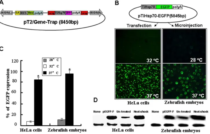
Although there are several versions of transposon-based gene trapping vectors that are used in various vertebrate systems [53,54,55,56], there is room for improvement to increase gene tag and mutation efficiency. Accordingly, we constructed a novel trapping vector pT2/Gene-Trap (Figure 1A) that is mediated by the SB transposon system. This vector contains two functional cassettes necessary for improving the efficiency of gene trapping and mutagenesis. In the improved efficiency cassette, expression of SB11 transposase gene was driven by aHsp70promoter from the tilapia genome (TiHsp70) [57], which can be activated at 37uC heat treatment. The inducible expression of SB11 conditionally controls the remobilization of integrated trapping cassettes to new target sites in a genome and reduces the cytotoxic effects ofSB
transposase on cells and tissues [50]. In the mutation cassette, a SA signal essential for the proper splice of the carpb-actinexon1 and exon2 [58] was inserted upstream of the IRES-Reporter gene such as a neomycin (Neo) or an enhanced green fluorescence protein (EGFP), which is derived from a commercial vector pIRES2-EGFP. Three stop codons required for different reading frames (TGA ATT AGT GA) were introduced following the SA signal to efficiently truncate the translation of trapped endogenous gene. The utilization of an EMCV/IRES element from Clontech can independently initiate the translation of Neo/EGFP gene once an endogenous gene is trapped and a fusion transcript is formed. This strong splice acceptor signal combined with the polyadenylation signal and transcriptional termination element in our gene-trap vector disrupted the transcription of trapped endogenous genes efficiently.
Inducible Activity of the TilapiaHSP70Promoter
To determine the response of tilapiaHSP70promoter to a mild heat shock at 37uC, SB11 gene in the improved efficiency cassette was substituted by an EGFP to generate a pTiHsp70-EGFP plasmid (Figure 1B). HeLa cells growing at 32uC were transfected with the pTiHsp70-EGFP. As shown in Figure 1B and 1C, strong fluorescent signals were found in 84% of transfected cells after heat induction for 1 h at 37uC and recovery at 32uC for another 2 h. In contrast, weak signals were seen in about 5% of transfected cells growing at 32uC. To test the in vivo activity of tilapia Hsp70
promoter, zebrafish embryos at one-cell stage were microinjected with pTiHsp70-EGFP and strong EGFP expression was found in 95% of embryos after heat induction for 1 h at 27 hpf, while weak EGFP signals were found in about 10% of non-induced embryos.Western blot analysis was further performed to detect EGFP in both heat shock-treated and un-treated cells (transfected with EGFP) and embryos (injected with pTiHsp70-EGFP). As shown in Figure 1D, the expression of EGFP was greatly increased in heat shock-treated cells and embryos, while it was subtle in both un-treated cells and embryos (Figure 1D), indicating that tilapia Hsp70 promoter is of minimal activity in both cultured cells and zebrafish embryos without heat shock. These data indicate that the tilapiaHsp70promoter is sensitive to the mild heat induction at 37uC and is suitable for the conditional control of gene expressionin vitroandin vivo.
Activity of the Mutation Cassette in an Intron
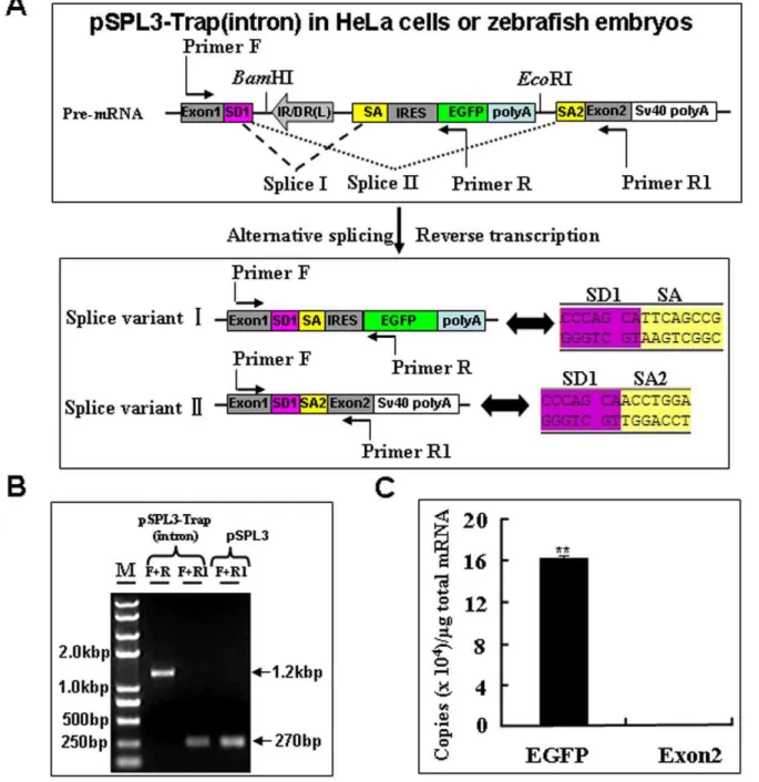
The success of gene trapping mainly depends on whether the trap cassette is landed in an endogenous gene or not. The activity of the mutation cassette was examined by subcloning it into the intron of pSPL3 vector, which is originally designed to search genomic DNA for potential exon sequences [59]. As shown in
Figure 2, a BamHI/EcoRI fragment containing the mutation cassette in Figure 1A was subcloned into the multiple cloning sites (MCS) of pSPL3 to generate a pSPL3-Trap(intron) vector, which was then used for HeLa cells transfection and zebrafish embryo microinjection. Images were taken under a fluorescence micro-scope after transfection for 24 h and 88% of transfected cells displayed strong EGFP expression. Similarly, strong EGFP expression was found in 92% (n = 1425) of pSPL3-Trap(intron)-injected embryos at 24 hpf.
Since the splice acceptor in gene trap vectors is essential for the formation of fusion transcripts to disrupt the expression of trapped genes, we examined further whether the EGFP expression in HeLa cells and developing embryos (Figure 2) resulted from the proper fusion of EGFP transcript to its upstream exon in the pSPL3. Total mRNA was isolated from HeLa cells transfected with the pSPL3-Trap(intron) vector. Two potential transcript variants I and II, and Sequencing trace files representing the splicing of SD1 with SA and SA1 were shown, respectively (Figure 3A) were analyzed using reverse transcription PCR (RT-PCR). As shown in Figure 3B, a 1.2-kb band was always shown in RT-PCR products of pSPL3-Trap(intron)-transfected HeLa cells
and a 270 bp band was occasionally detected by RT-PCR from the same sample. Sequencing results indicate that the 1.2-kb band represents the splice variant I containing exon1, IRES and partial EGFP, and that the 270 bp band is derived from the proper splicing of exon1 with exon2 in pSPL3. Similar results were obtained in zebrafish embryos injected with the pSPL3-Trap(in-tron) or pSPL3 vector (data not shown).Therefore, the SA signal in our trap vector is able to efficiently direct the proper splicing of reporter gene with an upstream exon in a trapped gene.
To examine further the efficiency of the mutation cassette in disruption of the expression of trapped genes, absolute quantitative real-time PCR assays (qRT-PCR) were performed to determine the copy numbers of transcripts including EGFP and exon2 after reverse transcription of total mRNA from pSPL3-Trap(intron)-transfected HeLa cells [60,61,62]. As shown in Figure 3C, the copy numbers of EGFP transcripts is about 1000 times higher than that of exon2 transcript. Similar results were obtained from zebrafish embryos injected with the pSPL3-Trap(intron) vector (data not shown). These data indicate that the mutation cassette in our gene trap vector is able to efficiently disrupt the expression of trapped gene when inserted into an intron.
Figure 1. Inducible activity of the tilapiaHsp70promoter (TiHsp70) at 37uC.(A) A novel gene-trap vector mediated bySleeping Beauty
transposon. IR/DR(L) and IR/DR(R), left and right inverted repeat/directed repeat of the SB transposon; SA, splice acceptor; IRES, internal ribosome entry site; Neo, kanamycin resistance gene; poly(A), poly(A) signal; TiHsp70, tilapiaHsp70promoter; SB11, SB11 transposase gene. (B) EGFP was used to monitor the inducible activity of TiHsp70 in vitroandin vivo.HeLa cells were transfected with pTiHsp70-EGFP at the density of about 80% confluence, treated in medium at 37uC for 1 h and recovered at 32uC for 2 h after transfection. Images were taken under a Nikon TE2000 fluorescent microscope and cell numbers in three fields of view were counted. Zebrafish embryos at one-cell stage were microinjected with pTiHsp70-EGFP. Injected embryos at 24 hpf were incubated in rearing water at 37uC for 1 h and then recovered at 28uC for 2 h. Low magnification fluorescent imaging of zebrafish embryos was performed on a SteReo Lumar V12 microscope form Zeiss and total embryos in three dishes were counted. (C) Statistical analysis of EGFP-expressing cells or embryos in (B). Data are given as means6standard Deviation (n = 3). * indicateP,0.05 versus the corresponding control. (D) Western blot analysis of EGFP in HeLa cells and zebrafish embryos. Heatshock-treated and -untreated cells (transfected with pTiHsp70-EGFP) and embryos (injected with pTiHsp70-EGFP) samples were undergone western blot and the pEGFP-F plasmid was used as a positive control. The expression of reference geneb-actinwas also analyzed in these samples.
Activity of the Mutation Cassette in an Exon
To examine the activity of the mutation cassette after its integration into an exon of trapped genes, an exon from the carp b-actingene [58] was subcloned into the MCS of pSPL3 vector to generate a pSPL3-E3 vector (Figure 4). The mutation cassette was introduced at theBamH I/EcoR I site in the exon of pSPL3-E3 to obtain the pSPL3-E3/Trap(exon), which was then used for HeLa cell transfection and zebrafish embryo microinjection. Images were taken under a fluorescence microscope after transfection for 24 h and 85% of transfected cells displayed strong EGFP expression. Similarly, strong EGFP expression was seen in about
90% of embryos injected with the pSPL3-E3/Trap(exon) vector. To investigate whether the EGFP expression resulted from the proper activity of the carp b-actin SA signal, total mRNA was isolated from pSPL3-E3/Trap(exon)-transfected HeLa cells and three potential transcript variants (I+III, II+III and IV) in Figure 5A were analyzed using RT-PCR. As shown in Figure 5B, two bands (1950 bp and 1500 bp) were amplified using the primer pair (F+R) and three bands (2421 bp, 1971 bp and 270 bp) using the primer pair (F+R1) from the cDNAs of pSPL3-E3/ Trap(exon)-transfected HeLa cells. In addition, two PCR products (411 bp and 270 bp) were obtained from the pSPL3-E3-transfect-Figure 2. Activity of the trapping cassette in an intron of pSPL3 vector.The trapping cassette was subcloned into theBamHI/EcoRI site of pSPL3 vector to generate the pSPL3-Trap(intron), which was used for transient transfection of HeLa cells at 80% confluence. Images were taken under a Nikon TE2000 fluorescent microscope at 48 h after transfection and cell numbers from three independent transfections were counted. Zebrafish embryos at one-cell stage were microinjected with the pSPL3-Trap(intron). Injected embryos at 24 hpf were imaged under a SteReo Lumar V12 microscope form Zeiss and total embryos in three dishes were counted. The ectopic expression of EGFP in one embryo was enlarged and shown in a merged image. SD1, splice donor for exon1; IR/DR(L) and IR/DR(R), left and right inverted repeat/directed repeat of the SB transposon; SA, splice acceptor; IRES, internal ribosome entry site; EGFP, enhanced green fluorescence protein gene; poly(A), poly(A) signal; SA2, splice acceptor for exon2. doi:10.1371/journal.pone.0044123.g002
ed HeLa cells. Sequencing data indicate that the 2421 bp and 1950 bp bands are derived from the fusion splice variant I +III, the 1971 bp and 1500 bp bands from the fusion transcript II+III, the 411 bp band from the proper splicing of exon1, exon3 and exon2 in pSPL3-E3, and the 270 bp band from the proper splicing of exon1 and exon2 in pSPL3 and pSPL3-E3. Similar results were obtained from zebrafish embryos injected with pSPL3-E3/
Trap(exon) and pSPL3-E3 vectors (data not shown). Thus, the SA signal in our trap vector can wield the proper splicing of reporter gene with an upstream splice donor signal (SD) in a trapped endogenous gene. In addition, the insertion of the trap cassette completely abolished the proper expression of trapped exons.
Figure 3. Transcriptional analysis of the trapping cassette in an intron of pSPL3 vector.(A) Potential splice variants I and II from the pSPL3-Trap(intron). Sequencing trace files representing the splicing of SD1 with SA and SA1 were shown, respectively. SD1 aSD1, splice donor for exon1; IR/DR(L) and IR/DR(R), left and right inverted repeat/directed repeat of the SB transposon; SA, splice acceptor; IRES, internal ribosome entry site; EGFP, enhanced green fluorescence protein gene; poly(A), poly(A) signal; SA2, splice acceptor for exon2. (B) RT-PCR analysis of transcripts from pSPL3-Trap(intron)-transfected HeLa cells. Sequencing results indicate the 1.2-kb band is derived from the splice variant I, which contains exon1, IRES and EGFP, and the 270 bp band results from splice variant II, which represents the proper spicing of exon1 with exon2 in pSPL3. (C) The absolute quantification of cDNA using real-time PCR was employed to determine the copy numbers of transcripts including EGFP (F+R, E = 96.1%, R2= 0.9981) and exon2 (F+R1, E = 97.2%, R2= 0.9989). Data are given as means6standard deviation (n = 3). ** indicateP,0.01 versus the exon2 expression level in pSPL3-Trap(intron)-transfected cells.
To detect further whether the mutation cassette can disrupt the expression of its downstream exon in trapped gene, absolute qRT-PCR was performed to determine the copy numbers of distinct transcripts in pSPL3-E3/Trap(exon)-transfected HeLa cells [60,61,62]. As shown in Figure 5C, the copy numbers of EGFP transcript is about five times higher than that of exon2 transcript, suggesting that insertion of the mutant cassette in an exon markedly inhibited the expression of downstream exons. Similar results were obtained from zebrafish embryos injected with the pSPL3-E3/Trap(exon) vector (data not shown).
Taken together, these data indicate that the mutation cassette in our gene trap vector is able to efficiently knockdown the
expression of trapped genes by direct disruption of trapped exon and inhibition of downstream exon splicing.
An IRES Element is Required for Independent Expression of Reporter Gene
Although the splice acceptor signal plays an important role in disrupting endogenous gene expression, an IRES element is indispensable to the selective/reporter gene expression. IRES-based vectors are able to capture a wide range of genes expressed in a variety of tissues and embryos at different developmental stages [52]. The ECMV/IRES has been shown to function in developing zebrafish [63]. To determine the efficiency of the ECMV/IRES in driving the expression of reporter gene in our Figure 4. Activity of the trapping cassette in an exon of pSPL3-E3 vector.pSPL3-E3 was generated by insertion of an exon from carp beta-actin gene (Exon3). The trapping cassette was then sucloned into theBamHI/EcoRI site of pSPL3-E3 vector to generate the pSPL3-E3/Trap(exon), which was used for transient transfection of HeLa cells at 80% confluence. Images were taken under a Nikon TE2000 fluorescent microscope at 48 h after transfection and cell numbers in three independent transfections were counted. Zebrafish embryos at one-cell stage were microinjected with the pSPL3-E3/Trap(exon). Injected embryos at 24 hpf were imaged under a SteReo Lumar V12 microscope form Zeiss and total embryos in three dishes were counted. The ectopic expression of EGFP in one embryo was enlarged and shown in a merged image. SD1, splice donor for exon1; SA3, splice acceptor for exon3; IR/DR(L) and IR/DR(R), left and right inverted repeat/directed repeat of the SB transposon; SA, splice acceptor; IRES, internal ribosome entry site; EGFP, enhanced green fluorescence protein gene; poly(A), poly(A) signal; SD3, splice donor for exon3; SA2, splice acceptor for exon2.
doi:10.1371/journal.pone.0044123.g004
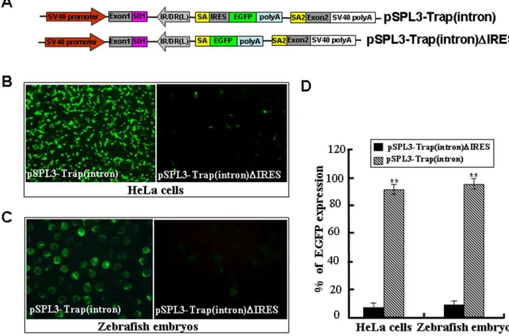
gene trap vector, we deleted the ECMV/IRES element from the pSPL3-Trap(intron) vector to generate the pSPL3-Trap(intron)-DIRES (Figure 6A). Then, the Trap(intron) or pSPL3-Trap(intron)DIRES were used for HeLa cell transfection and zebrafish embryo microinjection. As shown in Figure 6B-D, weak GFP signal was found in less than 10% of HeLa cells or embryos introduced with pSPL3-Trap(intron)DIRES, but strong GFP expression was seen in more than 90% of HeLa cells or embryos
carrying the pSPL3-Trap(intron). Similarly, deletion of the ECMV/IRES of pSPL3-E3/Trap(exon) in Figure 4 markedly reduced the ratio of EGFP-expressing HeLa cells or embryos. These data demonstrate that the ECMV/IRES element in our trap vector works well for independent expression of reporter gene once a gene is trapped and a fusion transcript formed.
Analysis of Chromosomal Integration Sites
To examine the gene structure at a locus of trapped chromosomes, a modified splinkerette PCR approach was used to amplify genomic DNA fragments adjacent to integrated trapping cassettes from G418-selected individual cell colonies (Figure S2). Eighteen transposition events were obtained from distinct cell colonies (Table 1). Blasting the human genome in the NCBI and ENSEMBL database with these junction sequences indicated that seventeen of them landed in an intron and one of them integrated in an exon at active genomic loci (Table 1). These data are in consistence with the fact that exons and introns comprise 1.5% and 24% of human genome, respectively [64]. Thus, our gene trap vector appears to insert into endogenous genes and the reporter Neo gene are properly expressed to maintain the survival of HeLa cells in medium containing G418. The success of an insertional mutagenesis approach mainly depends on whether the expression of endogenous genes is efficiently disrupted. Previous studies have shown that insertion of foreign DNA into most locations on a vertebrate genome has little or no effect on any gene or gene product [28,29]. To examine the effect of our trap vector on disruption of endogenous gene expression at the insertion site, RT-PCR was performed with
mRNA samples isolated from four cell colonies growing in medium containing G418 (Figure S2). One trapped gene locus in each cell colony was selected and analyzed using a pair of primers (F+R or F+R1) in Figure 7A. As shown in Figure 7B, a small DNA fragment (100–250 bp) was detected in normal HeLa cells (N/ET) and in each of cell colonies (C1/ET to C4/ET), while a large DNA fragment (,1-kb) was only obtained from each of
colonies (C1/FT to C4/FT). Sequencing results indicate that the small fragments represent the endogenous transcript ofINPP5B,
HERC2,TRIOorCPZand the large fragments are derived from the fusion transcript ofINPP5B,HERC2,TRIOorCPZ with the Neo gene. These data indicate that the reporter gene in our gene trap vector is correctly spliced to the trapped endogenous gene in each of these cell colonies and that the trapped gene in each cell colony is heterozygotic at the insertion site.
To further examine whether the expression of four trapped genes was affected by the insertion of a trap cassette, qRT-PCR was conducted by using mRNA samples from normal HeLa cells (N) and cell colonies (C1–C4). The expression levels of these four trapped genes reduced to 30%–45% of those in normal HeLa cells and was significantly lower than those in normal Hela cells and other cell colonies (P,0.01 or ,0.05 in all cases) (Figure 7C). Figure 6. An IRES sequence is required for independent expression of the reporter gene.(A) The ECMV/IRES element in the pSPL3-Trap(intron) vector was deleted to generate the pSPL3-pSPL3-Trap(intron)DIRES. SD1, splice donor for exon1; IR/DR(L) and IR/DR(R), left and right inverted repeat/directed repeat of the SB transposon; SA, splice acceptor; IRES, internal ribosome entry site; EGFP, enhanced green fluorescence protein gene; poly(A), poly(A) signal; SA2, splice acceptor for exon2. (B) pSPL3-Trap(intron) and pSPL3-Trap(intron)DIRES constructs were transfected into HeLa cells at 80% confluence, respectively. Images were taken under a Nikon TE2000 fluorescent microscope at 48 h after transfection and cell numbers in three independent transfections were counted. (C) Zebrafish embryos at one-cell stage were microinjected with the pSPL3-E3/Trap(exon). Injected embryos at 24 hpf were imaged under a SteReo Lumar V12 microscope form Zeiss and total embryos in three dishes were counted. (D) Statistical analysis of EGFP-expressing cells in (B) or embryos in (C). Each construct was tested three times and each experiment was done in triplicate. Data are given as means6standard Deviation. ** indicateP,0.01 versus the corresponding control.
doi:10.1371/journal.pone.0044123.g006
Therefore, ourSB-based gene trapping system is suitable for the monitor and interruption of gene expression and functions.
Remobilization of Gene Trap Cassettes
Theoretically, two approaches can be used to improve the efficiency ofSB transposon-based gene trapping: 1) increase the numbers of gene trap cassettes in a target genome; 2) allow the remobilization of integrated trap cassettes to new genomic sites [51]. Since less than 10 copies of transposons are usually found in the genome of transgenic animals, we tried to improve the gene trapping efficiency by inducting the remobilization of integrated trap cassettes in individual cell colony. Five cell colonies named
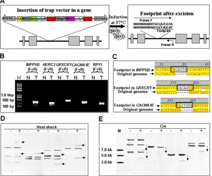
INPP5B,HERC2,GRXCR1,CACNA1EandRP11growing at 32uC (N) were induced at 37uC (T) for at least 48 hours to allow the expression of SB11 transposases. Total genomic DNA of cells was isolated and subjected to an excision assay as described previously [65,66], using a reverse primer (R) against the target genomic DNA and a forward primer (F) containing the typicalSBfootprints TAC(A/T)GTA at its 39-terminus and the target genome sequence (Figure 8A). The excision of a SB transposon from its original integration site gave rise to a PCR product in cells at 37uC, but not in cells at 32uC (Figure 8B). Sequencing results indicate that all of PCR products from the excision sites contain a typical TA-flanked footprint sequence TAC[A/T]GTA (Figure 8C), which is consistent with the activity ofSB-mediated DNA cleavage [66]. The remobilization of trap inserts was further analyzed with southern blotting. As shown in Figure 8D, DNA bands representing new trap inserts were detected from digested genome of colonies named INPP5B, HERC2, GRXCR1and
CACNA1E after heat shock at 37uC for 48 h and recovery at 32uC for 5 days, indicating that the integrated gene-trap inserts are excised from the original integration site and new integration patterns have happened after the heat-induced expression of SB11
transposase. These findings suggest that the induced remobiliza-tion of trap inserts in a target genome would provide new opportunities for excised trap inserts to reintegrate into other coding regions. The relocation of trap inserts from non-coding regions would significantly improve the efficiency of our gene-trapping vector in animal models.
To avoid the bio-safety concerns and the remobilization of trap inserts that may lead to the normal expression of trapped genes, the Cre recombinase was transiently expressed in G418-selected cell colonies named INPP5B, CACNA1E, RP11and HERC2 to delete the LoxP-TiHsp70-SB11-poly(A)-LoxP cassette in our gene-trap vector. As shown in Figure 8E, southern hybridization with the Neo probe indicated the proper reduction in size of DNA inserts after transient expression of Cre recombinase. These data suggest that the SB11 cassette can be conditionally deleted to stabilize the phenotypes of mutants.
The Activities of pT2/Gene-Trap in Transgenic Zebrafish A stable transgenic zebrafish line was established and splinker-ette PCR was performed to detect the transposable events in F1 genome of this line. As shown in Figure 9A, themamdc2a gene consisting of fourteen exons and thirteen introns was mutated by a canonical transposable element from pT2/Gene-Trap and the Gene-Trap cassette has landed in the intron7 ofmamdc2a gene. The integrity of the gene-trap cassette at the insertion site was then determined by PCR with different primers (Figure 9B). Next, RT-PCR analysis was conducted with mRNA isolated from wild-type (+/+), heterozygous (+/2) and homozygous (2/2) F2 embryos. The endogenous transcripts of mamdc2a gene were detected in wild-type and heterozygous but not in homozygous embryos, while the fusion transcripts of EGFP with exon7 ofmamdc2agene were found in heterozygous and homozygous but not in wild-type embryo (Figure 9C). The fusion transcript derived from EGFP and Table 1.Trapped endogenous genes from G418-resistant HeLa cells.
Junction sequences Gene name Accession number Chromosomename
Exon
number Insertion sites
gtgtctctcctatcTAcagttgaag IPNN5B NM_005540.2 Chr:1 18 intron1
ttggattttatacaTAcagttgaag APAF1 NG_029094.1 Chr:12 27 intron16
aaactctttacataTAcagttgaag HERC2 NM_004667.4 Chr:15 93 intron66
ccctttttgttaacTAcagttgaag MPP2 NM_005374.3 Chr:17 13 intron3
tctctttcacaaacTAcagttgaag TRIO NT_006576.16 Chr:5 57 intron2
tgacagaacatagaTAcagttgaag C4orf32 NM_152400.2 Chr: 4 2 intron1
atacatcttatagtTAcagttgaag ABCB11 NG_007374.1 Chr: 2 15 intron5
gcacccactgtacaTAcagttgaag CPZ NM_001014447.2 Chr:4 11 intron3
tccaacaccacatcTAcagttgaag CACNA1E NM_001205293.1 Chr:1 48 intron6
agaatattctgcatTAcagttgaag RP11-553P9.3 NT_016354.19 Chr:4 59UTR
gttccttcccaaccTAcagttgaag GRXCR1 NG_027718.1 Chr:4 4 intron1
tagcattggggagcTAcagttgaag U6snRNA NW_001838848.1 Chr: 2 N/A N/A
tttataatgacttaTAcagttgaag TMEM26 NM_178505.6 Chr:10 7 intron1
tgaggggaaaaataTAcagttgaag TPST2 NM_001008566.1 Chr:22 7 exon7
tactatcttgttacTAcagttgaag IMMT NM_006839.2 Chr:2 15 intron3
tctaagaattcaccTAcagttgaag RPL2I NT_024524.14 Chr:13 6 intron1
ttcagctgagtacaTAcagttgaag Unknown AL139023.6 Chr:14 N/A N/A
taaaatcaatcttaTAcagttgaag DENNEB NM_144977.4 Chr: 1 20 intron17
Junction sequences were obtained by the splinkerette PCR. Partial trap cassette sequences in italic were shown on the right of TA and partial genome sequences in regular on the left of TA.
exon7 of gene mamdc2a was confirmed by DNA sequencing (Figure 9D). Additionally, qRT-PCR was performed with primers E7F/E8R and mRNA isolated from wild-type, heterozygous and homozygous embryos at 72 hpf. Themamdc2atranscriptional levels in heterozygous and homozygous were significantly lower than that in wild-type (Figure 9E).
Apart from the molecular analysis of the trapped gene, the remobilization of gene-trap cassette from the intron7 ofmamdc2a
gene was tested after heat shock for 3 h of F2 embryos from the intercross ofmamdc2a-transgenic F1 fish. As shown in Figure 9F, the SB11 expression in heat-treated F2 embryos was significantly higher than that in non-treated embryos; however, we didn’t find any novel inserts in heat-treated embryos by southern blot analysis due to the short-term treatment that may not allow the generation of detectable new integration events (data not shown). Moreover, the capped Cre reombinase mRNA was microinjected into wild-type, heterozygous and homozygous embryos to determine whether the Cre/Loxp system in our trap vector can be excised for the stabilization of a disrupted endogenous gene. As shown in Figure 9G, ectopic expression of Cre recombinase in developing F1 and F2 embryos at 72 hpf led to the deletion of TiHsp-SB11
cassette from its original insertion site and the generation of smaller hybridization bands in heterozygous and homozygous embryos.
Discussion
A gene trap cassette generally consists of an upstream splice acceptor, a promoterless reporter or selectable marker and a downstream polyadenylation signal. When inserted into an intron of expressed genes, the trap cassette is transcribed from the promoter of trapped genes and is spliced to form a fusion transcript containing endogenous gene exon(s) upstream of the insertion site and the reporter [21]. Since the transcription is terminated prematurely at the inserted polyadenylation site, the translation of fusion transcript will give rise to a truncated and nonfunctional version of cellular protein and the reporter. Thus, the success of a gene trap cassette allows disrupting and recapitulating the expression of trapped genes and provides a DNA tag for rapid identification of disrupted endogenous genes. In this study, we have developed a novel gene-trap system that proved to be very effective in disrupting gene expression in HeLa Figure 7. Integration of the SB-based gene trap efficiently disrupts the expression endogenous genes in HeLa cells.(A) Schematic representation of a gene trapping insertion in an endogenous gene and potential transcripts in Hela cells. Endogenous exons are boxed and arrows indicate the positions of primers used for transcript analysis. IR/DR(L) and IR/DR(R), left and right inverted repeat/directed repeat of the SB transposon; SA, splice acceptor; IRES, internal ribosome entry site; Neo, kanamycin resistance gene; poly(A), poly(A) signal; TiHSP70, tilapiaHsp70
promotor; SB11, SB11 transposase gene. (B) RT-PCR analysis of transcripts from a trapped endogenous gene (INPP5B,HERC2,TRIOorCPZ) in four cell colonies. N: Normal HeLa cells; C: cell colonies; ET: Endogenous transcript; FT: Fusion transcript. TheGAPDHis used as the control for equal amount of cDNA template in PCR reactions. (C) Assessing the mutagenicity of gene-trap insertions by qRT-PCR. Total RNA was isolated from each cell colony and subjected to qRT-PCR analysis. The mRNA expression levels of an endogenous gene in the normal Hela cells and two other colonies without insertion at the gene loci (the controls) were compared to that in a cell colony containing an insertion at the corresponding gene loci. N represent normal HeLa cells; C1 representsINPP5Bgene; C2 representsHERC2gene; C3 representsTRIOgene; C4 representsCPZgene. Data are given as Means6Standard Deviation (n$3). ** and * indicateP,0.01 andP,0.05 versus the controls, respectively.
doi:10.1371/journal.pone.0044123.g007
cells and transgenic zebrafish. This gene-trap vector is composed of a mutation cassette as well as a temperature inducible cassette used for improved efficiency, which is mediated by our newly developed SB transposon and SB11 transposase [34,67]. In the mutation cassette, the modified splice acceptor signal from the carp b-actin gene [58] can properly direct the splice of a downstream reporter gene with an upstream exon at the insertion site. The IRES element is able to initiate the translation of a downstream reporter gene effectively. The normal splicing and expression of trapped endogenous genes in HeLa cells and
developing zebrafish embryos are efficiently disrupted by the combination of all elements in the mutation cassette. In the temperature inducible cassette, theHsp70 promoter from tilapia genome [57] is used to drive the expression of SB11 transposase in an inducible manner, which could make the remobilization of integrated trap cassettes for the improvement of trapping efficiency and reduce the overproduction inhibition of theSBsystem. Thus, ourSB-based gene trap system appears to be suitable for efficient analysis of expression patterns and functions of genes in vertebrates.
Figure 8. Remobilization of trap inserts in the HeLa cell genome.(A) A schematic diagram of trap cassette remobilization and a canonical footprint left in the original insertion site. Arrows indicate the primer containing the footprint (F) and a gene-specific primer (R). IR/DR(L) and IR/DR(R), left and right inverted repeat/directed repeat of the SB transposon; SA, splice acceptor; IRES, internal ribosome entry site; Neo, kanamycin resistance gene; poly(A), poly(A) signal; TiHSP70, tilapiaHsp70promoter; SB11, SB11 transposase gene. (B) Individual cell colony containing a trap insertion at shown gene loci (INPP5B,HERC2,GRXCR1,CACNA1EorRP11) was cultured at 32uC in two 35 mm dishes wells. Cells on one dish (T) was subjected to heat induction at 37uC and cells on another dish (N) was kept at 32uC. Total genome DNA was isolated and subjected to PCR analysis using gene-specific primers (F+R) in Table S1. (C) Sequencing trace files of independent remobilization events in cell colonies. These sequencing trace files representing independent remobilization event in genes namedINPP5B,GRXCR1andCACNA1E.After the excision and remobilization of the gene-trap vector from the original insertion genome locus, footprint was generated as shown in bold box. (D) Southern hybridizations. Neo probes were used to detect the copy number of transposons in the genome of HeLa cells incubated at 32uC (-) and the gene-trap cassettes excised from their insertion sites after heat shock (+) at 37uC for 48 h and recovered at 32uC for 5 days. Arrows point to newly-generated inserts after heat shock. (E) Southern blotting analysis with Neo probes indicated that the size of genomic DNA fragments with the trap cassette (dashed arrows) reduced by the transient expression of the Cre recombinase (+).
Transposon-based gene trap vectors have become indispensable tools for insertional mutagenesis in model vertebrates. However, most of gene trap systems exhibit a relatively low efficiency because of the following reasons. First, the activity of functional elements in most of conventional trap vectors is not carefully examined so it is not sure whether an endogenous gene is disrupted or not even if a gene is trapped. Tissue- or cell-specific expression of reporter gene was detected in the first effort onSB -mediated gene trapping in zebrafish, but no mutants were finally obtained [28].Tol2-mediated gene trapping was successfully used to trap endogenous genes; however, the insertion of trapping vector failed to abolish the transcription of trapped genes [29]. A plausible work was performed to generate molecularly null mutations in both larval and adult of zebrafish by artificially testing functional cassettes in a gene breaking vector [49]. Second,
less than 10 copies of transposons are usually found in a target genome [28,29], so the performance of targeting a functional gene by low copy of gene trap cassettes remains quite difficult. The remobilization of integrated transposons can generate new insertions and mutations because transposon reinsertions tend to occur around the original insertion sites. Conditionally gene expression and insertional mutagenesis mediated bySBtransposon had been performed in mouse with the doxycycline-repressible Tet-Off (tTA) system or Cre/Loxp system [51,68], in which the transposase was conditionally supplied by another transgenic line that seems to be effective but laborious and less economical. In addition, Tol2 elements were induced to remobilize from their original insertion sites and thus new mutants were generated in zebrafish [69] and Xenopus [70]. In this study, this strategy was successfully employed to induce the remobilization of integrated Figure 9. Molecular analysis of the trapped genemamdc2ain transgenic zebrafish.(A) Schematic representation of the insertion of gene trap cassette in the intron7 of gene. The locations of primers used for molecular analysis are indicated. (B) Integrity analysis of gene trap cassette in transgenic fish. Genomic DNA was extracted from the trail of transgenic fish and subjected to PCR analysis with primes indicated in (A). (C) RT-PCR analysis of mamdc2a transcript in wild-type (+/+), heterozygous (+/2) and homozygous (2/2). Primers E7F/E8R were used to amplify the endogenous transcript and primers E7F/R3 were used to amplify the exon7-EGFP fusion transcript. Theb-actintranscript was amplified as an internal control. (D) The fusion transcript generated from the proper splicing of EGFP gene and its upstream exon inmamdc2agene. Sequences in red represent exon7 ofmamdc2agene. Sequences in yellow represent the SA signal. Sequences in gray indicate the IRES followed by the partial EGFP sequence in green. (E) qRT-PCR was performed with primers E7F/E8R to determine the expression of endogenousmamdc2agene in wild-type, heterozygous and homozygous embryos at 72 hpf. Themamdc2alevels were normalized to theb-actinmRNA levels. (F) qRT-PCR analysis of SB11 expression in 24 hpf embryos frommamdc2a-transgenic line after heat shock at 37uC for 3 h and recovery at 28uC for 2 h. (G) Detection of Cre-mediated TiHsp-SB11 excision by southern hybridization. Cre reombinase mRNA was microinjected into wild-type, heterozygous and homozygous embryos. ‘‘+’’ represents the presence of Cre recombinase and ‘‘2’’ represents the absence of Cre recombinase. Line1–2: DNA from wild-type. Line 3– 4: DNA from heterozygous. Line 5–6: DNA from homozygous.
doi:10.1371/journal.pone.0044123.g009
SBtranposons in HeLa cells. Last but not the least, many gene-trap vectors include a SA element in front of a reporter/selective marker, in which the reporter/selective marker can only be expressed correctly in case the trap vector insertion was just in correct frame with the endogenous gene’s open reading frame which means there is only one third chance of the reporter/ selective gene expression to represent a successfully gene-trapping event. To circumvent this problem, we introduced the IRES element in front of the reporter/selective marker gene, which have been shown greatly improve the number of detectable gene trap events [71]. Thus, several problems facing most of conventional gene trap vectors are largely solved by our gene trapping system, so this gene trap vector could serve as an alternative tool for the insertional mutagenesis in zebrafish and higher vertebrates.
During the past two decades, various gene trap vectors are developed and successfully used for creating libraries of embryonic stem cell lines that harbor mutations in a single gene and can be used for making mice [72]. Presently, approximately 70% of the protein-coding genes in the mouse genome have been disrupted by gene trap insertions [73]. However, the achievement of saturation mutagenesis in a target genome via current transposon-based gene trapping systems remains difficult due to their insertion site preferences and local hopping. The insertion of most transposons in a target genome is nonrandom because of their characteristic preferences for insertion sites at the primary DNA sequence level. For example, the Harbinger3_DR transposon preferentially inserts into a 15-bp consensus sequence AAACACCWGGTCTTT [74], the PiggyBac transposon targets the tetranucleotide sequence TTAA, and all of known Tc1/mariner transposons, including
SB, Frog Prince, Minos and Hsmar1, prefer to integrate into the TA dinucleotides [46]. By contrast, the Tol2 element does not appear to have a pronounced insertion preference for any primary DNA sequence [40]. In addition, integration of some transposons exhibits hotspots and cold regions on the target chromosomes. For instances, the PiggyBac demonstrates a higher preference for integrations in regions surrounding transcriptional start sites and within long terminal repeat elements [75], and theTol2transposon shows a pronounced preference for integration close to transcrip-tional start sites [40]. By contrast, Tc1/mariner elements exhibit no or weak preference for transcription units [46]. Additionally, one-fourth of the SB trap insertions were found to insert in transcriptional units, a rate that is commensurate with random integration [28]. In this study, we demonstrate that the SB -mediated gene trap system seems to randomly integrate into introns and exons of target genes. Moreover, local hopping, a phenomenon of chromosomal transposition in which transposons have a preference for landing into cis-linked sites in the vicinity of the donor locus, limits the chromosomal regions accessible to a transposon jumping out of a given chromosomal site, however it may be useful for saturation mutagenesis [76], which appears to be a shared feature of cut-and-paste transposons. It has been shown that the majority (83%) of Tol2 reinsertions are mapped on chromosomes other than the transposon donor chromosomes and that 9% of local hopping events mapped less than 300 kb away from the donor loci [69]. TheSBtranposon seems to have a much larger local transposition interval between 5 and 40 Mb [54,77]. Therefore, the low transposition activity and diverse insertion site preferences of available transposon systems need to be carefully considered before construction of efficient transposon-based trapping vectors for large-scale mutagenesis.
Another limitation of conventional gene trap systems is how to capture the low-level expressed endogenous genes. To address this problem, a novel vector was recently developed to facilitate the recovery of poorly expressed genes in mouse embryonic stem cells
by insertion of an osteopontin enhancer into several conventional gene trap vectors [78]. This strategy would be useful for us to improve our trap system for the genome-wide mutagenesis, and ourSB-based gene trap system can be further optimized by the combination with Gal4/UAS system or Tet-on system to trap endogenous genes expressed at very low levels. In addition, several poly(A) trap systems have been developed to decipher endogenous genes regardless of their expression patterns and levels [79]. However, most of poly(A) trap systems are able to trigger the nonsense-mediated mRNA decay (NMD) [80] and thus fail to disrupt the expression of endogenous genes at the 59end. We are developing anotherSB-based poly(A) trapping system, in which the NMD can be reduced using strategies from other trapping vectors [81]. In conclusion, ourSB-based gene-trap vector can be used as an alternative tool for large-scale mutagenesis in cells and vertebrates and could proved to be an ideal platform for further development of highly active trapping vectors.
Materials and Methods
Ethics Statement
The animal protocol for this research was approved by the Animal Care and Use Committee of Hubei Province in China and by the Institutional Animal Care and Use Committee of Institute of Hydrobiology (Approval ID: Keshuizhuan 0829).
Plasmids
Our gene-trap vector pT2/Gene-Trap has been designed to efficiently tag and break genes (Fig.1A, GenBank accession number: BankIt1516608 Seq1 JQ692169). These cassettes were sequentially subcloned into the second generation ofSB transpo-son pT2/HB. An exon-trapping plasmid pSPL3 was utilized for artificially testing the transcriptional and splicing activities of gene trapping cassettes. TheTiHsp70promoter was obtained from the tilapia genome using primers 59-CTT GCT AGC GAG CTC ACC GCG AGC ACT CTG-39and 59- GCA CCG GTC TTG ATT GCT TTG ACT TCG-39 [57], then inserted at theNheI/
AgeI site upstream of SB11 transposase gene. The SA sequences from carp b-actin gene were amplified using primers 59- CTT GCT AGC GAT TGC AGC ACG AAA CAGG-39and 59- ATG ACG TCG GTA TAC GTA CGT CACTAA TTC-39 with the incorporation of stop codes (TGA ATT AGT GA) for three different read frames in the exon sequence. The IRES element in our trap vector was amplified from the pIRES2-EGFP (Clontech) vector and was used to mediate the reporter translation independently. Sequencing data indicate that all of these components were correctly inserted into the pT2/HB vector.
Cells and Zebrafish Embryos
HeLa cells (ATCCH CCL-2TM) were cultured under atmo-spheric condition (95% air and 5% CO2) at 32uC in Dulbecco’s
modified Eagle medium from GIBCOTM-Invitrogen Corporation, containing fetal bovine serum (10%, w/w), penicillin (100 u/mL), streptomycin (100 mg/mL) and amphotericin B (2mg/mL). The culture medium was replaced 2 to 3 times per week. HeLa cells on 35 mm dish at 80% confluence (about 26105cells/dish) were transfected with 2mg DNA.
in zebrafish embryos, about 200 pg DNA was microinjected in each embryo.
Western Blot Assays
Cells were lysted in a buffer containing 1% Nonidet P-40, 0.5% Sodium deoxycholate, 1% SDS, 10 mM Sodium orthovanadate, 2 mM PMSF, 20mg/mL Leupeptin, 2mM Pepstatin A, and
20mg/mL of Aprotinin. Embryo samples were prepared accord-ing to our previous protocol [82] and western blot analysis was conducted using primary antibodies against GFP orb-actinand then probed with HRP-conjugated secondary antibody.
Transfection and Selection of G418-resistant Cell Colonies
One day prior to transfection, approximate 36105cells in 2 mL of culture medium were seeded on 35 mm culture dishes and cultured overnight. Culture cells at 70%–80% confluence were transfected with the LipofectamineTM2000 from Invitrogen. Two
days after transfection, the cells were trypsinized and 10% of these cells in selective medium containing 600mg/mL of G418 were
evenly seeded onto 10 cm dishes. The selective medium was replaced twice a week until the formation of cell colonies. Different cell colonies were separately harvested and expanded in medium containing 300mg/mL of G418 for further analysis.
Splinkerette PCR Assays
Splinkerette PCR assays as described in previous studies [67,83] were performed to obtain the flanking chromosomal DNA sequences around the insertion sites ofSBtransposons. Genomic DNAs were extracted from individual G418-resistant HeLa cell colonies. The purified genomic DNA was digested with Sau3AI and a linker from the annealing of two complementary oligos (Long stand: 59-CGA AGA GTA ACC GTT GCT AGG AGA GAC CGT GGC TGA ATG AGA CTG GTG TCG ACA CTA GTGG-39; Short stand: 59-GAT CCC ACT AGT GTC GAC ACC AGT CTC TAA TTT TTT TTT TCA AAA AAA-39) was then ligated to the ends of Sau3AI-digested genomic DNA. Primary PCR was performed with primers 59-CGA AGA GTA ACC GTT GCT AGG AGA GACC-39and 59- TTA AAG GCA CAG TCA ACT TAG TGT ATG TAA ACT TCT G-39under the following conditions: 1 cycle at 95uC for 1 min; 10 cycles at 95uC for 10 s and 70uC for 2 min, decrease 0.5uC/cycle; 20 cycles at 95uC for 10 s and 65uC for 2 min; 1 cycle at 70uC for 10 min. The first-round PCR products were diluted for the nest-PCR assays with primers 59-GTG GCT GAA TGA GAC TGG TGT CGAC-39and 59- TGA AAA ACG AGT TTT AAT GAC TCC AAC TTA AG- 39under the following conditions: 1 cycle at 95uC for 2 min; 30 cycles at 95uC for 20 s, 61uC for 30 s and 72uC for 2 min; 1 cycle at 72uC for 10 min. The nested PCR products were separated on the 1.5% agarose gel. Specific DNA bands were purified and cloned into pZero2/TA vector for sequencing. The DNA sequences were BLASTed against the human genome in the ENSEMBL and NCBI database.
Transcriptional Expression Analysis
Total RNA samples were prepared from transfected cells, embryos or individual cell colonies using the TRIZOL reagent from Invitrogen, and treated with RNase-free DNase at 37uC for 30 min and then at 85uC for 10 min. The first-strand cDNAs were reversely transcribed with oligo-dT primers in the RevertAidTM First Strand cDNA Synthesis Kit from Fermentas according to the manufacture’s instructions. Various fusion transcripts of genes from cells transfected with pSPL3-derived vectors and
G418-resistant cell colonies were examined under the conditions: 1 cycle at 95uC for 5 min; 30 cycles at 95uC for 30 s, 60uC for 30 s and 72uC for 3 min; 72uC for 10 min. The PCR products were subjected to 1% agarose gel electrophoresis and sequencing. All specific primers are listed in Table S1.
Real-time PCR analysis was performed to determine the copy numbers of transcripts from the components of pSPL3-derived vectors and the expression level of trapped genes in cell colonies using the SYBRH Green Real-time PCR Master Mix from Toyobo on the Bio-Rad iQ5 2.0 machine. Total RNA samples were digested with the RNase-free DNase (Promega) and then cDNAs were synthesized using oligo-dT primers and random primers and M-MuLV Reverse transcriptase from Fermentas. An absolute quantification method was used to measure the copy numbers of transcripts from pSPL3-derived vectors. A ten-fold dilution series containing 102–106 copies of molecules was prepared from a template sample of known concentration, such as pSPL3-Trap(intron) and pSPL3-E3/Trap(exon) respectively for intron test and exon test, which were shown in FigureS1. The serial ten-fold dilutions and samples were then assayed in the same run. A standard curve was obtained by plotting cycle threshold (Ct) values against log-transformed concentrations of serial ten-fold dilutions (Figure S1). The copy numbers of transcripts in each sample were calculated through a comparison of Ct values from the standard curve.
qRT-PCR was used for expression analysis of trapped genes in cell colonies. Gene-specific primers (22 to 25-mer, Table S1) that span at least one intron were designed to amplify a 150–200 bp fragment from the genomic DNA flanking theSBinsertion sites. PCR reactions were run in triplicates on 96-well plates and each reaction contains 5 ul of diluted (1:10) cDNA template from 2mg of total mRNA, 100 nM of each primer and 10 ul of 26SYBR
Green I Master Mix in a volume of 20ml. The reaction conditions are as follows: 1 cycle at 95uC for 3 min; 40 cycles at 95uC for 10 s and 60uC for 30 s; 26 cycles at 70uC for 30 s. The expression of
GAPDH was used as the reference to calculate the relative expression of trapped genes in HeLa cell colonies using the 2–DDCT method [84]. Primers used were listed in Table S1.
Southern Blot Assays
HeLa cells (36105) were harvested by centrifugation at 1200 r/ min for 5 min. The cell pellets were re-suspended in 300ml 16
PBS buffer (0.8% NaCl, 0.02% KCl, 0.144% Na2HPO4, 0.024%
KH2PO4) and then lysed at 65uC for 6 h by addition of 300ml
DNA extraction buffer containing 10 mM Tris (PH 8.0), 100 mM EDTA (PH 8.0), 0.5% SDS, and 400mg/mL proteinase K. The
total DNA was purified by using the E.Z.N.A.TMTissue/DNA Kit from OMEGA/Bio-tek. Total genomic DNA (,20mg) was digested with theEcoRI at 37uC overnight, separated on a 0.7% agarose gel and transferred onto a positively charged nylon membrane from Roche. The probes were obtained from Neo coding sequences by PCR amplification with primer pairs of NEO-F/NEO-R (Table S1), and labeled with the DIG High Prime DNA Labeling and Detection Starter Kit II from Roche. Hybridization and immunological detection were processed according to the manufacturer’s procedures.
Remobilization of Integrated Transposons
The remobilization ofSBtransposons integrated in the genome of HeLa cells was performed by placing the G418-resistant HeLa cells at 37uC for 3 to 5 days to induce the expression of SB11 transposase gene in the trap cassette. Since the newly formed transposable events occur in a small portion of cells and G418-resistant cells in the same colony may contain the trap cassettes at
different loci, we designed two forward primers containing seven base pair sequences (TACAGTA or TACTGTA) at their 39ends to amplify a DNA fragment around the footprint ofSBtransposon remobilization using a PCR-based method described previously [66]. PCR assays were performed under the conditions: 95uC for 5 min; 34 cycles at 95uC for 30 s, 58uC for 30 s and 72uC for 30 s; 72uC for 10 min. The PCR products were purified and sequence, primers used in this experiment are listed in Table S1.
Statistical Analysis
Data were expressed as means 6 standard deviation and student’s t test was performed using the SPSS version 15.0 for windows (Inc., Chicago, Illinois, USA) to determine the significant difference (P,0.05 orP,0.01) between two groups.
Supporting Information
Figure S1 Standard curves for absolute quantification of EGFP and exon2 transcripts from pSPL3-derived
vectors. A ten-fold dilution series containing 102–106copies of
molecules was prepared from a template sample of known concentration, pSPL3-Trap(intron) and pSPL3-E3/Trap(exon) respectively for intron test and exon test. A standard curve was obtained by plotting cycle threshold (Ct) values against log-transformed concentrations of serial ten-fold dilutions. In pSPL3-Trap(intron)-trap test, the primer amplification efficiencies for EGFP and exon2 are 96.1%, and 97.2%, R2 are 0.9981 and 0.9989, respectively(A,B). In pSPL3-E3/Trap(exon)-trap test, the primer amplification efficiencies for EGFP and exon2 are 98.1%
and 97.6%, R2 are 0.9994 and 0.9976, respectively(C,D). Copy numbers of transcripts in samples were calculated through a comparison of Ct values from the standard curve.
(TIF)
Figure S2 Schematic overview of the experimental
procedure for gene trap analysis in HeLa cells. Gene
trap vector pT2/Gene-Trap was transfected into HeLa cells and induced in medium at 37uC for 24 h before G418 selection. After being selected in medium containing 600mg/mLG418 for three to
four weeks, individual cell colonies were separated and expanded for integration site analysis. IR/DR(L) and IR/DR(R), left and right inverted repeat/directed repeat of the SB transposon; SA, splice acceptor; IRES, internal ribosome entry site; Neo, kanamycin resistance gene; poly(A), poly(A) signal; TiHSP70, tilapiaHsp70promotor; SB11, SB11 transposase gene.
(TIF)
Table S1 Primers used in this study.
(DOC)
Acknowledgments
We thank all other members of the Cui’s lab for helpful suggestions and technical assistance.
Author Contributions
Conceived and designed the experiments: ZC. Performed the experiments: GS QL YL QG. Analyzed the data: GS ZC. Contributed reagents/ materials/analysis tools: QL PBH. Wrote the paper: ZC GS.
References
1. Driever W, Solnica-Krezel L, Schier AF, Neuhauss SC, Malicki J, et al. (1996) A genetic screen for mutations affecting embryogenesis in zebrafish. Development 123: 37–46.
2. Justice MJ, Noveroske JK, Weber JS, Zheng B, Bradley A (1999) Mouse ENU mutagenesis. Hum Mol Genet 8: 1955–1963.
3. Smith AJ, De Sousa MA, Kwabi-Addo B, Heppell-Parton A, Impey H, et al. (1995) A site-directed chromosomal translocation induced in embryonic stem cells by Cre-loxP recombination. Nat Genet 9: 376–385.
4. Gaiano N, Amsterdam A, Kawakami K, Allende M, Becker T, et al. (1996) Insertional mutagenesis and rapid cloning of essential genes in zebrafish. Nature 383: 829–832.
5. Allende ML, Amsterdam A, Becker T, Kawakami K, Gaiano N, et al. (1996) Insertional mutagenesis in zebrafish identifies two novel genes, pescadillo and dead eye, essential for embryonic development. Genes Dev 10: 3141–3155. 6. Thorey IS, Muth K, Russ AP, Otte J, Reffelmann A, et al. (1998) Selective
disruption of genes transiently induced in differentiating mouse embryonic stem cells by using gene trap mutagenesis and site-specific recombination. Mol Cell Biol 18: 3081–3088.
7. Hrabe de Angelis MH, Flaswinkel H, Fuchs H, Rathkolb B, Soewarto D, et al. (2000) Genome-wide, large-scale production of mutant mice by ENU mutagenesis. Nat Genet 25: 444–447.
8. Haffter P, Granato M, Brand M, Mullins MC, Hammerschmidt M, et al. (1996) The identification of genes with unique and essential functions in the development of the zebrafish, Danio rerio. Development 123: 1–36. 9. Carlson CM, Largaespada DA (2005) Insertional mutagenesis in mice: new
perspectives and tools. Nat Rev Genet 6: 568–580.
10. Talbot WS, Hopkins N (2000) Zebrafish mutations and functional analysis of the vertebrate genome. Genes Dev 14: 755–762.
11. Misra RP, Duncan SA (2002) Gene targeting in the mouse: advances in introduction of transgenes into the genome by homologous recombination. Endocrine 19: 229–238.
12. Mikkola HK, Orkin SH (2005) Gene targeting and transgenic strategies for the analysis of hematopoietic development in the mouse. Methods Mol Med 105: 3– 22.
13. Li E, Bestor TH, Jaenisch R (1992) Targeted mutation of the DNA methyltransferase gene results in embryonic lethality. Cell 69: 915–926. 14. Chen Z, Friedrich GA, Soriano P (1994) Transcriptional enhancer factor 1
disruption by a retroviral gene trap leads to heart defects and embryonic lethality in mice. Genes Dev 8: 2293–2301.
15. Sauer B (1998) Inducible gene targeting in mice using the Cre/lox system. Methods 14: 381–392.
16. Xin HB, Deng KY, Shui B, Qu S, Sun Q, et al. (2005) Gene trap and gene inversion methods for conditional gene inactivation in the mouse. Nucleic Acids Res 33: e14.
17. Gossler A, Joyner AL, Rossant J, Skarnes WC (1989) Mouse Embryonic Stem-Cells and Reporter Constructs to Detect Developmentally Regulated Genes. Science 244: 463–465.
18. Hicks GG, Shi EG, Li XM, Li CH, Pawlak M, et al. (1997) Functional genomics in mice by tagged sequence mutagenesis. Nat Genet 16: 338–344.
19. Hansen J, Floss T, Van Sloun P, Fuchtbauer EM, Vauti F, et al. (2003) A large-scale, gene-driven mutagenesis approach for the functional analysis of the mouse genome. Proc Natl Acad Sci U S A 100: 9918–9922.
20. Skarnes WC, Auerbach BA, Joyner AL (1992) A gene trap approach in mouse embryonic stem cells: the lacZ reported is activated by splicing, reflects endogenous gene expression, and is mutagenic in mice. Genes Dev 6: 903–918. 21. Stanford WL, Cohn JB, Cordes SP (2001) Gene-trap mutagenesis: past, present
and beyond. Nat Rev Genet 2: 756–768.
22. Golling G, Amsterdam A, Sun Z, Antonelli M, Maldonado E, et al. (2002) Insertional mutagenesis in zebrafish rapidly identifies genes essential for early vertebrate development. Nat Genet 31: 135–140.
23. Medico E, Gambarotta G, Gentile A, Comoglio PM, Soriano P (2001) A gene trap vector system for identifying transcriptionally responsive genes. Nat Biotechnol 19: 579–582.
24. Amsterdam A, Hopkins N (2006) Mutagenesis strategies in zebrafish for identifying genes involved in development and disease. Trends Genet 22: 473– 478.
25. Amsterdam A, Varshney GK, Burgess SM (2011) Retroviral-mediated Insertional Mutagenesis in Zebrafish. Methods Cell Biol 104: 59–82. 26. Uren AG, Kool J, Berns A, van Lohuizen M (2005) Retroviral insertional
mutagenesis: past, present and future. Oncogene 24: 7656–7672.
27. Ellis J (2005) Silencing and variegation of gammaretrovirus and lentivirus vectors. Hum Gene Ther 16: 1241–1246.
28. Clark KJ, Geurts AM, Bell JB, Hackett PB (2004) Transposon vectors for gene-trap insertional mutagenesis in vertebrates. Genesis 39: 225–233.
29. Kawakami K, Takeda H, Kawakami N, Kobayashi M, Matsuda N, et al. (2004) A transposon-mediated gene trap approach identifies developmentally regulated genes in zebrafish. Dev Cell 7: 133–144.
30. Collier LS, Carlson CM, Ravimohan S, Dupuy AJ, Largaespada DA (2005) Cancer gene discovery in solid tumours using transposon-based somatic mutagenesis in the mouse. Nature 436: 272–276.
32. Balciunas D, Ekker SC (2005) Trapping Fish Genes with Transposons. Zebrafish 1: 335–341.
33. Ni J, Clark KJ, Fahrenkrug SC, Ekker SC (2008) Transposon tools hopping in vertebrates. Brief Funct Genomic Proteomic 7: 444–453.
34. Geurts AM, Yang Y, Clark KJ, Liu GY, Cui ZB, et al. (2003) Gene transfer into genomes of human cells by the sleeping beauty transposon system. Molecular Therapy 8: 108–117.
35. Zayed H, Izsvak Z, Walisko O, Ivics Z (2004) Development of hyperactive sleeping beauty transposon vectors by mutational analysis. Mol Ther 9: 292– 304.
36. Galla M, Schambach A, Falk CS, Maetzig T, Kuehle J, et al. Avoiding cytotoxicity of transposases by dose-controlled mRNA delivery. Nucleic Acids Res.
37. Venter JC, Adams MD, Myers EW, Li PW, Mural RJ, et al. (2001) The sequence of the human genome. Science 291: 1304–1351.
38. Yant SR, Wu X, Huang Y, Garrison B, Burgess SM, et al. (2005) High-resolution genome-wide mapping of transposon integration in mammals. Mol Cell Biol 25: 2085–2094.
39. Wang W, Lin C, Lu D, Ning Z, Cox T, et al. (2008) Chromosomal transposition of PiggyBac in mouse embryonic stem cells. Proc Natl Acad Sci U S A 105: 9290–9295.
40. Grabundzija I, Irgang M, Mates L, Belay E, Matrai J, et al. Comparative analysis of transposable element vector systems in human cells. Mol Ther 18: 1200–1209.
41. McClive P, Pall G, Newton K, Lee M, Mullins J, et al. (1998) Gene trap integrations expressed in the developing heart: insertion site affects splicing of the PT1-ATG vector. Dev Dyn 212: 267–276.
42. Voss AK, Thomas T, Gruss P (1998) Efficiency assessment of the gene trap approach. Developmental Dynamics 212: 171–180.
43. Sivasubbu S, Balciunas D, Amsterdam A, Ekker SC (2007) Insertional mutagenesis strategies in zebrafish. Genome Biology 8: -.
44. Ivics Z, Hackett PB, Plasterk RH, Izsvak Z (1997) Molecular reconstruction of Sleeping Beauty, a Tc1-like transposon from fish, and its transposition in human cells. Cell 91: 501–510.
45. Plasterk RH, Izsvak Z, Ivics Z (1999) Resident aliens: the Tc1/mariner superfamily of transposable elements. Trends Genet 15: 326–332.
46. Ivics Z, Izsvak Z (2010) The expanding universe of transposon technologies for gene and cell engineering. Mob DNA 1: 25.
47. Hackett PB, Ekker SC, Largaespada DA, McIvor RS (2005) Sleeping beauty transposon-mediated gene therapy for prolonged expression. Adv Genet 54: 189–232.
48. Davidson AE, Balciunas D, Mohn D, Shaffer J, Hermanson S, et al. (2003) Efficient gene delivery and gene expression in zebrafish using the Sleeping Beauty transposon. Developmental Biology 263: 191–202.
49. Sivasubbu S, Balciunas D, Davidson AE, Pickart MA, Hermanson SB, et al. (2006) Gene-breaking transposon mutagenesis reveals an essential role for histone H2afza in zebrafish larval development. Mech Dev 123: 513–529. 50. Largaespada DA (2009) Transposon mutagenesis in mice. Methods Mol Biol
530: 379–390.
51. Geurts AM, Wilber A, Carlson CM, D Lobitz P, Clark KJ, et al. (2006) Conditional gene expression in the mouse using a Sleeping Beauty gene-trap transposon. Bmc Biotechnology 6.
52. Bonaldo P, Chowdhury K, Stoykova A, Torres M, Gruss P (1998) Efficient gene trap screening for novel developmental genes using IRES beta geo vector and in vitro preselection. Experimental Cell Research 244: 125–136.
53. Miskey C, Izsvak Z, Kawakami K, Ivics Z (2005) DNA transposons in vertebrate functional genomics. Cell Mol Life Sci 62: 629–641.
54. Largaespada DA, Collier LS, Carlson CM, Ravimohan S, Dupuy AJ (2005) Cancer gene discovery in solid tumours using transposon-based somatic mutagenesis in the mouse. Nature 436: 272–276.
55. Jenkins NA, Dupuy AJ, Akagi K, Largaespada DA, Copeland NG (2005) Mammalian mutagenesis using a highly mobile somatic Sleeping Beauty transposon system. Nature 436: 221–226.
56. Rad R, Rad L, Wang W, Cadinanos J, Vassiliou G, et al. (2010) PiggyBac transposon mutagenesis: a tool for cancer gene discovery in mice. Science 330: 1104–1107.
57. Molina A, Di Martino E, Martial JA, Muller M (2001) Heat shock stimulation of a tilapia heat shock protein 70 promoter is mediated by a distal element. Biochem J 356: 353–359.
58. Liu ZJ, Zhu ZY, Roberg K, Faras A, Guise K, et al. (1990) Isolation and characterization of beta-actin gene of carp (Cyprinus carpio). DNA Seq 1: 125– 136.
59. Wang XQ, Luk JM, Leung PP, Wong BW, Stanbridge EJ, et al. (2005) Alternative mRNA splicing of liver intestine-cadherin in hepatocellular carcinoma. Clinical Cancer Research 11: 483–489.
60. Bustin SA (2000) Absolute quantification of mRNA using real-time reverse transcription polymerase chain reaction assays. Journal of Molecular Endocri-nology 25: 169–193.
61. Bjarnadottir H, Jonsson JJ (2005) A rapid real-time qRT-PCR assay for ovine beta-actin mRNA. J Biotechnol 117: 173–182.
62. Boeuf P, Vigan-Womas I, Jublot D, Loizon S, Barale JC, et al. (2005) CyProQuant-PCR: a real time RT-PCR technique for profiling human cytokines, based on external RNA standards, readily automatable for clinical use. Bmc Immunology 6.
63. Fahrenkrug SC, Clark KJ, Dahlquist MO, Hackett PB, Jr. (1999) Dicistronic Gene Expression in Developing Zebrafish. Mar Biotechnol (NY) 1: 552–561. 64. Lander ES, Linton LM, Birren B, Nusbaum C, Zody MC, et al. (2001) Initial
sequencing and analysis of the human genome. Nature 409: 860–921. 65. Yant SR, Kay MA (2003) Nonhomologous-end-joining factors regulate DNA
repair fidelity during Sleeping Beauty element transposition in mammalian cells. Mol Cell Biol 23: 8505–8518.
66. Liu G, Aronovich EL, Cui Z, Whitley CB, Hackett PB (2004) Excision of Sleeping Beauty transposons: parameters and applications to gene therapy. J Gene Med 6: 574–583.
67. Cui Z, Geurts AM, Liu G, Kaufman CD, Hackett PB (2002) Structure-function analysis of the inverted terminal repeats of the sleeping beauty transposon. J Mol Biol 318: 1221–1235.
68. Keng VW, Villanueva A, Chiang DY, Dupuy AJ, Ryan BJ, et al. (2009) A conditional transposon-based insertional mutagenesis screen for genes associated with mouse hepatocellular carcinoma. Nat Biotechnol 27: 264–274. 69. Urasaki A, Asakawa K, Kawakami K (2008) Efficient transposition of the Tol2
transposable element from a single-copy donor in zebrafish. Proc Natl Acad Sci U S A 105: 19827–19832.
70. Yergeau DA, Kelley CM, Kuliyev E, Zhu H, Sater AK, et al. Remobilization of Tol2 transposons in Xenopus tropicalis. BMC Dev Biol 10: 11.
71. Bonaldo P, Chowdhury K, Stoykova A, Torres M, Gruss P (1998) Efficient gene trap screening for novel developmental genes using IRES beta geo vector and in vitro preselection. Exp Cell Res 244: 125–136.
72. Nord AS, Chang PJ, Conklin BR, Cox AV, Harper CA, et al. (2006) The International Gene Trap Consortium Website: a portal to all publicly available gene trap cell lines in mouse. Nucleic Acids Res 34: D642–648.
73. De-Zolt S, Altschmied J, Ruiz P, von Melchner H, Schnutgen F (2009) Gene-trap vectors and mutagenesis. Methods Mol Biol 530: 29–47.
74. Sinzelle L, Kapitonov VV, Grzela DP, Jursch T, Jurka J, et al. (2008) Transposition of a reconstructed Harbinger element in human cells and functional homology with two transposon-derived cellular genes. Proc Natl Acad Sci U S A 105: 4715–4720.
75. Wilson MH, Coates CJ, George AL, Jr. (2007) PiggyBac transposon-mediated gene transfer in human cells. Mol Ther 15: 139–145.
76. Takeda J, Keng VW, Yae K, Hayakawa T, Mizuno S, et al. (2005) Region-specific saturation germline mutagenesis in mice using the Sleeping Beauty transposon system. Nature Methods 2: 763–769.
77. Izsvak Z, Ivics Z (2005) Sleeping Beauty hits them all: transposon-mediated saturation mutagenesis in the mouse germline. Nature Methods 2: 735–736. 78. Schnutgen F, Hansen J, De-Zolt S, Horn C, Lutz M, et al. (2008) Enhanced
gene trapping in mouse embryonic stem cells. Nucleic Acids Res 36: e133. 79. Tsakiridis A, Tzouanacou E, Rahman A, Colby D, Axton R, et al. (2009)
Expression-independent gene trap vectors for random and targeted mutagenesis in embryonic stem cells. Nucleic Acids Res 37: e129.
80. Baker KE, Parker R (2004) Nonsense-mediated mRNA decay: terminating erroneous gene expression. Curr Opin Cell Biol 16: 293–299.
81. Shigeoka T, Kawaichi M, Ishida Y (2005) Suppression of nonsense-mediated mRNA decay permits unbiased gene trapping in mouse embryonic stem cells. Nucleic Acids Res 33: e20.
82. Li Y, Li Q, Long Y, Cui Z (2011) Lzts2 regulates embryonic cell movements and dorsoventral patterning through interaction with and export of nuclear beta-catenin in zebrafish. J Biol Chem 286: 45116–45130.
83. Uren AG, Mikkers H, Kool J, van der Weyden L, Lund AH, et al. (2009) A high-throughput splinkerette-PCR method for the isolation and sequencing of retroviral insertion sites. Nat Protoc 4: 789–798.
84. Livak KJ, Schmittgen TD (2001) Analysis of relative gene expression data using real-time quantitative PCR and the 2(-Delta Delta C(T)) Method. Methods 25: 402–408.