Cells in Rheumatoid Arthritis and Healthy Controls
Paul Martin Schlegel
1,2*, Ingeborg Steiert
1, Ina Kötter
3, Claudia A. Müller
11 Section for Transplantation Immunology and Immunohematology, Department II, Medical Clinic, University Hospital, Tuebingen, Germany, 2 Department of Anesthesiology and Intensive Care Medicine, University Hospital, Tuebingen, Germany, 3 Department II, Medical Clinic, University Hospital, Tuebingen, Germany
Abstract
Secretion of the proinflammatory cytokine Interleukin-17A (IL-17A) is the hallmark of a unique lineage of CD4 T cells designated Th17 cells, which may play a crucial role in the pathogenesis of rheumatoid arthritis (RA) and many autoimmune diseases. Recently, IL-17-producing cells other than T cells have been described, including diverse innate immune cells. Here, we show that the cellular sources of IL-17A in RA include a significant number of non-T cells. Multicolour fluorescence analysis of IL-17-expressing peripheral blood mononuclear cells (PBMC) revealed larger proportions of IL-17+CDγ- non-T cells in RA patients than in healthy controls (constitutive, 1γ.6% vs. 8.4%, and
after stimulation with PMA/ionomycin 17.4% vs. 7.9% p < 0.001 in both cases). The source of IL-17 included CDγ
-CD56+ NK cells, CDγ-CD14+ myeloid cells as well as the expected CDγ+CD4+ Th17 cells and surprisingly a
substantial number of CDγ-CD19+ B cells. The presence of IL-17A-expressing B cells was confirmed by specific PCR
of peripheral MACS-sorted CD19+ B cells, as well as by the analysis of different EBV-transformed B cell lines. Here
we report for the first time that in addition to Th17 cells and different innate immune cells B cells also contribute to the IL-17A found in RA patients and healthy controls.
Citation: Schlegel PM, Steiert I, Kötter I, Müller CA (β01γ) B Cells Contribute to Heterogeneity of IL-17 Producing Cells in Rheumatoid Arthritis and Healthy Controls. PLoS ONE 8(1β): e8β580. doi:10.1γ71/journal.pone.008β580
Editor: Peter Rosenberger, University of Tübingen, Germany
Received July 5, β01γ; Accepted October β5, β01γ; Published December 5, β01γ
Copyright: © β01γ Schlegel et al. This is an open-access article distributed under the terms of the Creative Commons Attribution License, which permits unrestricted use, distribution, and reproduction in any medium, provided the original author and source are credited.
Funding: The authors have no support or funding to report.
Competing interests: The authors have declared that no competing interests exist. * E-mail: martin.schlegel@uni-tuebingen.de
Introduction
Since its first description in 199γ [1], IL-17A (also referred to as IL-17) has received much attention as an important proinflammatory cytokine with a critical role in immune defence against extracellular pathogens as well as in the pathogenesis of different autoimmune diseases. It was first isolated from a cytotoxic T cell hybridoma (CTLA8) and later recognized to belong to a cytokine family which includes five additional members IL-17B, IL-17C, IL-17D, IL-17E (also known as IL-β5) and IL-17F. IL-17A and IL-17F share the highest sequence homology and signal through a heterodimeric IL-17 receptor complex which comprises the two subunits IL-17RA and IL-17RC [β]. Members of this cytokine family, especially IL-17A, act in different arms of the adaptive immune response [γ], as well as in the coordinated regulation of innate immunity against bacterial and fungal infections [4].
IL-17A was first described to be a signature cytokine of a new CD4+ T cell subset designated Th17 [5,6] which expresses
the lineage-specific transcription factor retinoic acid receptor-related orphan receptor- t (ROR t ) and is distinct from the Th1 and Thβ subsets [7]. Differentiation of Th17 cells from
naïve T cells in vivo was shown to require the cytokines IL-6 and transforming growth factor [8-10]. Recently, it has been recognized that several other ROR t-expressing lymphocytes also secrete IL-17. In mice and/or humans, these include CD8+
α T cells [11], δ T cells[1β], LTi-like innate lymphoid cells (ILCs)[1γ], natural killer T cells (NKT) [14], and CDγ+ invariant
natural killer cells [15]. In addition, it is more and more accepted that diverse innate myeloid immune cells are able to produce IL-17. This has been reported for monocytes and macrophages in gut tissues of patients with Crohn´s disease and ulcerative colitis [16], for neutrophils in systemic vasculitis [17], for mast cells in psoriatic skin lesions [18]. Most recently also B cells in mice and humans have been shwon to produce IL-17 in response to infection with Trypanosoma cruzi [19].
patients with RA, exposure of synovium explants to IL-17 in vitro was demonstrated to induce molecular mechanisms of joint destruction [ββ]. However, conflicting results were reported on the level of IL-17 in patients' serum, synovial membranes and synovial fluid as well as on the frequency of Th17 cells in blood and inflamed tissues. Whereas several investigators reported that IL-17 levels in synovial fluids of early RA were higher than in serum [βγ-β6], there are conflicting data on the cellular source of IL-17 in the literature [β7-γ0]. Some authors [γ1,γβ] detected raised Th17 levels in PBMC in comparison to healthy controls, while Janduns et al. [γγ] found increased frequencies of Th17 cells only in patients with seronegative spondyloarthritis, but not in RA. Hueber et al. [γ0] reported that only 1-8% of IL-17+ cells were CDγ+ T cells in
synovial tissues. The same authors showed that mast cells in synovial tissues of patients with RA also express IL-17A and could substantially contribute to proinflammatory immune reactions in joints.
As mast cells belong to a heterogeneous group of innate immune cells which can produce IL-17, RA patients were further investigated for the frequency and phenotype of IL-17+
non-T cells in PBMC and compared to healthy controls in the present study. We show that, although the frequencies of Th17 cells in PBMC of RA patients were not significantly different from controls, there were significantly higher numbers of IL-17+
non-T cells in RA patients. These non-T cells were especially enriched in B cells, but also included NK cells and monocytes. Furthermore this study shows for the first time that B cells among the heterogeneous populations of IL-17+ non-T cells are
also capable of producing IL-17 in RA patients and healthy controls.
Materials and Methods
Patients and Controls
Peripheral blood samples were collected from β0 RA patients full-filling the 1991 revised criteria for the classification of RA of the American College of Rheumatology (ACR) [γ4], as well as from β0 healthy volunteer controls. RA patients consisted of 5 men and 15 women with a mean age of 56.7 years (range, β6-79 years) and a median duration of the disease of 10.β years (range, 6 months- γ6 years) at the time of blood drawing. The median disease activity score (DASβ8) was β.89 (range 1.0-4.1). 16 RA patients were treated with corticosteroids, 7 with methotrexate and 8 received other immunosuppressive therapies at the time of blood drawing. The group of healthy donors consisted of 1β men and 8 women, whose median age was β9.β years (range β0-47 years). All controls were healthy at the time of blood donation. The study was approved by the Ethics Committee of the Medical Faculty, University of Tübingen according to the Declaration of Helsinki principals (1γ/β005V). All patients and controls provided written informed consent.
B cell lines
The EBV-transformed B cell lines Olga (IHW9071), WT51 (IHW90β1), AMAI (IHW9010), Boleth (IHW90γ1), LDβB (IHW908γ) obtained from the 10th International
Histocompatibility Workshop (IHW) were expanded in RPMI-1640 supplemented with β mM L-glutamine, 100 μg/ml penicillin, 100 μg/ml streptomycin (Roche Diagnostics, Mannheim, Germany), and 10% heat inactivated FCS (Sigma Aldrich, Taufkirchen, Germany).
Mononuclear Cell Preparations
Peripheral blood mononuclear cells (PBMC) were isolated from blood samples (heparinized blood) by Ficoll-Hypaque (Amersham, Freiburg, Germany) density gradient centrifugation. Isolated cells were cryopreserved in RPMI-1640 medium (Invitrogen Life Technologies, Karlsruhe, Germany) with 10% dimethylsulfoxide, β0% fetal calf serum (FCS; Sigma Aldrich, Taufkirchen, Germany) and stored in liquid nitrogen until analysis.
106 isolated thawed PBMC were incubated either with 1β.5
ng phorbol-1β-myristate-1γ-acetate (PMA) (Gibco, Karlsruhe, Germany) and 0.5 µg ionomycin (i) (Sigma Aldrich, Taufkirchen, Germany) or with 5 µg phytohemagglutinin (PHA) (Invitrogen Life Technologies, Karlsruhe, Germany) or with a pool of βγ peptides of MHC-class II-restricted T cell epitopes of influenza A and B virus, cytomegalovirus, Epstein-Barr virus and tetanus toxin at a concentration of β µg/peptide (Panatecs, Tübingen, Germany) per 106 cells for β0 h at γ7° C and 5%
COβ in RPMI-1640 medium. Unstimulated cells were kept in
RPMI-1640 medium at 5% COβ and γ7° C also for β0 h as
negative controls. After 4 h of stimulation, cytokine secretion was blocked by the addition of 10 µg brefeldin A (Sigma Aldrich, Taufkirchen, Germany) to the cell cultures.
Multi-Parameter Flow Cytometry
Multi-colour flow cytometry, cell surface immunophenotyping of PBMC and of B cell lines was systematically performed in FACS buffer (PBS, 0.1% BSA, 0.00β5% NaNγ) with the
following optimally pre-titrated fluorochrome-conjugated monoclonal antibodies: CDγ-FITC, CD8-PerCP, anti-CD56-Alexa700, anti-IFN- -PE-Cy7 (BD, Heidelberg, Germany), CD4-Pacific- Blue, CDβ5-APC-Cy7, CDβ8-APC, CD56-Alexa700, CD19-PerCP, anti-CD11b-PE, anti-CD14-PE-Cy7, anti-CD19-BV4β1, anti-CDβ0-BV570,anti-CDβ4-PE-Cy7, anti-CDβ7-BV605, anti-CDγ8-BV711, anti-IgD-FITC (BioLegend, Biozol, Eching, Germany), anti-CDγ-Q.655 (Invitrogen Life Technologies, Karlsruhe, Germany) and anti-CDβ0-FITC (Immunotools, Friesoythe, Germany). In each sample, dead cells were discriminated from live cells by the addition of ethidium monoazide (EMA, Invitrogen, Karlsruhe, Germany). Before specific surface labelling, human immunoglobulin Gamunex (Bayer, Leverkusen, Germany) was added to each sample for Fc-receptor blockade. Subsequently, cells were washed, fixed and permeabilized using Cytofix/Cytoperm reagent (BD, Heidelberg, Germany) for intracellular analysis of IL-17 applying anti-IL-17-PE (Invitrogen, Karlsruhe, Germany) or anti-IL-17-PerCP-Cy5.5 (eBioscience, Frankfurt, Germany). All events (at least 105 cell events) were acquired directly after
For each experiment, CompBeads anti-mouse Ig κ-chain (BD Biosciences) were stained with the applied fluorescence-labelled murine monoclonal antibodies as positive controls and BD Comp Beads Negative Control (BD Biosciences) were used as negative control reagents for compensation. Unstained cells, treated under the same experimental conditions, also served as negative controls. In a subset of patients and controls, matched isotype controls (mouse IgG1 or IgGβb), IL-17 single stained controls and FMO controls were included.
The compensation corrections of spectral overlaps of the fluorochrome-labelled antibody sets applied was calculated by Flow Jo 7.β.β (TreeStar Inc, San Carlos, CA, USA).
For data analysis using the Flow Jo 7.β.β software, the populations were gated in sequence. EMA-negative cells were selected in FSC-A vs EMA dot-plots to exclude dead cells as shown in Figure 1A. Subsequently, lymphocytes were gated based on characteristic properties of the cells in forward and side scatter. T cell subpopulations were then selected in the CDγ/SSC plot by their bright positivity as the CDγ+ subset or by
their lack of reactivity as the CDγ- subset, and evaluated for the
expression of IL-17 and CD4. Alternatively, IL-17+ cells were
first gated in the IL-17/CDγ plots and analyzed as IL-17+CDγ+ T
cells or IL-17+CDγ- non-T cells for CD4+, CD8+, CD56+ or
CD19+ lymphocyte subsets as well as CD11b+ or CD14+
monocytes. The B cell gating was performed as previously described [γ5]. Different fluorochrome-stained populations were highlighted by XY-quadrants and gates to obtain the corresponding statistics including counted events, percentage of parent, percentage of total and means.
Immunomagnetic Cell Sorting of CD19+ B cells and
CD4+ Th cells
Th cells and B cells were positively isolated from fresh PBMC of healthy donors and freshly thawed PBMC of RA patients using CD4 and CD19 MicroBeads (Miltenyi, Bergisch Gladbach, Germany) with a MiniMacs Separator in combination with MS columns according to the manufacturer's instructions. The purity of the cells after separation was >87% as determined by FACS analysis.
Polymerase Chain Reaction
RNA isolation was performed using RNeasy mini kits (Qiagen, Hilden, Germany). cDNA synthesis was done from 500 µg RNA of each sorted cell population using SuperScript III (Invitrogen, Karlsruhe, Germany) according to the manufacturer's instructions. A nested PCR was applied to determine IL-17 mRNA expression with the following two primer sets: set I: forward, 5'-ATG ACT CCT GGG AAG ACC TCA TTG-γ`, reverse, 5'-TTA GGC CAC ATG GTG GAC AAT CGG-γ'; nested set II: forward, 5'-AAT CTC CAC CGC AAT GAG GA-γ', reverse, 5'-CGT TCC CAT CAG CGT TGA TG-γ'. IL-17A was amplified in γ7 cycles (56°C) with the primer set I followed by a nested PCR with the primer set II in γβ cycles. The Puc mix marker 8 (Fermentas, St. Leon-Rot, Germany) was used. The expected 94 bp amplification product was detected by agarose gel-electrophoresis and verified by sequencing.
Statistical Analysis
Statistical analysis and graphical display of the results were performed using GraphPad Prism v4.0γ (GraphPad Software, San Diego California USA). Mann-Whitney U-tests and Wilcoxon signed rank tests were used to compare two unpaired or paired data sets where appropriate. A p value of less than 0.05 was considered to be statistically significant. Spearman's rank correlation was used to quantify the extent of statistical dependence between IL-17 and clinical parameters.
Results
Heterogeneity of IL-17-producing T cells in RA patients and healthy controls
To evaluate the frequency and cellular source of IL-17 in RA, IL-17+CDγ+ T cells were compared upon reactivity to different
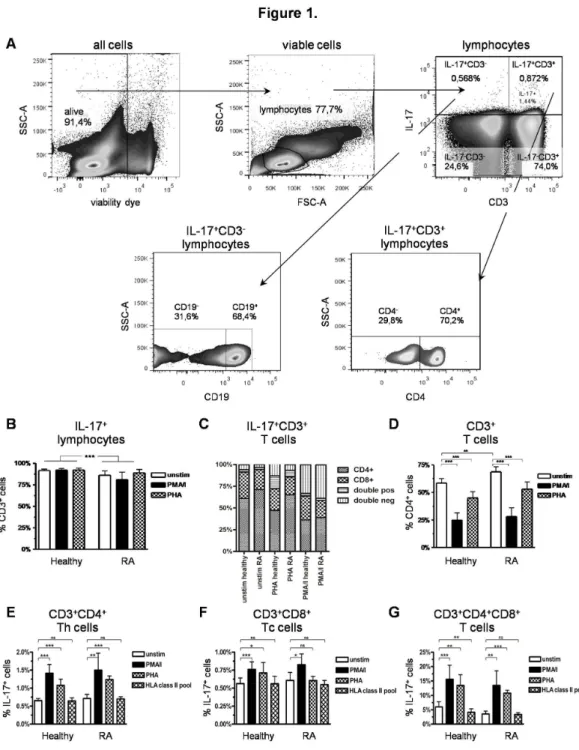
stimuli in RA patients and healthy controls. A representative example of the flow-cytometric gating strategy in viable PBMC of a RA patient is shown in Figure 1A. Most of the IL-17+
lymphocytes were present within the CDγ+ T cell subset in both
stimulated and unstimulated PBMC of RA patients and healthy controls (Figure 1B). The RA patients constitutively had a significantly higher proportion of IL-17+CDγ+CD4+ Th17 cells
than the healthy controls (Figure 1C), but also showed an overall significantly increased CD4+ T-helper cell fraction of the
CDγ+ T cells (Figure 1D). The higher percentages of
IL-17+CDγ+CD4+ Th17 cells thus appeared to reflect a general
increase of the CDγ+CD4+ T-helper cells in RA patients. On the
contrary, the fraction of IL-17-expressing CDγ+CD4+ T-helper
cells tended to be only slightly higher in RA patients than in the healthy controls (Figure 1E). These results are consistent with prevoius reports of other groups [β7,β9].
IL-17+CDγ+ T cells were also evaluated after stimulation with
PMA/ionomycin (PMA/i), PHA or a pool of peptides of MHC class II-restricted T cell epitopes. In RA patients and healthy controls, the proportion of CD4+ cells within IL-17+CDγ+ T cells
(Figure 1C) or CD4+ T-helper cells within CDγ+ T-lymphocytes
(Figure 1D) decreased significantly after PMA/i stimulation due to down-regulation of CD4 [γ6]. This was also observed after PHA activation. The fraction of IL-17+ cells was nevertheless
increased significantly among the CDγ+CD4+ helper T cells
after mitogen stimulation (Figure 1E). Stimulation with PMA/i led to an even higher IL-17+ cell fraction than PHA with no
significant difference between patients and controls.
Other IL-17-producing T cells were also identified, thus, small numbers of CDγ+CD8+ IL-17+ Tc cells (Figure 1C + F)
were present at similar frequencies in patients and controls. Presence of an IL-17+ CD8+ T cell subset had previously been
detected in lung tissue of patients with chronic obstructive pulmonary disease [γ7]. In the present study, also a small fraction of CDγ+CD4+CD8+ double-positive T cells was seen
after stimulation. These represented a notable proportion (β.γ%-14.9%) of the total IL-17-expressing CDγ+ T cell
population especially after PMA/i or PHA stimulation (Figure 1C). Double-negative CD4-CD8- IL-17+CDγ+ T cells which were
also seen in controls and patients (Figure 1C) probably included δ T cells or CD4-CD8- α T cells expressing IL-17, as
Figure 1. Gating strategy and IL-17+ T cells in RA patients and healthy controls. A) Exemplary gating strategy shown on
representative pseudo-colour dot-plots of unstimulated PBMC of a RA patient. Dead cells were excluded using EMA-viability dye. Lymphocytes were gated (FSC vs. SSC), followed by the subgating of CDγ+ and IL-17+ cells. IL-17+CDγ+ T cells and IL-17+CDγ
-non-T cells were further specified. Frequencies in each subgate are expressed as percentage of their parent population; B-D) Comparative analysis of CDγ+ lymphocytes in PBMC stimulated with PMA/i, PHA or incubated in RPMI-1640 without stimulation for
β0 h in RA patients and healthy controls; B) Percentage of CDγ+ T cells within the IL-17+ lymphocytes; C) Percentage of CD4+ and
CD8+ cells within the IL-17+CDγ+ T cell population, D) Percentage of CD4+ cells within CDγ+ T cells; E-F) Comparative analysis of
IL-17 expression in different T cell populations stimulated with PMA/i, PHA, a pool of βγ peptides of MHC-class II-restricted T cell epitopes or incubated in RPMI-1640 without stimulation for β0 h; E) Percentage of IL-17+ cells within CDγ+CD4+ Th cells; F)
Percentage of IL-17+ cells within CDγ+CD8+ Tc cells; G) Percentage of IL-17+ cells within CDγ+CD4+CD8+ double positive T cells.
Data from healthy controls (n = β0) and RA patients (n = β0). *p < 0.05; **p < 0.01, ***p < 0.001,ns = not significant, column represents the mean and bars indicate 95% CI.
after stimulation with PHA or PMA/I cannot be entirely excluded.
Chronic infection has been repeatedly suggested to play an important role in the development of RA [γ9]. We used a pool of MHC II-restricted peptides from viral proteins of EBV, CMV, influenza A, influenza B and tetanus toxin to stimulate IL-17 production in PBMC. These experiments did not reveal major changes of the IL-17+CDγ+ T cell frequencies in either RA
patients or controls (Figure 1E-G). The only change was a significant decrease of IL-17+ double-positive CDγ+CD4+CD8+ T
cells observed in healthy individuals after incubation with the peptide pool (Figure 1G).
IL-17 producing non-T cells
PBMC of RA patients revealed a significantly larger IL-17+CDγ- non-T cell fraction compared to healthy controls
(Figure βA, p < 0.01). The nature of these IL-17-producing non-T cells was next investigated. First, the previously described IL-17-producing CDγ-CD56+ NK cells [γ], were found to
represent a small proportion of the IL-17+CDγ- non-T cell
populations (Figure βB). There was a tendency towards higher proportions of these cells in healthy controls compared to RA patients. Within these CDγ-CD56+ NK cells, IL-17+ lymphocytes
increased significantly on stimulation with PMA/i (Figure βC), but to the same extent in both RA patients and healthy controls. A small proportion of the IL-17+ non-T cells also
appeared to be CDγ-CD14+ monocytes remaining within the
lymphocyte gate used for standardized evaluation in these analyses (Figure βD). Presence of an IL-17+ monocyte subset
in PBMC could be confirmed by specifically gating on monocytes in SSC vs. FSC. Healthy controls were found to possess an even larger proportion of these IL-17+CDγ-CD14+
monocytes than RA patients (Figure βE). In contrast to NK cells, PMA/i stimulation led to a significant decrease of IL-17+
cells in the monocyte gate (Figure βE), but also decreased the CD14+ cell fraction of the IL-17+CDγ- population significantly
(Figure βD).
Interestingly, up to 70% of all unstimulated IL-17+CDγ- non-T
cells expressed CD11b, the alpha chain of the heterodimeric integrin alpha-M beta-β (αM β) expressed on monocytes, macrophages, granulocytes, follicular dendritic cells, natural killer cells, and even some T/B cells. Upon PMA/i stimulation, this cell fraction also decreased significantly in healthy persons and in RA patients (Figure βF). Among these IL-17+CDγ-CD11b + cells, CD14+ monocytes, as well as CD19+ B cells, were
identified (data not shown). In addition to B cells and monocytes, a respectable number of IL-17+CDγ-CD11b+ non-T
cells could not be further characterized with the antibody sets used here.
IL-17 expressing B cells
A large population of IL-17+CDγ- non-T cells was surprisingly
found to consist of CD19+ B cells both in RA patients and
healthy controls. To our knowledge this is the first description of IL-17 producing B cells in PBMC. The B cell subset could account for up to a mean of β5% of the IL-17+CDγ- non-T cells
(Figure γA), although with highly variable percentages in healthy individuals (range 0.6% - 6β%, n = 8) as well as in RA
patients (range 0.γ% - 71.5%, n = 8) independent of their disease state and treatment after short term in vitro culture. The variability of this subset was much smaller when examined directly ex vivo, and most of the IL-17+CDγ- non-T cells were
found to express CD19 (Figure γB). Moreover, after stimulation with PMA/i, the B cell fraction of the IL-17+CDγ- non-T cells did
not change (Figure γA). The IL-17+ fraction of B-cells, however,
decreased considerably after ex vivo culture (Figure γC). We further stained γ healthy donors with B cell specific antibodies and IL-17 to further differentiate those IL-17 producing B cells. 96% of IL-17+CDγ-CD19+ B cells expressed CDβ0+ (data not
shown). More than half of the of IL-17+CDγ-CD19+ B cells were
CDβ7-IgD+ naïve B cells while the rest accounted for up to β1%
of CDβ7-IgD+ non-switched memory B cells and β1% of CDβ4+ +CDγ8++ regulatory B cells (Figure γD). IL-17 expression was
similarly investigated in five established EBV-transformed B cell lines. A subset of IL-17+ cells (0.7γ% - 1.17%) was also
observed in all analyzed B cell lines (Figure γE), which concomitantly expressed CDβ0 and CD11b (data not shown).
To further confirm IL-17 mRNA expression in B cells subsets, IL-17 RT-PCR was performed on EBV-transformed B cell lines as well as on CD4 and CD19-specific immunomagnetic bead-enriched lymphocytes from PBMC of healthy donors and RA patients (Figure γF). The highest amount of IL-17 transcripts was detected within the CD4+ sorted Th cells, followed by the
CD19+ sorted B cells with no major differences between RA
patients and healthy donors. Only a small amount of IL-17 mRNA was detected in those negatively enriched remaining peripheral blood cells. The presence of IL-17 mRNA in B cell lines was also confirmed by RT-PCR for the tested EBV-transformed B cell lines Olga and AMAI (Figure γF). The IL-17 specificity of the detected amplification products was verified by sequencing.
Influence of in vitro culture on the differentiation of IL-17+ cells
Because in vitro culture could influence detection of IL-17+
cells, we compared the different IL-17-expressing cell populations described above in PBMC ex vivo to those cultured for β0 h in RPMI-1640 medium with the addition of brefeldin A for 16h. After in vitro culture, a significant increase of the percentages of IL-17+CDγ+ T cells (Figure 4A),
IL-17+CDγ+CD4+ Th cells (Figure 4B) and IL-17+CDγ-CD11b+
cells (Figure 4C) was observed, which appeared to correspond to a significant decrease of the IL-17+CDγ-CD19+ B cells
(Figure γB) and IL-17+CDγ-CD56+ NK cells (Figure 4D)
subsets. No influence of in vitro culture was seen on the frequencies of the IL-17+CDγ-CD14+ monocytes (data not
shown).
Discussion
conclusions on the cellular source and pathogenetic role of IL-17 in human RA. The present study shows that in RA patients IL-17 is also derived from a substantial number of cells other than the Th17 subset which include NK cells, other innate immune cells and in particular B lymphocytes in peripheral blood. The present analysis also provides data that differences in the detection of IL-17+ cells in former studies might be due to
differences in the applied in vitro cultures.
In accordance with previous analyses [β7,4β], we found only slightly raised frequencies of IL-17+ lymphocytes in PBMC of
RA patients relative to healthy controls. Furthermore, neither the observed IL-17+CDγ+ T cell populations in general nor the
included CD4+ Th17 cells although slightly elevated in RA were
significantly different in patients and controls as had been previously reported [β7]. The Th17 cell fractions were most
likely concomitantly augmented as a result of the observed elevation of the whole subset of CDγ+CD4+ T lymphocytes in
RA patients. The IL-17+ cells also could be significantly
expanded with mitogens such as PMA/i or PHA, but not with a viral peptide pool, in both RA patients and healthy individuals, as might be expected. These findings suggest no major abnormalities in the differentiation of the Th17 cells in the RA patients.
Most patients included in this study suffered from disease of long duration with well-controlled low or moderate activity as reflected by the level of C-reactive protein, leukocyte count, and DASβ8 score. This could account for the minor differences seen in the circulating IL-17 populations between patients and controls and may explain results of other authors at variance with our study. Analysis of treatment-naïve patients with early
Figure 2. IL-17+ non-T cells. A-F) Comparative analysis of PBMC stimulated with PMA/i for β0h or incubated in RPMI-1640
medium with the addition of Brefeldin A for 16 h in RA patients and healthy controls; A) Percentage of CDγ- non-T cells within IL-17+
lymphocytes (RA n = β0, healthy controls n = β0); B) Percentage of CD56+ NK cells within IL-17+CDγ- non-T cells; C) Percentage of
IL-17+ cells within CDγ-CD56+ NK cells; D) Percentage of CD14+ monocytes in IL-17+CDγ- non-T cells within the lymphocyte gate
and within the monocyte gate (FSC vs. SSC); E) Percentage of IL-17+ cells within CDγ-CD14+ monocytes within the monocyte gate;
F) Percentage of CD11b+ cells within IL-17+CDγ- lymphocytes.
Data B)-F) PMBC of healthy controls (n = 8) and RA patients (n = 8).
*p < 0.05, **p < 0.01, ***p < 0.001, ns = not significant, column represents the mean and bars indicate 95% CI.
RA had shown significantly higher percentages of peripheral Th17 cells compared to healthy controls [γ1]. In addition, most of the patients in the present study were also treated with corticosteroids and/or methotrexate, which are known to decrease IL-17 levels [4γ-45]. Moreover, peripheral Th17 levels also failed to correlate with other clinical parameters of disease activity and severity (data not shown) as had been observed
previously [β7,β9,4β]. These findings indicate that IL-17+CDγ+ T
lymphocyte levels including Th17 cells in peripheral blood in particular may not be informative of the disease status later during RA.
In the present study we also focused on in vitro alterations of IL-17 producing cells. After short-term culture in which the lymphocyte responses to different stimuli were tested, a
Figure 3. IL-17 expressing B cells. A) Percentage of CD19+ B cells within IL-17+CDγ- lymphocytes in RA patients (n = 8) and
healthy controls (n = 8) in unstimulated PBMC or after stimulation with PMA/i for β0 h; B+C) freshly thawed PBMC or PBMC cultured for β0 h in RPMI-1640 medium with the addition of Brefeldin A for 16h (n = 5 and n = 8, respectively) in RA patients and healthy controls; B) Percentage of CD19+ B cells within IL-17+CDγ- lymphocytes; C) Percentage of IL-17+ cells within CDγ-CD19+ B
cells; D) Percentage of IgD+CDβ7- naïve B cells, IgD+CDβ7+ non-switched memory B cells and CDβ4++CDγ8++ regulatory B cells of
IL-17+CDγ-CD19+ B cells of freshly thawed PBMC of healthy controls (n=γ); E) Percentage of IL-17+ cells in EBV-transformed B cell
lines without stimulation; F) Representative gel-electrophoresis of amplificates of IL-17A RT-PCR of unstimulated freshly thawed CD4+ and CD19+ MACS-sorted cells of a healthy donor: CD4+ T cells (lane 1), CD19+ B cells (lane β), CD4-CD19- cells (lane γ); of a
RA patient: CD4+ T cells (lane 4), CD19+ B cells (lane 5), CD4-CD19- cells (lane 6); and of the EBV-transformed B cell lines: Olga
(lane 7), AMAI (lane 8); upper level: amplificates of IL-17A, lower level: corresponding control amplificates of -actin, Marker: Puc 8 mix ladder; The data shown are representative results of RT-PCR of γ healthy donors and γ RA patients.
*p < 0.05; **p < 0.01, ns = not significant; columns represent the mean and bars indicate 95% CI.
significantly higher proportion of CDγ+ T cells and CDγ+CD4+
Th17 cells within the IL-17+ lymphocyte population, but a
relative decline of IL-17+CDγ- non-T cells was observed in
unstimulated control samples of PBMC, in comparison to direct ex vivo analysis. Thus, in our study in vitro culture might have even led to an overestimation of the amount of CDγ+ T cells
and CDγ+CD4+ Th17 subsets expressing IL-17, but
underestimation of in vivo frequencies of IL-17+CDγ- non-T
lymphocytes, such as the B cells, monocytes and NK cells. Similar culture systems have been used frequently in previous analyses of IL-17+ cells in RA and could also have influenced
the predominant and/or variable detection of T cells as major
IL-17-producing cells in diseased and healthy individuals in previous studies.
In contrast to the Th17 cells, we observed that IL-17 expressing CDγ- non-T cells were significantly elevated in
PBMC of RA patients compared to healthy controls. This applied to the directly ex vivo tested PBMC as well as to the short-term cultured samples. Recent reports of local effector cells in joints of established RA have provided first evidence that innate immune cells, particularly mast cells may serve as another major source of IL-17A in RA and contribute to pro-inflammatory effector responses in the arthritic synovium [γ0,40]. IL-17 producing T cells, however, were variably and
Figure 4. Influence of in vitro culture on IL-17+ cells. A-D) Comparison of freshly thawed cells and cells cultured for β0 h in
RPMI-1640 medium with the addition of Brefeldin A for 16 h (n = 5 and n = 8, respectively) in RA patients and healthy controls; A) Percentage of CDγ+ T cells within IL-17+ lymphocytes; B) Percentage of CD4+ cells within IL-17+CDγ+ T cells; C) Percentage of
CD11b+ cells within IL-17+CDγ- lymphocytes; D) Percentage of CD56+ NK cells within IL-17+CDγ- lymphocytes.
*p < 0.05, **p < 0.01,***p < 0.001, ns = not significant, columns represent the mean and bars indicate 95% CI.
often rarely observed in the inflamed human synovium [β9,γ0,40]. These unexpected findings were related to the plasticity of Th17 cells and their phenotypic changes upon tissue entry and/or over the course of the disease. In the present study, up to γ0% of IL-17+ PBMC represented
IL-17+CDγ- non-T cell subpopulations in the peripheral blood of
RA patients, significantly different to healthy individuals. Among these, NK cells, monocytes and, rather surprisingly, B cells were identified. Therefore, these data also suggest that different adaptive and innate immune cells can all contribute to IL-17A production in RA. Our observations thus further support the view that additional innate immune cells are important effectors of IL-17- driven chronic inflammatory pathways in RA. However, it still needs to be determined whether, such IL-17+CDγ- non-T cells preferentially localize to synovial tissue
and participate in the pathology of the inflamed joints at various stages of the disease. To the best of our knowledge, in this study we show for the first time that subsets of CD19+ B cells
express IL-17A in PBMC of RA patients and healthy individuals. Although the signalling pathways and interactions of B cells in RA are still incompletely understood, they are known to have a pivotal role in the pathogenesis of RA in humans and animal models [46]. It has been shown that autoantigen-specific B cells can act as antigen-presenting cells equally as efficiently as dendritic cells, and thus drive activation of auto-reactive CD4+ T cells in RA [47-50]. In this study, about
96% of the IL17+ producing CDγ-CD19+ B cells coexpressed
CDβ0, a specific surface molecule of mature naïve and antigen-reactive, but not plasma B cells, which is effectively used as a target structure for therapeutic modulation and/or depletion of B cells by monoclonal antibodies such as Rituximab. Most of the IL-17+ B cells (54%) in the healthy
controls of this study seemed to represent CD19+CDβ7-IgD+
naïve B cells. Naïve B cells were shown to produce proinflammatory cytokines in response to B cell receptor and CD40 stimulation [51]. Another large fraction of the IL-17+ B cells (β1%) were CD19+CDβ4++CDγ8++ regulatory B cells which
have been shown to differentiate into naïve B cells, switched and non-switched memory B cells upon CpG stimulation via TLR9 [5β]. Regulatory B cells are capable to suppress Th1 and Th17 differentation [5γ]. Flores-Borjaet et al. showed that in RA patients regulatory B cells failed to suppress differentiation of Th17 cells [54]. Furthermore, CD19+IgD+CDβ7+ non-switched
memory B cells also accounted for up to β1% of the IL-17+ B
cells of this study. Non-switched B cells have been reported to accumulate in the synovium of RA patients and to respond to TNF-α treatment [55].
Unfortunately, we could not clarify stimulation of IL-17 in B cells in this study. PMA/i failed to stimulate IL-17+CDγ-CD19+ B
cells in vitro, but led to an increase of IL-17 in CDγ+CD4+ Th17
cells. So far the bacterium T. cruzi today is the only known trigger of IL-17 production in B cells [19]. It was shown to
depend on a parasite trans-sialidase activity which modifies the cell-surface CD45 and thus initiates a new signalling program independent of ROR t, RORα or AhR for the stimulation of IL-17. Neither IL-6, IL-βγ nor MyD88-associated TLR signalling were relevant for this IL-17 production in B cells [56]. The B cell activating factor activating factor (BAFF), a member of the tumor necrosis superfamily, has been recognized to positively influence proinflammatory cytokine production in B cells and might also be capable to stimulate IL-17 production in B cells. BAFF seems to play a pivotal role in different autoimmune diseases including RA [57]. In RA, increased BAFF levels have been observed to correlate with high levels of the rheumatoid factor [58], with clinical disease activity, and response to treatment in early rheumatoid arthritis [59]. In a clinical phase II trial the blockade of BAFF with a neutralising antibody called Tabalumab provided promising results in patients with active rheumatoid arthritis (RA) [60]. BAFF gene silencing has been shown to lead to a significant reduction of IL-17 and Th17 cells [61]. In addition, had Doreau et al. observed that IL-17 alone, but together with BAFF even with higher efficiency, could promote proliferation and differentiation of B-cells and prolong their survival [6β]. These findings suggest complex mutual interactions of BAFF and IL-17 on the regulation of B cell differentiation and function.
Further studies are needed to elucidate signalling pathways for the expression of IL-17A in B cells.
Our data suggest that B cells could also participate in dysregulated IL-17A production and enhance chronic inflammatory innate immune responses as well as erosive arthritis in RA. Thus, clinical effects of today´s B cell-directed therapies in RA could be also mediated by influences on IL-17 production and levels in peripheral blood and tissues. Known beneficial clinical responses of B cell depletion with Rituximab (anti-CDβ0-antibody) in several clinical trials have been reviewed by Korhonen et al. [6γ] recently.
In conclusion, our study provides evidence that in addition to Th17 cells, a substantial number of other innate and adaptive immune cells, in particular also B cells participate in the IL-17production during established RA and may thus influence disease activity and joint destruction.
Acknowledgements
We thank Prof. G. Pawelec for the critical review of this manuscript.
Author Contributions
References
1. Rouvier E, Luciani MF, Mattéi MG, Denizot F, Golstein P (199γ) CTLA-8, cloned from an activated T cell, bearing AU-rich messenger RNA instability sequences, and homologous to a herpesvirus Saimiri gene. J Immunol 150: 5445-5456. PubMed: 8γ905γ5.
β. Wright JF, Bennett F, Li B, Brooks J, Luxenberg DP et al. (β008) The human IL-17F/IL-17A heterodimeric cytokine signals through the IL-17RA/IL-17RC receptor complex. J Immunol 181: β799-β805. PubMed: 18684971.
γ. Weaver CT, Hatton RD, Mangan PR, Harrington LE (β007) IL-17 Family Cytokines and the Expanding Diversity of Effector T Cell Lineages. Annu Rev Immunol β5: 8β1-85β. doi:10.1146/ annurev.immunol.β5.0ββ106.141557. PubMed: 17β01677.
4. Umemura M, Yahagi A, Hamada S, Begum MD, Watanabe H et al. (β007) IL-17-mediated regulation of innate and acquired immune response against pulmonary Mycobacterium bovis bacille Calmette-Guerin infection. J Immunol 178: γ786-γ796. PubMed: 17γγ9477. 5. Harrington LE, Hatton RD, Mangan PR, Turner H, Murphy TL et al.
(β005) Interleukin 17-producing CD4+ effector T cells develop via a lineage distinct from the T helper type 1 and β lineages. Nat Immunol 6: 11βγ-11γβ. doi:10.10γ8/ni1β54. PubMed: 16β00070.
6. Park H, Li Z, Yang XO, Chang SH, Nurieva R et al. (β005) A distinct lineage of CD4 T cells regulates tissue inflammation by producing interleukin 17. Nat Immunol 6: 11γγ-1141. doi:10.10γ8/ni1β61. PubMed: 16β00068.
7. Ivanov II, McKenzie BS, Zhou L, Tadokoro CE, Lepelley A et al. (β006) The orphan nuclear receptor RORgammat directs the differentiation program of proinflammatory IL-17+ T helper cells. Cell 1β6: 11β1-11γγ. doi:10.1016/j.cell.β006.07.0γ5. PubMed: 169901γ6.
8. Veldhoen M, Hocking RJ, Atkins CJ, Locksley RM, Stockinger B (β006) TGFbeta in the context of an inflammatory cytokine milieu supports de novo differentiation of IL-17-producing T cells. Immunity β4: 179-189. doi:10.1016/j.immuni.β006.01.001. PubMed: 1647γ8γ0.
9. Mangan PR, Harrington LE, O'Quinn DB, Helms WS, Bullard DC et al. (β006) Transforming growth factor-beta induces development of the T(h)17 lineage. Nature 441: βγ1-βγ4. doi:10.10γ8/nature04754. PubMed: 166488γ7.
10. Bettelli E, Carrier Y, Gao W, Korn T, Strom TB et al. (β006) Reciprocal developmental pathways for the generation of pathogenic effector TH17 and regulatory T cells. Nature 441: βγ5-βγ8. doi:10.10γ8/ nature0475γ. PubMed: 166488γ8.
11. Kondo T, Takata H, Matsuki F, Takiguchi M (β009) Cutting edge: Phenotypic characterization and differentiation of human CD8+ T cells producing IL-17. J Immunol 18β: 1794-1798. doi:10.4049/jimmunol. 0801γ47. PubMed: 19β018γ0.
1β. O'Brien RL, Roark CL, Born WK (β009) IL-17-producing gammadelta T cells. Eur J Immunol γ9: 66β-666. doi:10.100β/eji.β008γ91β0. PubMed: 19β8γ718.
1γ. Takatori H, Kanno Y, Watford WT, Tato CM, Weiss G et al. (β009) Lymphoid tissue inducer-like cells are an innate source of IL-17 and IL-ββ. J Exp Med β06: γ5-41. doi:10.1084/jem.β007β71γ. PubMed: 19114665.
14. Bird L (β008) NKT cells: NKT cells join the IL-17 gang. Nature Reviews Immunology 8: γβ4. doi:10.10γ8/nriβγβ5.
15. Michel JJ, Turesson C, Lemster B, Atkins SR, Iclozan C et al. (β007) Identification of an IL-17-producing NK1.1(neg) iNKT cell population involved in airway neutrophilia. J Exp Med β04: 995-1001. doi:10.1084/ jem.β0061551. PubMed: 17470641.
16. Fujino S, Andoh A, Bamba S, Ogawa A, Hata K et al. (β00γ) Increased expression of interleukin 17 in inflammatory bowel disease. Gut 5β: 65-70. doi:10.11γ6/gut.5β.1.65. PubMed: 1β47776β.
17. Hoshino A, Nagao T, Nagi-Miura N, Ohno N, Yasuhara M et al. (β008) MPO-ANCA induces IL-17 production by activated neutrophils in vitro via classical complement pathway-dependent manner. J Autoimmun γ1: 79-89. doi:10.1016/j.jaut.β008.0γ.006. PubMed: 18501β96. 18. Lin AM, Rubin CJ, Khandpur R, Wang JY, Riblett M et al. (β011) Mast
cells and neutrophils release IL-17 through extracellular trap formation in psoriasis. J Immunol 187: 490-500. doi:10.4049/jimmunol.11001βγ. PubMed: β1606β49.
19. Bermejo DA, Jackson SW, Gorosito-Serran M, Acosta-Rodriguez EV, Amezcua-Vesely MC et al. (β01γ) Trypanosoma cruzi trans-sialidase initiates a program independent of the transcription factors ROR t and Ahr that leads to IL-17 production by activated B cells. Nat Immunol 14: 514-5ββ. doi:10.10γ8/ni.β569. PubMed: βγ56γ688.
β0. Lubberts E, Joosten LA, Oppers B, van den Bersselaar L, Coenen-de Roo CJ et al. (β001) IL-1-independent role of IL-17 in synovial inflammation and joint destruction during collagen-induced arthritis. J Immunol 167: 1004-101γ. PubMed: 11441109.
β1. Lubberts E, Koenders MI, van den Berg WB (β005) The role of T-cell interleukin-17 in conducting destructive arthritis: lessons from animal models. Arthritis Res Ther 7: β9-γ7. doi:10.1186/ar1550. PubMed: 1564β151.
ββ. Chabaud M, Lubberts E, Joosten L, van den Berg W, Miossec P (β001) IL-17 derived from juxta-articular bone and synovium contributes to joint degradation in rheumatoid arthritis. Arthritis Res γ: 168-177. doi: 10.1186/arβ94. PubMed: 11β99057.
βγ. Shahrara S, Huang Q, Mandelin AM βnd, Pope RM (β008) TH-17 cells in rheumatoid arthritis. Arthritis Res Ther 10: R9γ. doi:10.1186/arβ477. PubMed: 18710567.
β4. Ziolkowska M, Koc A, Luszczykiewicz G, Ksiezopolska-Pietrzak K, Klimczak E et al. (β000) High levels of IL-17 in rheumatoid arthritis patients: IL-15 triggers in vitro IL-17 production via cyclosporin A-sensitive mechanism. J Immunol 164: β8γβ-β8γ8. PubMed: 106791β7. β5. Chabaud M, Durand JM, Buchs N, Fossiez F, Page G et al. (1999)
Human interleukin-17: A T cell-derived proinflammatory cytokine produced by the rheumatoid synovium. Arthritis Rheum 4β: 96γ-970. doi:10.100β/15β9-01γ1(199905)4β:5. PubMed: 10γβγ45β.
β6. Kotake S, Udagawa N, Takahashi N, Matsuzaki K, Itoh K et al. (1999) IL-17 in synovial fluids from patients with rheumatoid arthritis is a potent stimulator of osteoclastogenesis. J Clin Invest 10γ: 1γ45-1γ5β. doi: 10.117β/JCI570γ. PubMed: 10ββ5978.
β7. Church LD, Filer AD, Hidalgo E, Howlett KA, Thomas AMC et al. (β010) Rheumatoid synovial fluid interleukin-17-producing CD4 T cells have abundant tumor necrosis factor-alpha co-expression, but little interleukin-ββ and interleukin-βγR expression. Arthritis Res Ther 1β: R184. doi:10.1186/arγ15β. PubMed: β09β95γ6.
β8. Appel H, Maier R, Wu P, Scheer R, Hempfing A et al. (β011) Analysis of IL-17(+) cells in facet joints of patients with spondyloarthritis suggests that the innate immune pathway might be of greater relevance than the Th17-mediated adaptive immune response. Arthritis Res Ther 1γ: R95. doi:10.1186/arγγ70. PubMed: β168940β.
β9. Yamada H, Nakashima Y, Okazaki K, Mawatari T, Fukushi JI et al. (β008) Th1 but not Th17 cells predominate in the joints of patients with rheumatoid arthritis. Ann Rheum Dis 67: 1β99-1γ04. PubMed: 1806γ670.
γ0. Hueber AJ, Asquith DL, Miller AM, Reilly J, Kerr S et al. (β010) Mast cells express IL-17A in rheumatoid arthritis synovium. J Immunol 184: γγγ6-γγ40. doi:10.4049/jimmunol.090γ566. PubMed: β0β00β7β. γ1. Colin EM, Asmawidjaja PS, van Hamburg JP, Mus AMC, van Driel M et
al. (β010) 1,β5-dihydroxyvitamin Dγ modulates Th17 polarization and interleukin-ββ expression by memory T cells from patients with early rheumatoid arthritis. Arthritis Rheum 6β: 1γβ-14β. doi:10.100β/art. β504γ. PubMed: β00γ94β1.
γβ. Shen H, Goodall JC, Hill Gaston JS (β009) Frequency and phenotype of peripheral blood Th17 cells in ankylosing spondylitis and rheumatoid arthritis. Arthritis Rheum 60: 1647-1656. doi:10.100β/art.β4568. PubMed: 19479869.
γγ. Jandus C, Bioley G, Rivals J-P, Dudler J, Speiser D et al. (β008) Increased numbers of circulating polyfunctional Th17 memory cells in patients with seronegative spondylarthritides. Arthritis Rheum 58: βγ07-βγ17. doi:10.100β/art.βγ655. PubMed: 18668556.
γ4. Hochberg MC, Chang RW, Dwosh I, Lindsey S, Pincus T, et al. (199β) The American College of Rheumatology 1991 revised criteria for the classification of global functional status in rheumatoid arthritis. Arthritis Rheum γ5: 498-50β
γ5. Morbach H, Eichhorn EM, Liese JG, Girschick HJ (β010) Reference values for B cell subpopulations from infancy to adulthood. Clin Exp Immunol 16β: β71-β79. doi:10.1111/j.1γ65-ββ49.β010.04β06.x. PubMed: β0854γβ8.
γ6. Weyand CM, Goronzy J, Fathman CG (1987) Modulation of CD4 by antigenic activation. J Immunol 1γ8: 1γ51-1γ54. PubMed: γ1006γ8. γ7. Chang Y, Nadigel J, Boulais N, Bourbeau J, Maltais F et al. (β011) CD8
positive T cells express IL-17 in patients with chronic obstructive pulmonary disease. Respir Res 1β: 4γ. doi:10.1186/1465-99β1-1β-4γ. PubMed: β1477γ50.
γ8. Roark CL, Simonian PL, Fontenot AP, Born WK, O'Brien RL (β008) gammadelta T cells: an important source of IL-17. Curr Opin Immunol β0: γ5γ-γ57. doi:10.1016/j.coi.β008.0γ.006. PubMed: 184γ9808. γ9. Hitchon CA, El-Gabalawy HS (β011) Infection and rheumatoid arthritis:
still an open question. Curr Opin Rheumatol βγ: γ5β-γ57. doi:10.1097/ BOR.0b01γeγβ8γ477b7b. PubMed: β15γβ48γ.
osteoarthritis synovium. Arthritis Res Ther 1γ: R150. doi:10.1186/ arγ466. PubMed: β19γγγ91.
41. Chen D-Y, Chen Y-M, Chen H-H, Hsieh C-W, Lin C-C et al. (β011) Increasing levels of circulating Th17 cells and interleukin-17 in rheumatoid arthritis patients with an inadequate response to anti-TNF-α therapy. Arthritis Res Ther 1γ: R1β6. doi:10.1186/arγ4γ1. PubMed: β18014γ1.
4β. Aerts NE, De Knop KJ, Leysen J, Ebo DG, Bridts CH et al. (β010) Increased IL-17 production by peripheral T helper cells after tumour necrosis factor blockade in rheumatoid arthritis is accompanied by inhibition of migration-associated chemokine receptor expression. Rheumatology (Oxford) 49: ββ64-ββ7β. doi:10.109γ/rheumatology/ keqββ4. PubMed: β07β44γγ.
4γ. Chakir J, Shannon J, Molet S, Fukakusa M, Elias J et al. (β00γ) Airway remodeling-associated mediators in moderate to severe asthma: effect of steroids on TGF-beta, IL-11, IL-17, and type I and type III collagen expression. J Allergy Clin Immunol 111: 1β9γ-1β98. doi:10.1067/mai. β00γ.1557. PubMed: 1β789βγβ.
44. Liu X, Yang P, Lin X, Ren X, Zhou H et al. (β009) Inhibitory effect of Cyclosporin A and corticosteroids on the production of IFN-gamma and IL-17 by T cells in Vogt-Koyanagi-Harada syndrome. Clin Immunol 1γ1: γγγ-γ4β. doi:10.1016/j.clim.β008.1β.007. PubMed: 19β00788. 45. Miranda-Carús ME, Benito-Miguel M, Balsa A, Cobo-Ibáñez T, Pérez
de Ayala C et al. (β006) Peripheral blood T lymphocytes from patients with early rheumatoid arthritis express RANKL and interleukin-15 on the cell surface and promote osteoclastogenesis in autologous monocytes. Arthritis Rheum 54: 1151-1164. doi:10.100β/art.β17γ1. PubMed: 16575870.
46. Marston B, Palanichamy A, Anolik JH (β010) B cells in the pathogenesis and treatment of rheumatoid arthritis. Curr Opin Rheumatol ββ %W http://view.ncbi.nlm.nih.gov/pubmed/β00905β6: γ07-γ15
47. Takemura S, Klimiuk PA, Braun A, Goronzy JJ, Weyand CM (β001) T cell activation in rheumatoid synovium is B cell dependent. J Immunol 167: 4710-4718. PubMed: 1159180β.
48. O'Neill SK, Shlomchik MJ, Glant TT, Cao Y, Doodes PD et al. (β005) Antigen-specific B cells are required as APCs and autoantibody-producing cells for induction of severe autoimmune arthritis. J Immunol 174: γ781-γ788. PubMed: 15749919.
49. Gavanescu I, Benoist C, Mathis D (β008) B cells are required for Aire-deficient mice to develop multi-organ autoinflammation: A therapeutic approach for APECED patients. Proc Natl Acad Sci U S A 105: 1γ009-1γ014. doi:10.107γ/pnas.0806874105. PubMed: 18755889. 50. Wilson CL, Hine DW, Pradipta A, Pearson JP, van Eden W et al. (β01β)
Presentation of the candidate rheumatoid arthritis autoantigen aggrecan by antigen-specific B cells induces enhanced CD4(+) T helper type 1 subset differentiation. Immunology 1γ5: γ44-γ54. doi: 10.1111/j.1γ65-β567.β011.0γ548.x. PubMed: ββ18β481.
51. Duddy ME, Alter A, Bar-Or A (β004) Distinct profiles of human B cell effector cytokines: a role in immune regulation? J Immunol 17β: γ4ββ-γ4β7. PubMed: 15004141.
5β. Capolunghi F, Cascioli S, Giorda E, Rosado MM, Plebani A et al. (β008) CpG drives human transitional B cells to terminal differentiation and production of natural antibodies. J Immunol 180: 800-808. PubMed: 18178818.
5γ. Mauri C, Bosma A (β01β) Immune regulatory function of B cells. Annu
Rev Immunol γ0: ββ1-β41.
doi:10.1146/annurev-immunol-0β0711-0749γ4. PubMed: ββββ4776.
54. Flores-Borja F, Bosma A, Ng D, Reddy V, Ehrenstein MR et al. (β01γ) CD19+CDβ4hiCDγ8hi B cells maintain regulatory T cells while limiting TH1 and TH17 differentiation. Sci Transl Med 5: 17γra1βγ. PubMed: βγ4β7β4γ.
55. Souto-Carneiro MM, Mahadevan V, Takada K, Fritsch-Stork R, Nanki T et al. (β009) Alterations in peripheral blood memory B cells in patients with active rheumatoid arthritis are dependent on the action of tumour necrosis factor. Arthritis Res Ther 11: R84. doi:10.1186/arβ718. PubMed: 19500γγ5.
56. León B, Lund FE (β01γ) IL-17-producing B cells combat parasites. Nat Immunol 14: 419-4β1. doi:10.10γ8/ni.β59γ. PubMed: βγ598γ88. 57. Szodoray P, Jonsson R (β005) The BAFF/APRIL system in systemic
autoimmune diseases with a special emphasis on Sjogren's syndrome. Scand J Immunol 6β: 4β1-4β8. doi:10.1111/j.1γ65-γ08γ.β005.01688.x. PubMed: 16γ056γ8.
58. Mariette X, Roux S, Zhang J, Bengoufa D, Lavie F et al. (β00γ) The level of BLyS (BAFF) correlates with the titre of autoantibodies in human Sjogren's syndrome. Ann Rheum Dis 6β: 168-171. doi:10.11γ6/ ard.6β.β.168. PubMed: 1β5β5γ88.
59. Bosello S, Youinou P, Daridon C, Tolusso B, Bendaoud B et al. (β008) Concentrations of BAFF correlate with autoantibody levels, clinical disease activity, and response to treatment in early rheumatoid arthritis. J Rheumatol γ5: 1β56-1β64. PubMed: 185β8969.
60. Genovese MC, Fleischmann RM, Greenwald M, Satterwhite J, Veenhuizen M et al. (β01γ) Tabalumab, an anti-BAFF monoclonal antibody, in patients with active rheumatoid arthritis with an inadequate response to TNF inhibitors. Ann Rheum Dis 7β: 1461-1468. doi: 10.11γ6/annrheumdis-β01β-β0β775. PubMed: βγβ68γ67.
61. Kwan Lam Lai, King Hung Ko O, Zheng BJ, Lu L (β008) Local BAFF gene silencing suppresses Th17-cell generation and ameliorates autoimmune arthritis. Proc Natl Acad Sci U S A 105: 1499γ-14998. doi: 10.107γ/pnas.0806044105. PubMed: 188β00γβ.
6β. Doreau A, Belot A, Bastid J, Riche B, Trescol-Biemont MC et al. (β009) Interleukin 17 acts in synergy with B cell-activating factor to influence B cell biology and the pathophysiology of systemic lupus erythematosus. Nat Immunol 10: 778-785. doi:10.10γ8/nrmβ786. PubMed: 1948γ719. 6γ. Korhonen R, Moilanen E (β010) Anti-CDβ0 antibody rituximab in the