Cells: Lesson from
NLPR3
Mutated Patients
Denise Lasiglie`1,2, Elisabetta Traggiai1, Silvia Federici1, Maria Alessio3, Antonella Buoncompagni1, Andrea Accogli1, Sabrina Chiesa1, Federica Penco1, Alberto Martini2, Marco Gattorno1*
1Rheumatology Unit, Second Division of Pediatrics ‘‘G. Gaslini’’ Institute, Genoa, Italy,2Laboratory of Immunology of Rheumatic diseases, Department of Pediatrics, University of Genoa, Genoa, Italy,3Department of Pediatrics, Federico II Hospital, Naples, Italy
Abstract
Background:T helper 17 cells (TH-17) represent a lineage of effector T cells critical in host defence and autoimmunity. In
both mouse and human IL-1bhas been indicated as a key cytokine for the commitment to TH-17 cells. Cryopyrin-associated
periodic syndromes (CAPS) are a group of inflammatory diseases associated with mutations of the NLRP3 gene encoding the inflammasome component cryopyrin. In this work we asked whether the deregulated secretion of IL-1bsecondary to mutations characterizing these patients could affect the IL-23/IL-17 axis.
Methodology/Principal Findings:A total of 11 CAPS, 26 systemic onset juvenile idiopathic arthritis (SoJIA) patients and 20 healthy controls were analyzed. Serum levels of IL-17 and IL-6 serum were assessed by ELISA assay. Frequency of TH17 cells
was quantified upon staphylococcus enterotoxin B (SEB) stimulation. Secretion of IL-1b, IL-23 and IL-6 by monocyte derived dendritic cells (MoDCs), were quantified by ELISA assay. A total of 8 CAPS and 11 SoJIA patients were also analysed before and after treatment with IL-1bblockade. Untreated CAPS patients showed significantly increased IL-17 serum levels as well as a higher frequency of TH17 compared to control subjects. On the contrary, SoJIA patients displayed a frequency of TH17
similar to normal donors, but were found to have significantly increased serum level of IL-6 when compared to CAPS patients or healthy donors. Remarkably, decreased IL-17 serum levels and TH17 frequency were observed in CAPS patients
followingin vivoIL-1bblockade. On the same line, MoDCs from CAPS patients exhibited enhanced secretion of IL-1band IL-23 upon TLRs stimulation, with a reduction after anti-IL-1 treatment.
Conclusion/Significance:These findings further support the central role of IL-1bin the differentiation of TH17 in human
inflammatory conditions.
Citation:Lasiglie` D, Traggiai E, Federici S, Alessio M, Buoncompagni A, et al. (2011) Role of IL-1 Beta in the Development of Human TH17 Cells: Lesson fromNLPR3 Mutated Patients. PLoS ONE 6(5): e20014. doi:10.1371/journal.pone.0020014
Editor:Joseph El Khoury, Massachusetts General Hospital and Harvard Medical School, United States of America
ReceivedFebruary 7, 2011;AcceptedApril 8, 2011;PublishedMay 26, 2011
Copyright:ß2011 Lasiglie` et al. This is an open-access article distributed under the terms of the Creative Commons Attribution License, which permits unrestricted use, distribution, and reproduction in any medium, provided the original author and source are credited.
Funding:The work was partially supported by Telethon Italia (Grant NGGP09127) and Ricerca Corrente Ministeriale. No additional external funding received for this study. The funders had no role in study design, data collection and analysis, decision to publish, or preparation of the manuscript.
Competing Interests:The authors have declared that no competing interests exist.
* E-mail: [email protected]
Introduction
T helper 17 (TH17) is a subset of effector CD4+T cells crucial
for the response to fungal and extracellular bacteria [1,2]. They also play a pathogenetic role in several animal models of autoimmune diseases [1,3], as well as in human chronic inflammation [4–5]. IL-17 is known to induce the mobilization, recruitment, and activation of neutrophils [5]. Moreover, it is able to stimulate the expression of several proinflammatory cytokines and chemokines by a broad range of cellular targets, including epithelial cells, endothelial cells and macrophages [5].
Human TH-17 cells are defined by the expression of surface
markers such as CCR6 and CCR4 [6], CD161 [4] and IL-23R [7], as well as by the production of different proinflammatory cytokines such as IL-17A and IL-17F, IL-21, IL-22, TNF–a, IL-9 , IL-10 and IL-26 [5,8,9].
The differentiation of naı¨ve T cells into pro-inflammatory TH17
is dependent on the extracellular environment in which T cells are activated.
TGF-band IL-6 have been indicated as the two key cytokines for the in vitro differentiation of murine TH17 cells [10–12].
However, when the development of TH17 cells has been analyzed
in humans, a number of evidences showed the predominant role of IL-1b(alone or in synergy with other cytokines, i.e. 23 and IL-6) [4,6,7], raising the hypothesis that a dichotomy between mice and humans exists [5].
actual role of IL-1b in the generation of TH17 cells in humans.
Here we report that exaggerated IL-1b secretion due toNLPR3 mutations affects the IL-23/IL-17 axis in CAPS patients, providing further evidence of the role of IL-1b in TH17
differentiation.
Methods
A total of 11 CAPS (6 CINCA, 5 MWS) were enrolled in the study (Table 1). Twenty-six systemic onset juvenile idiopathic arthritis (SoJIA) patients with active disease and 20 aged-matched healthy individuals were used as disease-and healthy -controls, respectively. Samples were taken after informed consent approved by ‘‘G. Gaslini’’ Ethical board.
CAPS patients displayed the following mutations of NLRP3 gene: T348M (2 patients), D303N (2 patients), E304K (2 patients), M406I, N477K, E525K, I572F, R260W [18]. At the time of the enrolment in the study, all CAPS patients were naı¨ve from anti-IL-1 and displayed an active phase of their disease, in terms of presence of disease-related clinical manifestations and elevation of acute phase reactants. Disease activity in SoJIA patients was defined by the presence of at least two of the main clinical manifestations (fever, arthritis, rash) and elevation of acute phase reactants despite ongoing therapy with non steroidal anti-inflammatory drugs, oral steroids and/or methotrexate. Peripheral blood mononuclear cells (PBMCs) were isolated by density gradient centrifugation using Fycoll from LympholyteH, Ceder-lane. Eight CAPS patients and 11 SoJIA patients were evaluated the day before and 7–10 days after anti-IL-1 treatment (Anakinra).
Flow cytometry
Thawed PBMCs were stained with the following antibodies: allophycocyanin conjugated anti human CD4 (APC Beckton Dickinson, BD), fluorescein isothiocyanate conjugated anti human CD45RA (FITC Beckman Coulter), phycoerythrin conjugated anti human CCR6 (PE, BD) and phycoerythrin conjugated anti human CD161 (Beckman Coulter). Cells were incubated with 50ml of PBS 1% FCS for 20 min at 4uC in the dark and acquired with a FACSCanto cytometer. Data were analyzed with Flow-Jo software. Quantification of circulating memory CCR6+
and CD161+ was determined electronically gating on alive
lympho-cytes and expressed as the absolute number (number6103/mm3) multiplying the relative percentage of the different subsets by the peripheral lymphocytes count.
Quantification of TH17 producing cells
Freshly isolated or thawed PBMCs were stimulated overnight with staphylococcus enterotoxin B (SEB, 100 ng/ml, Sigma Aldrich), anti-CD28 (1 mcg/ml, BD), anti-CD49d (1 mcg/ml, BD) Mabs in the presence of Brefeldin A (BFA 1 mcg/ml, Sigma Aldrich), as described previously [19–21]. Surface staining of CD3 (PeCy5, BD), CD8 (APC-Cy7, BD) and intracellular staining of
IL-17 (eBiosciences) and IFN-c(BD) were performed according to manifacturer’s instructions.
Generation and stimulation of monocyte-derived DCs [MoDCs]
Peripheral blood CD14+ monocytes were positively isolated
with CD14-specific micro-beads (Miltenyi Biotech). DCs were obtained from monocytes after culture with granulocyte-macro-phage colony-stimulating factor (GM-CSF, 50 ng/ml, Peprotech) and IL-4 (20 ng/ml Peprotech). At day 7, DCs were cultured for additional 48 h in 96-well flat bottom plates with Lypopolisaccaride (LPS, 1mg/ml, Sigma Aldrich), Staphylococcus Aureus (S.a 108/ml, Invivogen) and Zymosan A (50mg/ml, Sigma Aldrich).
Serum and supernatant cytokine detection
Levels of IL-1b, IL-23, IL-17 and IL-6 in culture supernatants or sera were quantified by ELISA assay (eBiosciences) according to the manufacturer’s instructions.
Statistical analysis
Differences among groups were evaluated using the non parametric Kruskal-Wallis test. Post-hoc analysis was performed with non-parametric U Mann-Whitney test. Comparison of variables before and after treatment was performed with Wilcoxon test.
Results
IL-17 and IL-6 serum levels and ex-vivo phenotype of circulating CCR6+and CD161+memory CD4+T cells
Serum IL-17 was measured in 10 activeNLPR3-mutated CAPS patients compared to 20 healthy controls and 20 SoJIA patients. As shown in Figure 1A, IL-17 was significantly higher in CAPS patients (median 5,1 pg/ml range:0–14.4) when compared to healthy controls [0.4 pg/ml, 0–6.5 pg/ml) (p = 0.04). A slight elevation of serum IL-17 was also observed in some active SoJIA patients (1.9 pg/ml, 0–8.6 pg/ml), with no statistical differences with either CAPS patients or healthy controls (Figure 1A).
CAPS patients displayed higher serum levels of IL-6 (median 8.4 pg/ml, 0–38 pg/ml) as well when compared to healthy controls (median 2.5 pg/ml, 0–13.5 pg/ml) (P = 0.02). Differently from what observed for IL-17, active SoJIA patients showed a significant elevation of serum IL-6 (median 54.3 pg/ml range:15– 230) with respect to CAPS patients and healthy controls (P,0.001) (Figure 1B).
CCR6 and CD161 have been recently indicated as additional markers to define TH17 producing cells [22] [4]. Thus, CCR6 and
CD161 expression was evaluated in circulating memory CD4+
T cells (CD4+
CD45RA2) from 7NLPR3-mutated CAPS patients, 10
healthy controls and 12 active SoJIA. The number of circulating CCR6+
memory CD4+
T cells inNLPR3-mutated CAPS patients
Table 1.Clinical characteristics of CAPS and SoJIA patients at the moment of the study.
Male/Female
Age (years; range)
Disease duration (years; range)
CRP mg/dL
(mean, range) Rash
Active
Arthritis Fever
CAPS (n = 11)
7/4 11.7 (2.6–43) 10.6 (0.6–22.8) 5.2 (1.9–10.2) 10/11 8/11 6/11
SoJIA (n = 26)
16/10 7.6 (1.5–19.3) 3.6 (0.3–10.9) 12.3 (1.3–25) 11/26 23/26 16/26
doi:10.1371/journal.pone.0020014.t001
(Figure 1C) tent to be higher than that found in the other two subgroups, although the difference was not statistically significant. Conversely, the absolute number of CD161+
memory T cells was comparable among the three subgroups (Figure 1D). The same data were observed when the percentage of alive T lymphocytes were evaluated (not shown).
Frequency of IL-17 and IFN-cCD4+T cells in CAPS patients
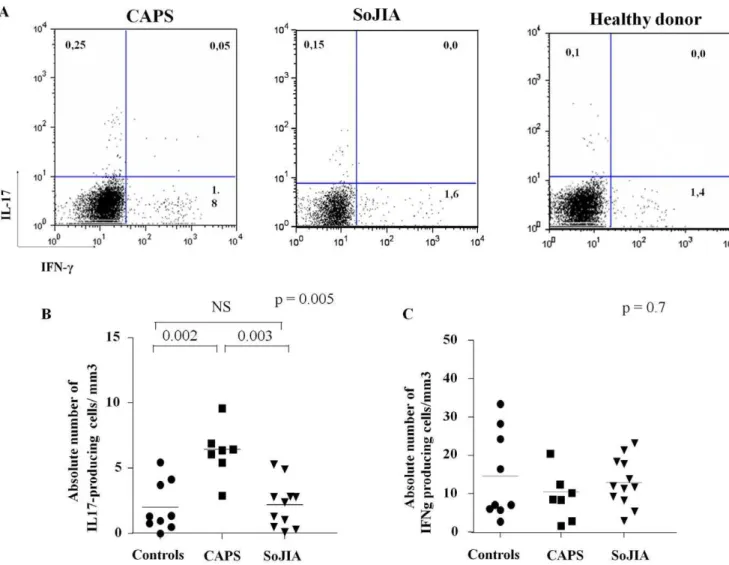
The frequency of IL-17 and IFN-cproducing cells upon anti-CD28, anti-CD49d and SEB stimulation was analyzed according to the experimental protocol recently described by Milner et al. [16]. As shown in Figure 2B, active CAPS patients displayed a significantly higher absolute number (median 7,5 cells/mm3, range 2,9–9,5) of IL-17 producing cells compared to healthy controls (median 1,34 cells/mm3, range 0,4–5,4) (p = 0.002) and active SoJIA patients (median 1,31 cells/mm3, range 0,3–5,2) (p = 0.003). The same results were also obtained when percentage
of IL-17 producing cells was assessed (CAPS: median 0,37%, range 0,2–0,73; healthy donors: median 0,1%, range 0–0,28; SoJIA: median 0,06% range 0,0–0,36) (P = 0.01, see also Figure 3D). Conversely, no differences in the absolute numbers (Figure 2C) and percentages (not shown) of IFN-cproducing T cells was observed among the three subgroups.
Effect of anti-IL-1 treatment on serum IL-17 and frequency of IL-17 producing cells
Most of the CAPS patients analyzed in the present study were treated with IL-1 blockers, namely IL-1 receptor antagonist (Anakinra). As previously shown, the treatment was able to dramatically control the clinical manifestations of all treated patients with a complete normalization of laboratory parameters (hemogram and acute phase reactants) within two weeks [17].
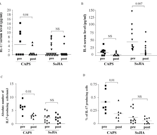
The effect of anti IL-1 treatment on IL-17 serum concentra-tions and on the frequency of IL-17 producing cells is shown in Figure 3. A significant decrease of IL-17 serum concentration was
Figure 1. Analysis of IL-17 and IL-6 serum concentrations and peripheral CD4+T cell phenotype.IL-17 (panel A) and IL-6 (panel B) serum levels were measured in 10 active CAPS patients, 20 active SoJIA patients and 20 healthy controls by ELISA. C–D)Ex-vivoanalysis of circulating CCR6+ (panel B) and CD161+
(panel C) memory T cells (CD4+
CD45RA2) in the same three subgroups. Heterogeneity test among groups was evaluated using
the non parametric Kruskal-Wallis test (upper right of each graph). Post-hoc analysis with non-parametric U Mann-Whitney test revealed the difference among the three subgroups.
observed in all CAPS patients after 7–10 days of treatment (Figure 3A). A slight, albeit non significant, decrease was also observed when analyzing IL-6 serum concentration (Figure 3B). Accordingly, treatment with IL-1 blockers was able to normalize the frequency of IL-17 producing cells after SEB stimulation in NLPR3-mutated CAPS patients, either when evaluated as absolute number [Figure 3C) or percentage of CD4+ T cells (Figure 3D).
In the present study, 11 SoJIA patients were analyzed before and after anti-IL-1 treatment. After 7–10 days of treatment a significant reduction of IL-6 was observed in anti-IL-1 treated patients (Figure 3B). As previously observed, the long term effect of IL-1 blockade in SoJIA patients is rather variable [23]. In the subsequent follow-up, 7 SoJIA patients displayed a complete response, with a dramatic and persistent control of systemic and articular manifestations, whereas the remaining 5 patients were incomplete or non-responders [23]. Despite a few SoJIA responder patients displayed a decrease serum levels of IL-17 and frequency of IL-17 producing cells after anti-IL-1 treatment, a retrospective evaluation was not able to find any significant differences in the serum levels of IL-17 and IL-6, nor in the frequency of IL-17
producing cells before anti-IL-treatment betweenresponderandnon responderSoJIA patients (data not shown).
Monocytes-derived dendritic cells from CAPS patients display an increased secretion of IL-1band IL-23
Since CAPS is associated with a primary defect of the innate immune system, it is conceivable that the TH17 skewed
phenotype observed in CAPS patients could be related to an aberrant influence on T cell differentiation by antigen-presenting cells rather than by an intrinsic T cells defect [24]. Thus, we analyzed thein vitroIL-1b, IL-23 and IL-6 secretion by MoDCs cells after stimulation with TLR ligands in 4 active CAPS patients (2 CINCA and 2 MWS) withNLPR3mutation, 4 active SoJIA patients and 9 healthy controls. After stimulation with single TLR-ligand (e.g. LPS), DCs produced variable amount of IL-6 but little of IL-1band IL-23 either in healthy controls, CAPS and SoJIA patients (data not shown) [25]. Nonetheless, Zymosan, which simultaneously binds to TLR2 dectin-1 and dectin 2 [26– 27] was able to induce the secretion of detectable amounts of all three cytokines.
Figure 2. CAPS patients show an higher absolute number of IL-17 producing cells afterin vitroexpansion.A) Dot plot electronically gated on alive CD4+T cells of one representative CINCA patient, one systemic onset juvenile idiopathic arthritis (SoJIA) patient and one healthy control B–C) Absolute number of IL-17 (panel B) and IFN-c(panel C) producing cells in 7 NLPR3-mutated CAPS and 12 SoJIA active patients and in 9 age-matched healthy controls. Heterogeneity test among groups was evaluated using the non parametric Kruskal-Wallis test (upper right of each graph). Post-hoc analysis with non-parametric U Mann-Whitney test revealed the difference among the three subgroups.
doi:10.1371/journal.pone.0020014.g002
As shown in Figure 4, MoDc from 4 CAPS patients produced significantly higher amount of IL-1b and IL-23 compared to 10 healthy controls and 4 SoJIA patients, whereas no relevant differences were observed in IL-6 secretion among the three subgroups.
Remarkably, after 7 days of treatment with IL-1 blockers, in all CAPS patients we observed a marked reduction of both IL-1b
(pre-treatment median 83 ng/ml, range 28.5–148; post-treatment median 24.2, range 20–104) and IL-23 (pre-treatment median 1348.6 ng/ml, range 261–2048; post-treatment median 459.4, range 106–1038) secretion by MoDCs (data not shown).
Discussion
Given the growing evidence supporting the central role of the IL-1b in the commitment of IL-17 producing cells [6], in the present study we analyzed the IL-23/IL-17 axis in CAPS patients in order to explore a possible involvement of IL-1b in TH-17
differentiation in this context.
The protein mutated in CAPS, namely Cryopyrin is a key protein of the inflammasome, a multi-protein complex responsible for activation of the IL-1 converting enzyme (ICE) (or Caspase-1),
which in turn converts pro-IL-1b to the mature, active 17 kDa form [28]. In response to a broad range of stimuli, cryopyrin oligomerizes and binds the adaptor protein ASC (Apoptosis associated Speck-like protein containing a CARD). This associa-tion activates directly two molecules of Caspase-1 which, in turn, convert pro-IL-1bto the mature, active 17 kDa form. Therefore, activated cryopyrin induces the release of the active form of IL-1b
[14,28,29].
NLPR3mutations are associated with its gain of function with a consequent excessive production of IL-1b[14,30] even in absence of a second signal, such as extracellular adenosine triphosphate (ATP) [17]. Due to the pleiotrophic proinflammatory effect of IL-1b, the specific inhibition of IL-1bis able to dramatically dampen the severe systemic inflammatory picture of CAPS patients [15,16].
In contrast to what previously observed in mice models [10,11], studies in humans have pointed out the potential pivotal role of IL-1b, together with IL-23 and IL-6, in the differentiation of human naı¨ve T cells into IL-17 producing cells [4,6,7]. This hypothesis has been disputed by other authors supporting the crucial role of TGF-bin the differentiation of TH-17 in humans [31].
Figure 3. Effect of anti-IL-1 treatment on IL-17 and IL-6 serum levels and Th17 frequency.Variation of IL-17 (panel A) and IL-6 (panel B) serum levels after anti-IL-1 treatment in 8 CAPS patients and 11 SoJIA patients (7 responders and 5 non responders). The decrease frequency of IL-17 producing T cells after anti-IL-1 treatment is reported either as absolute number (Panel C) and percentage of alive lymphocytes (Panel D). Statistical analysis was performed using non-parametric Wilcoxon-Test.
In this scenario CAPS, as the prototype of an IL-1 driven human monogenic diseases, represents a powerful tool to investigate the possible involvement of IL-1b in the commitment
of naı¨ve T cells towards IL-17 producing cells during the course of chronic inflammatory diseases.
In the present study, several evidences suggest the existence of a TH17 skewed phenotype in NLPR3-mutated CAPS patients: i)
CAPS patients showed increased levels of serum IL-17, ii) stimulated PBMCs displayed higher frequency of IL-17 producing cells compared to healthy controls; iii) monocytes-derived dendritic cells from CAPS patients displayed increased production of IL-1band IL-23. Notably, either IL-17 serum concentrations or the increased number of IL-17 producing cells observed in CAPS patients were clearly down-modulated by anti IL-1b treatment, suggesting a possible IL-1 dependency of the TH17 skewed
phenotype observed in CAPS patients. Moreover, when mono-cyte-derived DCs from the same CAPS patients were evaluated 1 week after anti-IL-1btreatment, a clear reduction of the secretion of both IL-1band IL-23 was found, as already observed for IL-1b
in NLRP3-mutated monocytes after in vivo treatment with IL-1 blockers [17].
The inhibitory role of anti-IL-1 treatment in the TH17
differentiation has been already observed in previous studies. Thein vitrouse of IL-1ra was able to decrease the amount of IL-17 secreted by naı¨ve T cells polarized with the supernatants of MoDcs upon 48 h of Zymosan stimulation [32]. Moreover, PBMCs from celiac patients showed an evident down-modulation of IL-23 in presence of IL-1Ra [33]. This latter observation, supports the hypothesis that the over-expression of IL-23 observed in CAPS patients could be related to an IL-1bdependent mechanism, likely associated to the activation of the inflammasome. In our study the pattern of IL-6 secretion by MoDcs, another cytokine which is classically considered to be crucial to the TH17 differentiation, did
not show the same behavior observed for IL-1band IL-23. Even if IL-6 is considered to be down-stream o IL-1b, its expression is not influenced by the activation of the inflammasome. It is therefore conceivable that, at least in this experimental setting, NLRP3-mutated MoDC’s did not show an over secretion of IL-6, as also observed in circulating monocytes from CAPS patients (Carta et al., manuscript in preparation).
Our study performed in a genetically driven IL-1b mediated disease showed that, at least in CAPS, IL-1band IL-23 may play the major role in the differentiation of TH17 cells.
The different behavior observed in an other systemic inflam-matory disease such as SoJIA, let us make the hypothesis that the above mentioned findings are likely secondary to the genetic defect of theNLRP3gene rather than to a non-specific influence of the ongoing inflammation.
SoJIA is a multifactorial, inflammatory disease that share a number of clinical features (systemic inflammation, arthritis, rash, persistent elevation of acute phase reactants) with CAPS. The pathogenesis of SoJIA is still rather controversial even taking into account the likely heterogeneity of this condition. A number of experimental evidences supports the pivotal pathogenic role of IL-6 in SoJIA [34–37]. These findings are in line with the efficacy of IL-6 blockade observed in this condition [38,39].
However, as observed in other IL-6 mediated disease such as Castelman’s disease and smoldering multiple myeloma [40,41], a variable percentage of these patients (from 40 to 87% according to the different studies) also shows a dramatic and persistent response to anti-IL-1 blockade, with the rest of SoJIA patients being resistant to such a treatment [23,42,43],
The different rate of response to anti IL-1 treatment observed in SoJIA patients may explain the variability of the findings concerning the actual pathogenic role of IL-1 in this latter disease. In some studies, gene expression analysis revealed the presence of a prevalent IL-1b signature in SoJIA [42,44]. The same picture
Figure 4. elevated level of IL-1band IL-23 secreted by MoDCs of CAPS patients upon Zymosan stimulation.Secretion of IL-1b
(A), IL-23 (B) and IL-6 (C) by MoDs upon 48 hours of challenge with or without Zymosan in 4 CAPS patients, 10 healthy controls and 4 active SoJIA patients. Bold horizontal lines represent median values. Boxes contain the 50% of values falling between the 25th and 75th percentiles, whiskers lines that extend from the boxes represent the highest and lowest values for each subgroups. Statistical analysis was performed using non-parametric U Mann-Whitney test.
doi:10.1371/journal.pone.0020014.g004
was not found in other independent studies [45–47], that however confirmed the prevalent role of genes of innate immunity in this condition. Similar variability was also observed when the pattern of secretion of IL-1bfrom SoJIA PBMCs and/or monocytes was compared to healthy controls [23,42,48]. In any case, when compared to active NLRP3-mutated CAPS patients, monocytes from active SoJIA secrete lower amounts of IL-1b [17,23] and displayed a different kinetics of secretion [49], independently from the pattern of clinical response to IL-1b blockade [23]. These latter observations do not exclude the possibility that in SoJIA the paracrine secretion of IL-1b in privileged sites can induces different target cells to produce large amounts of IL-6, thus explaining the good response to IL-1 blockade, as observed in other ‘‘IL-6-driven’’ diseases [40,41,50].
In the present study the serum concentrations of IL-6 in CAPS patients, that we found similar to those reported by Goldbach-Mansky et al. [16], were significantly lower when compared to active SoJIA patients. This finding is in agreement with the normal secretion of IL-6 observed in CAPS MoDc’s and monocytes (Carta et al. manuscript in preparation) and may explain the anecdotal poor response to IL-6 blockade in CAPS patients [51].
In the present study, SoJIA patients showed a less evident elevation of IL-17 during the active phase of disease and no significant increase in the frequency of IL-17 producing cells. Our observation do not excludeper sethe possible contribution of the IL-17/IL-23 axis in.
SoJIA. Indeed, as already observed in RA and other forms of JIA [5,52], SoJIA patients display an increased frequency of IL-17 producing cells in synovial fluid when compared to peripheral blood (not shown). The finding of a TH17 skewed phenotype in a
pure IL-1 driven disease such as CAPS further confirms the pivotal role of IL-1bin the TH17 differentiation in human inflammatory
diseases. Interestingly, recent evidences coming from different studies have pointed out the pivotal role of IL-1bin thein vivoand in vitrodifferentiation of naı¨ve T cells into TH-17 cells also in mice [24,53,54], disputing the existence of a actual dichotomy between mice and humans [54]. Even if CAPS represents the example of an
inflammatory diseases predominantly driven by a primary deregulation of the innate arm of the immune response, our study illustrate its possible consequences on the adaptive response. Whether the expansion of TH17 phenotype observed in CAPS
patients is simply an epiphenomenon secondary to the over-secretion of IL-1bdue toNLPR3mutations or it may contribute to the maintenance of chronic inflammation, likely through the influence of IL-17 in neutrophils mobilization and recruitment, is still unclear.
Two recent parallel studies using NLRP3 mutant knock-in mouse strains were not able to clarify this issue. In fact, theNLPR3 A352V and L353Pknockinmouse were characterized by an early lethality and by a minimal involvement of T cells and IL-17 and IL-22 expression in either spleen and skin infiltrate [55]. Conversely, the generation of the NLPR3-R258W mice was associated with a more prolonged survival and with the development of a spontaneous skin inflammation with a prevalent neutrophilic infiltration, but with an evident lymphoid component that was characterized by a TH17 cytokine predominance [56].
In conclusion, the present study performed in a genetically driven IL-1bmediated disease supports the pivotal role of IL-1bin TH17 differentiation in humans, and suggest the hypothesis that a
continuous cross-talk between innate and adaptive immunity may contribute to the development and maintenance of chronic inflammation in these conditions.
Acknowledgments
We thank A. Rubartelli for the critical reading of the paper and helpful suggestions.
Author Contributions
Conceived and designed the experiments: DL ET MG. Performed the experiments: DL FP ET. Analyzed the data: DL ET AM MG SC. Contributed reagents/materials/analysis tools: SF MA AA AB SC. Wrote the paper: MG DL.
References
1. Cua DJ, Sherlock J, Chen Y, Murphy CA, Joyce B, et al. (2003) Interleukin-23 rather than interleukin-12 is the critical cytokine for autoimmune inflammation of the brain. Nature 421: 744–8.
2. Harrington LE, Hatton RD, Mangan PR, Turner H, Murphy TL, et al. (2005) Interleukin 17-producing CD4+effector T cells develop via a lineage distinct from the T helper type 1 and 2 lineages. Nat Immunol 6: 1123–32. 3. Murphy CA, Langrish CL, Chen Y, Blumenschein W, McClanahan T, et al.
(2003) Divergent pro- and antiinflammatory roles for IL-23 and IL-12 in joint autoimmune inflammation. J Exp Med 198: 1951–7.
4. Annunziato F, Cosmi L, Santarlasci V, Maggi L, Liotta F, et al. (2007) Phenotypic and functional features of human Th17 cells. J Exp Med 204: 1849–61.
5. Miossec P, Korn T, Kuchroo VK (2009) Interleukin-17 and type 17 helper T cells. N Engl J Med 361: 888–98.
6. Acosta-Rodriguez EV, Napolitani G, Lanzavecchia A, Sallusto F (2007) Interleukins 1beta and 6 but not transforming growth factor-beta are essential for the differentiation of interleukin 17-producing human T helper cells. Nat Immunol 8: 942–9.
7. Wilson NJ, Boniface K, Chan JR, McKenzie BS, Blumenschein WM, et al. (2007) Development, cytokine profile and function of human interleukin 17-producing helper T cells. Nat Immunol 8: 950–7.
8. Nowak EC, Noelle RJ (2010) Interleukin-9 as a T helper type 17 cytokine. Immunology 131: 169–73.
9. Hirota K, Martin B, Veldhoen M (2010) Development, regulation and functional capacities of Th17 cells. Semin Immunopathol 32: 3–16. 10. Bettelli E, Carrier Y, Gao W, Korn T, Strom TB, et al. (2006) Reciprocal
developmental pathways for the generation of pathogenic effector TH17 and regulatory T cells. Nature 441: 235–8.
11. Mangan PR, Harrington LE, O’Quinn DB, Helms WS, Bullard DC, et al. (2006) Transforming growth factor-beta induces development of the T(H)17 lineage. Nature 441: 231–4.
12. McGeachy MJ, Bak-Jensen KS, Chen Y, Tato CM, Blumenschein W, et al. (2007) TGF-beta and IL-6 drive the production of IL-17 and IL-10 by T cells and restrain T(H)-17 cell-mediated pathology. Nat Immunol 8: 1390–7. 13. Neven B, Callebaut I, Prieur AM, Feldmann J, Bodemer C, et al. (2004)
Molecular basis of the spectral expression of CIAS1 mutations associated with phagocytic cell-mediated autoinflammatory disorders CINCA/NOMID, MWS, and FCU. Blood 103: 2809–15.
14. Agostini L, Martinon F, Burns K, McDermott MF, Hawkins PN, et al. (2004) NALP3 forms an IL-1beta-processing inflammasome with increased activity in Muckle-Wells autoinflammatory disorder. Immunity 2004 20: 319–25. 15. Hawkins PN, Lachmann HJ, McDermott MF (2003) Interleukin-1-receptor
antagonist in the Muckle-Wells syndrome. N Engl J Med 348: 2583–4. 16. Goldbach-Mansky R, Dailey NJ, Canna SW, Gelabert A, Jones J, et al. (2006)
Neonatal-onset multisystem inflammatory disease responsive to interleukin-1beta inhibition. N Engl J Med 355: 581–92.
17. Gattorno M, Tassi S, Carta S, Delfino L, Ferlito F, et al. (2007) Pattern of 1beta secretion in response to lipopolysaccharide and ATP before and after interleukin-1 blockade in patients with CIASinterleukin-1 mutations. Arthritis Rheum 56: 3interleukin-138–48. 18. Lepore L, Paloni G, Caorsi R, Alessio M, Rigante D, et al. (2010) Follow-up and
Quality of Life of Patients with Cryopyrin-Associated Periodic Syndromes Treated with Anakinra. J Pediatr 157: 310–315.
19. Veldhoen M, Hirota K, Christensen J, O’Garra A, Stockinger B (2009) Natural agonists for aryl hydrocarbon receptor in culture medium are essential for optimal differentiation of Th17 T cells. J Exp Med 206: 43–9.
20. Waldrop SL, Pitcher CJ, Peterson DM, Maino VC, Picker LJ (1997) Determination of antigen-specific memory/effector CD4+T cell frequencies by flow cytometry: evidence for a novel, antigen-specific homeostatic mechanism in HIV-associated immunodeficiency. J Clin Invest 99: 1739–50.
22. Acosta-Rodriguez EV, Rivino L, Geginat J, Jarrossay D, Gattorno M, et al. (2007) Surface phenotype and antigenic specificity of human interleukin 17-producing T helper memory cells. Nat Immunol 8: 639–46.
23. Gattorno M, Piccini A, Lasiglie D, Tassi S, Brisca G, et al. (2008) The pattern of response to anti-interleukin-1 treatment distinguishes two subsets of patients with systemic-onset juvenile idiopathic arthritis. Arthritis Rheum 58: 1505–15. 24. Ben-Sasson SZ, Hu-Li J, Quiel J, Cauchetaux S, Ratner M, et al. (2009) IL-1
acts directly on CD4 T cells to enhance their antigen-driven expansion and differentiation. Proc Natl Acad Sci U S A 106: 7119–24.
25. Napolitani G, Rinaldi A, Bertoni F, Sallusto F, Lanzavecchia A (2005) Selected Toll-like receptor agonist combinations synergistically trigger a T helper type 1-polarizing program in dendritic cells. Nat Immunol 6: 769–76.
26. Lyakh L, Trinchieri G, Provezza L, Carra G, Gerosa F (2008) Regulation of interleukin-12/interleukin-23 production and the T-helper 17 response in humans. Immunol Rev 226: 112–31.
27. Saijo S, Ikeda S, Yamabe K, Kakuta S, Ishigame H, et al. (2010) Dectin-2 recognition of alpha-mannans and induction of Th17 cell differentiation is essential for host defense against Candida albicans. Immunity 32: 681–91. 28. Martinon F, Tschopp J (2004) Inflammatory caspases: linking an intracellular
innate immune system to autoinflammatory diseases. Cell 117: 561–74. 29. Mariathasan S, Monack DM (2007) Inflammasome adaptors and sensors:
intracellular regulators of infection and inflammation. Nat Rev Immunol 7: 31–40.
30. Hoffman HM, Rosengren S, Boyle DL, Cho JY, Nayar J, et al. (2004) Prevention of cold-associated acute inflammation in familial cold autoinflam-matory syndrome by interleukin-1 receptor antagonist. Lancet 364: 1779–85. 31. O’Garra A, Stockinger B, Veldhoen M (2008) Differentiation of human T(H)-17
cells does require TGF-beta! Nat Immunol 9: 588–90.
32. Gerosa F, Baldani-Guerra B, Lyakh LA, Batoni G, Esin S, et al. (2008) Differential regulation of interleukin 12 and interleukin 23 production in human dendritic cells. J Exp Med 205: 1447–61.
33. Harris KM, Fasano A, Mann DL (2008) Cutting edge: IL-1 controls the IL-23 response induced by gliadin, the etiologic agent in celiac disease. J Immunol 181: 4457–60.
34. de Benedetti F, Massa M, Pignatti P, Albani S, Novick D, et al. (1994) Serum soluble interleukin 6 (IL-6) receptor and IL-6/soluble IL-6 receptor complex in systemic juvenile rheumatoid arthritis. J Clin Invest 93: 2114–9.
35. Fishman D, Faulds G, Jeffery R, Mohamed-Ali V, Yudkin JS, et al. (1998) The effect of novel polymorphisms in the interleukin-6 (IL-6) gene on IL-6 transcription and plasma IL-6 levels, and an association with systemic-onset juvenile chronic arthritis. J Clin Invest 102: 1369–76.
36. de Benedetti F, Alonzi T, Moretta A, Lazzaro D, Costa P, et al. (1997) Interleukin 6 causes growth impairment in transgenic mice through a decrease in insulin-like growth factor-I. A model for stunted growth in children with chronic inflammation. J Clin Invest 99: 643–50.
37. de Benedetti F, Martini A (2005) Targeting the interleukin-6 receptor: a new treatment for systemic juvenile idiopathic arthritis? Arthritis Rheum 52: 687–93. 38. Woo P, Wilkinson N, Prieur AM, Southwood T, Leone V, et al. (2005) Open label phase II trial of single, ascending doses of MRA in Caucasian children with severe systemic juvenile idiopathic arthritis: proof of principle of the efficacy of IL-6 receptor blockade in this type of arthritis and demonstration of prolonged clinical improvement. Arthritis Res Ther 7: R1281–R1288.
39. Yokota S, Imagawa T, Mori M, Miyamae T, Aihara Y, et al. (2008) Efficacy and safety of tocilizumab in patients with systemic-onset juvenile idiopathic arthritis:
a randomised, double-blind, placebo-controlled, withdrawal phase III trial. Lancet 371: 998–1006.
40. Lust JA, Donovan KA (1999) The role of interleukin-1 beta in the pathogenesis of multiple myeloma. Hematol Oncol Clin North Am 13: 1117–25. 41. El-Osta H, Janku F, Kurzrock R (2010) Successful treatment of Castleman’s
disease with interleukin-1 receptor antagonist (Anakinra). Mol Cancer Ther 9: 1485–8.
42. Pascual V, Allantaz F, Arce E, Punaro M, Banchereau J (2005) Role of interleukin-1 (IL-1) in the pathogenesis of systemic onset juvenile idiopathic arthritis and clinical response to IL-1 blockade. J Exp Med 201: 1479–86. 43. Lequerre T, Quartier P, Rosellini D, Alaoui F, De BM, et al. (2008)
Interleukin-1 receptor antagonist (anakinra) treatment in patients with systemic-onset juvenile idiopathic arthritis or adult onset Still disease: preliminary experience in France. Ann Rheum Dis 67(3): 302–8.
44. Allantaz F, Chaussabel D, Stichweh D, Bennett L, Allman W, et al. (2007) Blood leukocyte microarrays to diagnose systemic onset juvenile idiopathic arthritis and follow the response to IL-1 blockade. J Exp Med 204: 2131–44.
45. Fall N, Barnes M, Thornton S, Luyrink L, Olson J, et al. (2007) Gene expression profiling of peripheral blood from patients with untreated new-onset systemic juvenile idiopathic arthritis reveals molecular heterogeneity that may predict macrophage activation syndrome. Arthritis Rheum 56: 3793–804.
46. Barnes MG, Grom AA, Thompson SD, Griffin TA, Pavlidis P, et al. (2009) Subtype-specific peripheral blood gene expression profiles in recent-onset juvenile idiopathic arthritis. Arthritis Rheum 60: 2102–12.
47. Ogilvie EM, Khan A, Hubank M, Kellam P, Woo P (2007) Specific gene expression profiles in systemic juvenile idiopathic arthritis. Arthritis Rheum 56: 1954–65.
48. Frosch M, Ahlmann M, Vogl T, Wittkowski H, Wulffraat N, et al. (2009) The myeloid-related proteins 8 and 14 complex, a novel ligand of toll-like receptor 4, and interleukin-1beta form a positive feedback mechanism in systemic-onset juvenile idiopathic arthritis. Arthritis Rheum 60: 883–91.
49. Tassi S, Carta S, Delfino L, Caorsi R, Martini A, et al. (2010) Altered redox state of monocytes from cryopyrin-associated periodic syndromes causes accelerated IL-1beta secretion. Proc Natl Acad Sci U S A 107: 9789–94.
50. Dinarello CA (2009) Targeting the pathogenic role of interleukin 1beta in the progression of smoldering/indolent myeloma to active disease. Mayo Clin Proc 84: 105–7.
51. Matsubara T, Hasegawa M, Shiraishi M, Hoffman HM, Ichiyama T, et al. (2006) A severe case of chronic infantile neurologic, cutaneous, articular syndrome treated with biologic agents. Arthritis Rheum 54: 2314–20. 52. Nistala K, Adams S, Cambrook H, Ursu S, Olivito B, et al. (2010) Th17
plasticity in human autoimmune arthritis is driven by the inflammatory environment. Proc Natl Acad Sci U S A 107: 14751–6.
53. Chung Y, Chang SH, Martinez GJ, Yang XO, Nurieva R, et al. (2009) Critical regulation of early Th17 cell differentiation by interleukin-1 signaling. Immunity 30: 576–87.
54. Ghoreschi K, Laurence A, Yang XP, Tato CM, McGeachy MJ, et al. (2010) Generation of pathogenic T(H)17 cells in the absence of TGF-beta signalling. Nature 467: 967–71.
55. Brydges SD, Mueller JL, McGeough MD, Pena CA, Misaghi A, et al. (2009) Inflammasome-mediated disease animal models reveal roles for innate but not adaptive immunity. Immunity 30: 875–87.
56. Meng G, Zhang F, Fuss I, Kitani A, Strober W (2009) A mutation in the Nlrp3 gene causing inflammasome hyperactivation potentiates Th17 cell-dominant immune responses. Immunity 30: 860–74.