N E U T R O P H I L S
:
W A R R I O R S A N D C O M M A N D E R S I N I M M U N E M E D I A T E D I N F L A M M A T O R Y D I S E A S E SCascão R.
*Rosário H.S.,
**Fonseca J.E.
*,***due to their dual role as anti-infectious and pro-flammatory cells, being critical effectors in both in-nate and humoral immunity. Neutrophils genera-te chemotatic signals and cytokines that recruit, differentiate and activate B and T lymphocytes and program antigen presenting cells (APCs), thus es-tablishing a “bridge” between the innate and adap-tive immune system. Neutrophils are present in high numbers in areas of inflammation, where they constitute an important source of cytokines and other immune mediators, and can therefore parti-cipate in immune decision making.1Neutrophils’
rate of production and retention in the bone mar-row are in turn controlled through a reciprocal feedback mechanism which involves different mo-lecules produced by the adaptive immune system.1
This complex network of communication exists between innate and adaptive effectors cells throughout the immunological response and evol-ves until its resolution.1Neutrophils are important
decision-shapers in this complex system and fur-ther understanding of the specific roles of these cells may well help to answer one of the main ques-tions in the immune system domain: “What triggers an immune response?”.1
With this question in mind this review will focus on the characteristics, functions and pathogenic properties of neutrophils and their contribution to the development of immune mediated inflamma-tory diseases such as Rheumatoid arthritis (RA).
Neutrophil Biology
Neutrophils have two important roles in the im-mune system: imim-mune surveillance and elimina-tion of microorganisms. These funcelimina-tions require a quick transition from a circulating/inactive to an adhesive/active phenotype to allow migration to-wards inflamed tissues. In order to achieve their main goals, neutrophils must pass through diffe-rent and complex phases, as described below.
*Rheumatology Research Unit, Instituto de Medicina Molecular, Faculdade de Medicina da Universidade de Lisboa, Lisboa. **Microvascular Biology and Inflammation Unit, Instituto de Medicina Molecular, Faculdade de Medicina da Universidade de Lisboa, Lisboa.
***Department of Rheumatology and Bone Metabolic Diseases, Santa Maria Hospital, CHLN, Lisboa.
Abstract
Neutrophils are critical effector cells in the immu-ne system. They not only play crucial roles in pa-thogenic defense but are also able to modulate the function of other immune cells and consequently contribute to the immune response fate. The herein review is focused in neutrophil biology in a gene-ral perspective and its contribution to the course of immune mediated inflammatory diseases. Keywords: Neutrophil; Inflammation; Trafficking; Activation; Apoptosis; Rheumatoid arthritis.
Resumo
Os neutrófilos são células efectoras centrais no sis-tema imune. Estas células além de serem cruciais para a defesa do organismo contra agentes patogé-nicos, são também capazes de controlar as funções de outras células do sistema imune e, consequen-temente, contribuir para a resolução da resposta imune. Este artigo de revisão apresenta a biologia dos neutrófilos sob uma perspectiva geral e a sua contribuição para o desenvolvimento de doenças inflamatórias autoimunes.
Palavras-chave: Neutrófilos; Inflamação; Recruta-mento; Activação; Apoptose; Artrite reumatóide.
Introduction
N E U T R O P H I L S: WA R R I O R S A N D C O M M A N D E R S
Granulopoiesis
Neutrophils differentiate from myeloid precursors through a process named granulopoiesis, which comprises the promyelocyte, myelocyte and me-tamyelocyte stages. This is a multistage process which results in the continuous production of high numbers of mature neutrophils from a small num-ber of hematopoietic stem cells.2Granulopoiesis is
closely regulated by several cytokines and intrinsic myeloid transcription factors. Recently, it has been shown that lymphoid enhancer-binding factor (LEF)-1 is an important transcription factor in the re-gulation of proliferation and differentiation of gra-nulocytes,3specifically in the differentiation of
mye-loid progenitors to mature neutrophils under granu-locyte-colony stimulating factor (G-CSF) control.2
Expression of G-CSF by macrophages, fibroblasts and endothelial cells is stimulated by inflammatory cytokines, such as interleukin (IL)-1, IL-6, IL-17 and tumor necrosis factor (TNF).4Also important for
gra-nulopoiesis are β-integrins, particularly β2-integrin
that supports cell retention and β1-integrin which is
necessary for neutrophils release.5
In normal conditions, only a small percentage of neutrophils are released from the bone marrow into circulation, where they have a very short half-life and are rapidly cleared by constitutive apoptosis. This process is crucial to keep neutrophil produc-tion balanced.6In a situation of inflammation the
production of neutrophils increases and these cells are mobilized both in mature and immature stages. This response is designated as “emergency or stress granulopoiesis”.7Once activated, neutrophils
con-tribute to the recruitment of immune cells, ampli-fication of inflammation and tissue damage by ge-neration of reactive oxygen species (ROS) and se-cretion of proteases, chemokines and cytokines. Upon resolution of inflammation, neutrophils die by apoptosis and are phagocyted by macrophages, downregulating IL-23. Consequently, a suppressi-on of IL-17 expressisuppressi-on occurs, which results in a decreased G-CSF level and therefore reduces neu-trophil production and release. Another relevant molecule is SDF-1 (CXCL12), which is a chemoat-tractant for neutrophils8that binds to CXCR4
recep-tor and is present constitutively at high concentra-tions in the bone marrow, where it provides a sig-nal for cell retention.5G-CSF can disrupt
SDF-1/CXCR4 signaling, thus contributing to neutrophil mobilization.5On the other hand, CXCR4
expres-sion increases during neutrophil ageing leading to
the preferential homing of senescent cells into the bone marrow.8,9Therefore, SDF-1/CXCR4 signaling
has a dual role in the regulation of cell retention and homing, being an important player in neutrophil homeostasis.5
Trafficking
Neutrophils in the peripheral blood are present in two pools: a circulating pool in large blood vessels and axial stream of small vessels, and a marginating pool.10In the absence of inflammation the
margi-nating pool comprises granulocytes transiently ar-rested in narrow capillaries, mainly in the lungs.10
Circulating neutrophils contact and transiently in-teract with endothelial cell surface molecules in a roll-and-release tumbleweed-like motion.11 Rolling
In case of inflammation, rolling constitutes the first step of neutrophil recruitment and allows for tight interactions with endothelial cells that conse-quently lead to the migration to tissues and sites of inflammation.11The main participants in the rolling
process are selectins.11These molecules are
ty-pe 1 membrane-glycoproteins characterized by a NH2-terminal C-type lectin and an EGF-like
doma-in. L-selectin, present on the surface of neutrophils, interacts with endothelial cells and other neutro-phils via P-selectin glycoprotein ligand (PSGL)-1.12
Endothelial cells express P-selectin (present in the Weibel-Palade granules) just a few minutes after stimulation by thrombin, histamine or ROS and E-selectin one to two hours following cell stimula-tion with IL-1, TNF or lipopolysaccharide (LPS) which cause increased gene transcription.13
lectin counter-receptors include PSGL-1 and E-se-lectin ligand (ESL)-1, both located on neutrophil microvilli.14In opposite to P- and E-selectins
recep-tors, which are only expressed on endothelial cell after activation, L-selectin is constitutively presen-ted on the leukocyte surface and its binding capa-city is increased upon cell activation, possibly through receptor oligomerization.15PSGL-1, which
binds L-selectin, P-selectin and E-selectin, is also important for the triggering of intracellular signa-ling pathways upon ligand engagement, leading to neutrophil activation, as well as activation of β2
-in-tegrins,16 tyrosine phosphorilation, secretion of
cytokines,17transcriptional activation,18and
is a counter receptor for leukocyte L-selectin, some studies revealed that neutrophil roll on previously adherent cells via L-selectin,20and this adhesion
could synergistically enhance leukocyte accumula-tion on inflamed endothelium.10
Adhesion
After rolling on activated endothelium in areas of inflammation, the response of neutrophils to a che-moattractant gradient is tight stationary adhesion.11
Neutrophil adhesion to endothelial cells or extra-cellular matrix is performed via the β2-integrin
fa-mily. These receptors are composed by variable
α subunits, such as CD11a, CD11b, CD11c, and a
constant β subunit, named CD18, which has cyto-solic domains that interact with the cytoskeleton, allowing for cell adhesion stabilization and provi-ding a framework for signaling proteins.11The most
important β2-integrins on neutrophil surface are
CD11a/CD18 (LFA-1), which has intercellular adhe-sion molecule (ICAM)-1 and ICAM-2 as counter-li-gands, and CD11b/CD18 (MAC-1 or CR3), that binds to ICAM-1, ICAM-2, fibrinogen, complement fragment iC3b and heparin, among others. Che-moattractants displayed on the endothelial mem-brane rapidly activate a complex network of intra-cellular events in neutrophils, leading to β2
-me-diated adhesion,11thus providing a mechanism
that controls acute and chronic inflammation.10
MAC-1 activation can be induced by chemoattrac-tants such as IL-8, platelet-activating factor (PAF) and complement C5a, cytokines (e.g. TNF), growth factors such as granulocyte macrophage-colony sti-mulating factor (GM-CSF) and microbial molecu-les like formylated peptides and LPS.10Adhesion
participants constitute targets for a number of the-rapeutic agents with anti-inflammatory actions, such as corticosteroids, which diminish the expres-sion of adheexpres-sion molecules on the endothelium21
and neutrophil surface,22or salicylates, which block
the activation of MAC-1 leading to the inhibition of neutrophil adhesion.23
Diapedesis
Diapedesis of neutrophils occurs on tight junction’s discontinuities of endothelial cell borders, with ne-cessary modifications of these adherent junctions. In fact, VE-cadherin, β-catenin, and plakoglobin become disorganized in regions of firm adhesion between neutrophil and the endothelial layer.24In
leukocytes the process of diapedesis involves two adhesion molecules of the Ig-superfamily (CAMs):
platelet endothelial cell adhesion molecule-1 (PE-CAM-1 or CD31) and junctional adhesion molecu-le (JAM).25,26JAM is not present on neutrophils cell
surface, being instead concentrated at inter-endo-thelial tight junctions.10Contrarily, PECAM-1 forms
homophilic interactions (PECAM-1/PECAM-1) and is expressed on neutrophils membrane and at en-dothelial cell junctions.10This molecule also
trans-duces intracellular signals and its dimerization in-creases CD11b/CD18 binding capacity.27
However, not only protein-protein interactions regulate leukocyte trafficking, but also locally gene-rated lipid mediators are involved.28For example,
recently it has been shown that lipoxins (LX), in particular LXA4, function as an innate “stop signal”,
controlling local inflammatory mechanisms.29In
addition, the synthesis of “resolvins” by macropha-ges upon apoptotic cell inmacropha-gestion blocks neutrophil recruitment, controlling the initiation of inflam-mation resolution.30
Neutrophil transepithelial migration is driven by chemokines such as IL-8, which is secreted by in-fected epithelial cells on their basolateral face31and
formyl peptides secreted by bacteria which are transported across epithelial cells.32These
chemo-tactic factors trigger the complex adhesive and de-adhesive mechanisms that make crossing through the epithelium layer possible.10
Migration
The third step in neutrophil response is the migra-tion to inflamed tissues due to immobilized chemo-attractant gradients.10Chemoattractants can be
re-leased by microorganisms, necrotic, stromal and epithelial cells present in locals of inflammation, and they tend, upon release, to bind to the extra-cellular matrix elements due to their negative char-ge.10Crosstalk between chemoattractant receptors
and the activated signaling pathways might cause desensitization to one attractant by another, al-lowing for leukocyte recruitment through their fi-nal target within a tissue.10Migration is mediated
by β2-integrin in concert with β1- and β3-integrins
that are mainly packed in neutrophil granules and present on their plasmatic membrane upon che-moattractant activation and during migration.33,34
The formation of new adhesion interactions at the front of the cell and cell rear detachment from the adhesive substrate are also required for migrati-on.35De-adhesion is facilitated by anti-adhesive
membrane molecules (for example, CD43) that cluster on the cell rear.36
Chemokine signaling
Neutrophils produce several chemokines, like che-mokine ligand (CXCL)1, CCL3, CCL4, CXCL8 (IL-8), CXCL9 and CXCL10 in response to stimulation with LPS, TNF, interferon (IFN)-γ and G-CSF.37However,
the production of cytokines such as IL-1β, IL-6 and TNF is still a contradictory issue.38,39In order to
con-trol the production of chemoattractants, neutro-phils require a relatively selective combination of stimuli.37In addition, neutrophils are able to
regu-late the production of both chemokines and cytoki-nes by other immune cells.37One example is that
IL-8 can release TNF-related apoptosis-inducing ligand (TRAIL) from interferon-activated neutro-phils,40which plays an immunoregulatory function
on activated T cells.37At sites of inflammation
the-re athe-re several chemokines (such as IL-8) that athe-re the- re-levant players in the modulation of neutrophil function.11In fact, neutrophils express various
structurally related receptors for these molecules allowing for the triggering of functional responses such as adhesion, migration, degranulation and oxidative burst. The chemokine receptors, which are G-protein-coupled seven-transmembrane glycoproteins (“serpentines”), expressed by these cells are CCR1, CCR2, CXCR2, CXCR4 and CCR6.37
Cytokine production and receptors expression
Neutrophils express several cytokine receptors (e.g. for IL-1 and TNF) which lead to the amplification of many of their functions, like adhesion and ROS pro-duction. On the other hand, neutrophils are able to synthesize (constitutively or inducibly), and secre-te pro- or anti-inflammatory cytokines, as well as other cytokine types and growth factors.10These cellsdo not produce a wide range of cytokines, never-theless they are an important cytokine source once present in a higher number than other leukocytes. The production of cytokines is regulated by different molecules, such as other cytokines as well as bacte-rial endotoxins (LPS). It is a highly controlled pro-cess dependent on the agonist and, in some cases, necessary co-stimulation by at least two agonists (e.g. the secretion of IL-12 requires both IFN-γ and LPS).41The secretion of cytokines is also regulated by
previous accumulation of mRNA. Although neu-trophils have low transcriptional activity, data have pointed out that regulation of cytokine expression can be performed at the level of mRNA stability in addition to a post-transcriptional mechanism of
re-N E U T R O P H I L S: WA R R I O R S A N D C O M M A N D E R S
gulation. For example, IL-10 inhibits IL-8 gene trans-cription and activates IL-8 mRNA degradation whi-le IL-1Ra under specific experimental conditions is controlled at the translational level.10
Neutrophil Granules
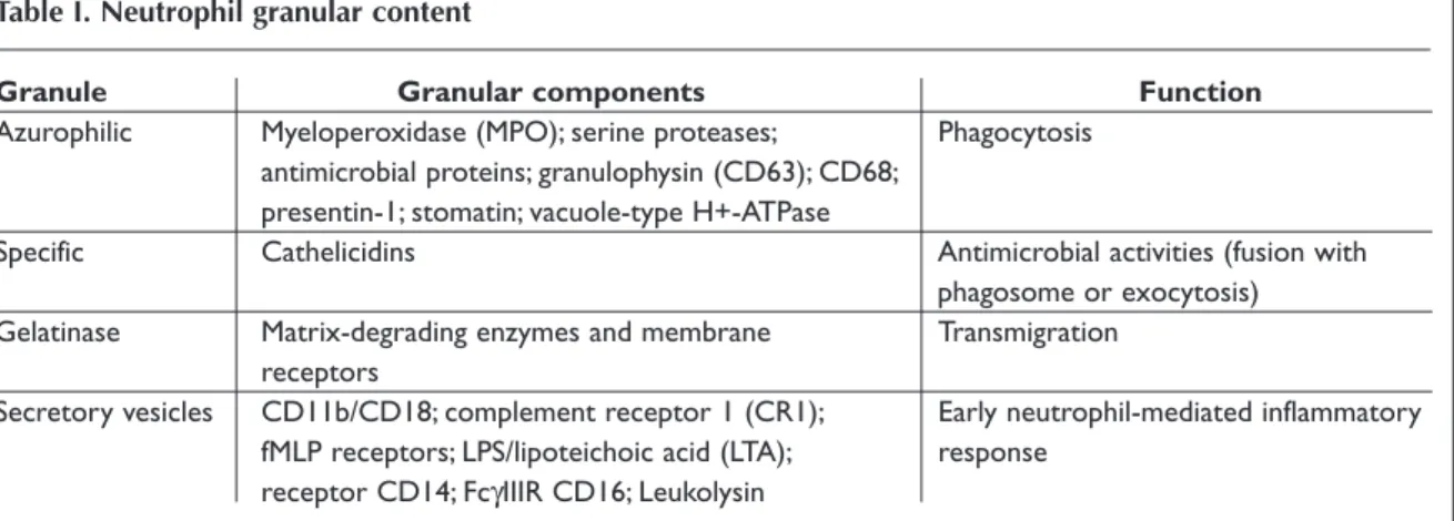
The majority of neutrophil functions, such as adhe-sion and phagocytosis, require the mobilization of cytoplasm granules and secretory vesicles, which contain antimicrobial proteins, enzymes, compo-nents of the respiratory burst oxidase and diverse membrane-bound receptors42(see Table I.).
Granu-les are formed in a process named granulopoiesis that follows myeloid cell differentiation, beginning at early promyelocyte state when immature trans-port vesicles arise from the Golgi apparatus and fuse together.43Specifically, neutrophils harbor four
types of granules, named azurophilic, specific and gelatinase granules, and secretory vesicles that appear sequentially during different granulopoiesis stages. Although granules share common structu-ral features, such as a phospholipidic bilayer mem-brane and an intra-granular matrix containing pro-teins for exocytosis or phagosome delivery, their protein content is quite different.42This difference
can be explained by the “targeting-by-timing” hypo-thesis, which proposes that the targeting of proteins into granules is determined by the time of their bi-osynthesis44,45and their targeting efficiency.46
Addi-tionally, gene expression is highly regulated by com-bination of myeloid transcription factors present at specific stages of cell development.45,47
The ability for exocytosis is different among gra-nules. Secretory vesicles have the highest propen-sity for exocytosis followed by gelatinase, specific and azurophilic granules.48A higher ability for
exocytosis is related to a higher density of vesicle-associated membrane protein (VAMP)-2, a fusoge-nic protein.49Besides the functions mentioned in
the table above, some granule components, such as defensins, azuricidin and human cathelicidin hCAP-18, also have the ability to induce CD4+ and
CD8+ T cells chemotaxis,50revealing the capacity of
neutrophils to participate in the amplification of the inflammatory response and to communicate with adaptive immune cells.
Phagocytosis
pha-The phagocytosis pathway via C3bi-opsonized targets, which is performed by complement recep-tor 3 (CR3), is different from FcγR via. One differen-ce is the fact that ingestion is independent of a rise of cytosolic-free Ca2+and of increased inositol
phosphate production.55Moreover, phagocytosis is
not accompanied by respiratory burst activation and arachidonic metabolites and cytokine produc-tion.56Finally, it also does not involve Rac or
Cdc42;57instead it is Rho activation, which follows
complement receptor stimulation, that leads to membrane protrusions extending over the surface of the opsonized particle, forming the “phagocytic cup”.58,59
Oxidative burst
Neutrophils have dependent and oxygen--independent microbicidal weapons.10
Oxygen-de-pendent pathways lead to the production of ROS by NADPH oxidase complex, in a process named oxi-dative or respiratory burst.
NADPH oxidase is an enzymatic complex com-posed of membrane and cytosolic components (see Figure 1.). After neutrophil activation, cytosolic components present in a heterotrimeric complex p47phox-p67phox-p40phoxare phosphorylated by
kina-ses, such as p38 MAPK and phosphatidylinositol--3-OH-kinase (PI3K),60and are translocated to the
plasma membrane, allowing its interaction with the membrane and with other oxidase proteins.61
Additionally, specific and azurophilic granules and secretory vesicles fuse with the plasma membrane to form the phagosome, thus allowing for gp91phox
and p22phoxinteraction with the membrane.62In
addition, phorbol 12-myristate 13-acetate (PMA) gocytes that eliminate pathogens and cellular
de-bris. The phagocytosis of opsonized particles has two different pathways: through Fcγ receptors (FcγRs) for immunoglobulin (IgG)-coated particles and complement receptors for complement-coa-ted particles. Human neutrophils constitutively ex-press low-affinity FcγRIIA (CD32) and FcγRIIIB (CD16b) receptors. Additionally, interferon-primed neutrophils express FcγRI (CD64), a high-affinity receptor.51After Fcγ receptor binding, pseudopods
are formed to surround and engulf the particle.52
Concomitantly, particles coated with complement fragment C3bi bind to activated CD11b/CD18 with ingestion occurring by “sinking” into the cell.52
Binding of IgG-opsonized particles to FcγRs trig-gers downstream activation of signaling pathways, which contribute for membrane extension over the particle, fusion and final closure of the “phagocytic cup”. After formation of the phagosome microbici-dal functions must be acquired, such as enzymes, vacular (V)-ATPases and NADPH oxidase complex. This maturation process occurs by the fusion with granules and secretory vesicles, as well as removal of components by vesicular fission, processes that require cell signaling. Contrarily to macrophages, phagosomes of neutrophils are not very acidic even with fusion of acidic granules and, consequently, of V-ATPases insertion (which pump H+into the
lu-men of the phagosome). This is due to NADPH oxi-dase action that alters the pH of phagosome by con-suming luminal H+ and producing ROS, thereby
di-minishing the efficiency of granule fusion and de-creasing its permeability to H+.53Altogether,
NADPH oxidase shows a more relevant role in pha-gosome acidification that is thought to be required for optimal phagosome maturation.54
Table I. Neutrophil granular content
Granule Granular components Function
Azurophilic Myeloperoxidase (MPO); serine proteases; Phagocytosis antimicrobial proteins; granulophysin (CD63); CD68;
presentin-1; stomatin; vacuole-type H+-ATPase
Specific Cathelicidins Antimicrobial activities (fusion with
phagosome or exocytosis) Gelatinase Matrix-degrading enzymes and membrane Transmigration
receptors
Secretory vesicles CD11b/CD18; complement receptor 1 (CR1); Early neutrophil-mediated inflammatory fMLP receptors; LPS/lipoteichoic acid (LTA); response
activation leads to p47phox, p67phoxand Rac2
trans-location to the plasma membrane as well as to spe-cific granules where, in complex with membrane components, they can produce O2-for a short
peri-od of time.63In granules NADPH oxidase activity is
dependent on protein kinase C (PKC)δ and PI3K for proper assembly.64,65NADPH oxidase complex
accepts electrons from reduced NADPH at the cyto-solic surface of the membrane and transfers them to O2on the extracellular surface of the membrane
leading to the downstream production of O2and
H2O or hypochlorous acid (HOCl). HOCl
constitu-tes a strong microbicidal agent as it oxidaconstitu-tes seve-ral bacterial molecules.66 However, this molecule
also damages most tissues.11Indeed, HOCl
media-tes the activation of pro-collagenases and pro-ge-latinases67and the production of cholesterol
chlo-rohydrins,68leading to tissue injury. Inappropriate
NADPH oxidase assembly and activation can be re-gulated and prevented by neutrophils through dif-ferential location of its components. Production of O2-by NADPH oxidase is only possible after certain
events, such as phosphorylation, translocation, and multiple conformational changes.69Neutrophils
can also regulate gene expression of NADPH oxida-se proteins at the transcriptional level by oxida-several transcription factors depending on cytokines and other inflammatory mediators.70
Some studies suggest that ROS and MPO activity are not enough for microbicidal capacity; instead, the proteases activated by the respiratory burst pro-cess are actually responsible for destroying inva-ding agents.71Additionally, ROS is implicated in the
regulation of cellular signaling pathways related with homeostasis, proliferation, differentiation, in-flammatory and immune responses.72It is also
teresting to refer that ROS have characteristics of in-tracellular messenger such as diffusibility and ra-pid turnover, allowing for spatial and temporal sig-naling specificity73,74in a nontoxic concentration.75
Intracellularly, ROS can alter redox state and oxidi-ze proteins.76Alteration of redox state can regulate
signaling pathways at many levels including recep-tor functions, enzymatic activity, transcription
fac-N E U T R O P H I L S: WA R R I O R S A N D C O M M A N D E R S
tors and gene expression patterns.73,77ROS can also
participate in pathways triggered by pro-inflam-matory cytokines and chemokines.75For example,
neutrophil apoptosis can be increased by activation of inositol phosphatase (SHIP) by the tyrosine ki-nase Lyn (of the Srk-family) via NADPH oxidase-de-rived ROS.78Another example is that ROS can
acti-vate transcription factor nuclear factor (NF)-kB that is involved in cytokine and chemokine expression by neutrophils in an inflammatory milieu.79
Addi-tionally, ROS can also modulate signaling pathways in adjacent cells such as macrophages and endo-thelial cells.75In the case of inflammation, ROS can
be also released extracellularly leading to tissue da-mage.
Apoptosis
In the absence of activating stimulus, neutrophil stay in circulation approximately 6 to 18 hours and undergo constitutive apoptosis,6a systematic and
stereotyped programmed cell death.80Therefore,
neutrophils are cells characterized for having a very short half-life.
Apoptosis has two different pathways: extrinsic and intrinsic. The extrinsic pathway is triggered by the ligation of external pro-apoptotic molecules to neutrophil surface death receptors, such as Fas (Apo-1/CD95), TNFRs, TRAIL-R2 and TRAIL-R3.81
Death receptors are cell surface receptors which contain cysteine-rich extracellular domains and a cytoplasmic motif named “death domain” (DD).82,83
These domains allow for the interaction of recep-tors and intracellular molecules of the apoptotic process,81like pro-caspases (cysteine-dependent
aspartate-specific proteases) that become activa-ted. The intrinsic pathway is triggered by the rease of cytochrome (cyt) c from the mitochondria le-ading to caspase activation. Neutrophil possesses caspases-1, -3, -4, -6, -7, -8, -9, -10 and -14.84In the
intrinsic pathway pro-apoptotic Bcl2proteins are
able to localize in the outer mitochondrial mem-brane, altering its permeability. Then cyt c is relea-sed into the cytoplasm, where it forms a complex with apoptosis protease-activating factor (APAF)-1, present in high levels, and caspase-9. Ultimately, caspase-9 cleaves downstream caspases and ini-tiates apoptosis.85
In the setting of inflammation the apoptotic de-lay is an important factor for the accumulation of neutrophils in the place of injury. Actually, host and bacterial anti-apoptotic mediators are also able to
delay neutrophil apoptosis, converging on com-mon intracellular molecular pathways. The process of extravasation itself can mediate cell survival,86by
cell contact with activated endothelium87and
expo-sure to cytokines. In fact, neutrophil apoptosis can be delayed by IL-1β, IL-2, IL-6, IL-15, TNF, INF-γ, G-CSF, GM-CSF, LPS88and IL-8.89It is interesting to
refer that IL-1, IL-6 and TNF can be produced by activated neutrophils to regulate themselves.90
An-timicrobial human β-defensins (hBDs), particularly hBD-3, also prolong the lifespan of neutrophils through down-regulation of truncated Bid (tBid) and up-regulation of Bcl-xL.91Most interesting are
the recent data which reveal that cathepsin D (sto-red in azurophilic granules) activates caspase-8 in a caspase-independent but ROS-dependent man-ner.92However, under inflammatory conditions
ca-thepsin D is kept in granules and neutrophil apop-tosis became reduced.92On the other hand,
bacte-rial molecules, such as LPS and LTA, can delay cons-titutive apoptosis via engagement of Toll-like receptors (TLR) 4 and TLR 2, respectively.93,94Also,
TLRs 7, 8 and 9 affect neutrophils life span. Contra-rily, after neutrophil phagocyte bacteria, apoptosis is accelerated95by a process called
phagocytosis-in-duced cell death (PICD). However, bacterial inges-tion has also been shown to delay apoptosis.80
Re-garding this matter, it has been shown that enga-gement of β2-integrins, which are involved in PICD,
can both accelerate and delay constitutive apopto-sis depending on their activation state and the ba-lance between death and survival signals, some of which appear to be cell lineage specific.96
Neutrophil apoptosis is controlled by Bcl2family
proteins, which include anti-apoptotic proteins such as Mcl-1, A1 and Bcl-xL, and pro-apoptotic
proteins, such as Bax-α, Bid, Bak and Bad.6,97The
ra-tio established between anti- and pro-apoptotic molecules, for example, upregulation of Bcl-xL and
downregulation of Bax-α,90regulates cell death
de-lay.98Activated neutrophils produce high amounts
of ROS that can increase apoptosis.99Recent studies
suggest that death receptor clustering and the sub-sequent activation of caspase-8 are ROS dependent and may occur independently of Fas ligation in spontaneous apoptosis.100In addition, many cell
signaling pathways and cell molecules known to be important in the regulation of apoptosis are in-fluenced by the redox environment.85Some studies
pointed out that early mitochondrial dysregula-tion101,102is a critical step in the induction of
mi-tochondria permeability, ROS can be released into cytoplasm,103promoting alternative cell death
pa-thway.104Additionally, exogenous ROS can act upon
mitochondrial membrane leading to its depolariza-tion, thus constituting initial stimuli for the activa-tion of the intrinsic pathway.85Therefore, in
inflam-mation sites where activated neutrophils produce higher quantities of ROS there exists a mechanism of apoptotic induction acting as a potential nega-tive feedback in the inflammatory response.85
The resolution of inflammation requires at least two different processes, neutrophil apoptosis and clearance of apoptotic cells by macrophages, pre-venting the host tissue damage by inappropriate release of cell enzymes and proteases.105Apoptotic
neutrophils externalize phosphatidilserine (PS) and express CD35 and CD63 at the cell surface thus facilitating the recognition by macrophages.106-108
These are not the only clearance signaling mecha-nisms. Apoptotic neutrophils have different ways to signal for macrophage ingestion and clearance, such as recruitment signals,109membrane changes
and cell receptors (e.g. vβ3/CD36, CD14, CD31, CR3/CR4 among others).110On the other hand, the
ingestion of apoptotic neutrophils, as well as opso-nized particles, by macrophages promotes their re-lease of soluble Fas ligand (FasL) which reacts with its receptor (FasR) present on neutrophils,10leading
to apoptosis of the remaining neutrophils.111
Ma-crophages phagocyte apoptotic cells via their αvβ3 integrin-CD36 complex that binds to neutrophils through thrombospodin and other unknown li-gands present on the cell surface.105CD36 is also
im-portant for the recognition of PS on apoptotic cell surface.112Additionally, the phagocytosis of
apop-totic neutrophils can inhibit IL-1β, IL-8, IL-10, GM-CSF, TNF, leukotriene C4and thromboxane B2
production by macrophages,106thus suppressing
the secretion of inflammatory mediators and, consequently, leading to the resolution of inflam-mation.10
NETosis
In 2004 it was shown by Brinkmann et al that neu-trophils were able to form extracellular structures, named neutrophil extracellular traps (NETs).113
NETs are assembled in cells activated by different pathways and are composed by nuclear compo-nents, such as chromatin DNA, histones anchored to this molecular backbone, and cytoplasmic
com-ponents, such as granular peptides and enzymes. Upon activation of several receptors, such as TLRs and FcRs, there is a triggering of a signal transdu-cing cascade that induces the activation of NADPH oxidase and downstream leads to the assembly of NETs, suggesting that its formation is ROS-depen-dent.114Due to its composition, NETs function as a
web of high concentration of antimicrobial pro-teins that can trap and kill Gram-positive and -ne-gative bacteria, but also fungus.115Neutrophils die
upon release of these structures. However, this is a form of cell death different from apoptosis and ne-crosis, named “NETosis”.116NETs represent an
un-conventional form of immune defense, because neutrophils can trap microorganisms that had no direct contact with the cell and the structure re-mains active even after neutrophils’ death, thus prolonging the microbicidal response. Although NETs assume a role in sites of infection, the extra-cellular release of proteins, such as cathepsin G and elastase, can cause tissue damage.117In addition,
the presence of nucleic acid can contribute to the development of autoimmune diseases like systemic lupus erythematosus (SLE) in which there is an exa-cerbated reaction against the host DNA.115
Neutrophils and Rheumatoid arthritis:
a case study
Based on their characteristics and functions it is easy to conclude that neutrophils have an enor-mous potential for host defense. However, they also have a non-specific action being dependent on components of the immune system to distinguish between invading agents and host antigens.10
The-refore, if the shutting off of the recruitment of neu-trophils is impaired or if the acute insult is not re-solved, these cells can inflict injury to the tissues.10
Specifically, in RA, neutrophils are involved in joint damage.118RA is a chronic inflammatory disease
mainly characterized by synovial hyperplasia and joint destruction. Although the etiopathology of this condition is not completely understood, it is known that it is associated with misregulation of both the cellular immune system and the cytokine network.119,120Neutrophils are prominent
partici-pants in the joint inflammation of patients with RA. Insight from a mouse animal model of autoimmu-ne arthritis has suggested that autoimmu-neutrophils are the first immune cells to infiltrate the joint at the early disease stage, with the earliest signs of ankle joint
inflammation correlating with the presence of neu-trophils in the synovia.121,122In the same model,
abrogation of the synovial inflammatory response was achieved by previous neutrophil depletion,122
further strengthening the importance of the neu-trophil as an essential component of the initial im-mune response in RA. In the clinical setting, we have observed that neutrophils are present in high numbers in the synovial tissue during the initial stages of RA (unpublished data),123 as already
re-ported in previous studies,1and are described to
persist in the synovial fluid during the perpetuation of this disease.124A patient in an active disease
sta-te may have a synovial fluid cellular infiltrasta-te con-taining up to 90% of neutrophils.125However, it is
as-sumed that they are largely absent in joint tissues, except in cartilage and pannus-cartilage interface where in the early stages of disease cartilage dama-ge occurs due to the action of serine and metallo-proteinases (MMPs)126,127stored in their granules
and by the production of ROS and chlorinated oxi-dants.118Despite these findings, neutrophils are
ge-nerally an understudied cell in RA and are often considered simply as a terminally differentiated cell of the innate immune system, merely with cytoto-xic potential and capacity to inflict tissue damage, and lacking the potential to interfere with the ini-tiation and development of an autoimmune disea-se such as RA.
In RA, neutrophils are recruited into inflamed joints by chemoattractants present in synovial fluid, such as LTB4, C5a, IL-8 and TGF-β. When
the-se cells arrive at the joints, they become expothe-sed to a wide spectrum of pro-inflammatory cytokines and growth factors such as IL-1β, IL-6, TNF and GM-CSF.128In addition, IL-17 appears to stimulate
the production of cytokines that attract neutrophils to the site of inflammation, stimulate granulopoie-sis and/or induce production of chemokines.129,130
Other important activating factor appears to be IgG-containing immune complexes that trigger sti-mulation of the respiratory burst and degranula-tion.131,132In addition to cytokines, other stimuli,
such as adhesion, transmigration and hypoxia, are also able to activate gene expression.
There are many differences in protein expres-sion between synovial fluid and blood-derived neu-trophils in RA patients. Neuneu-trophils from synovial fluid have mobilized pre-formed molecules from intracellular stores to the cell surface and activated gene expression resulting from enhanced trans-cription and translation.133,134Consequently, several
gene products are up-regulated such as IL-8, CXCR4, TNFR and MMP-9, allowing not only the up-regulation of cell functions but also the develop-ment of new cellular responses, e.g. the antigen-presentation to T cell via activated major histocom-patibility complex (MHC) classe II expression.125In
fact, synovial fluid neutrophils were described to acquire an antigen-presenting function through cytokine stimulation, allowing T cell function mo-dulation, an important feature in RA pathology.134
This hypothesis is conflicting with the traditional view of neutrophils as a terminally differentiated cell.135
Moreover, recent data from animal studies show that neutrophils play a crucial role in the initiation and progression of arthritis. For example, in the K/BxN mice model the administration of a neu-trophil-depleting antibody before or simultaneous-ly with disease induction prevent its initiation. Also, its administration performed until 3 days after arthritis induction can revert its progression.122
Considering RA as a T cell driven pathology, some authors assumed that the differences betwe-en rheumatoid and non-rheumatoid circulating neutrophils are a consequence rather than an ini-tially event in RA.10However, therapies in use and
others being tested in clinical trials for RA are com-monly anti-inflammatory drugs that have profound effect on neutrophil functions, molecules (e.g. -adhesion) and activation (e.g. cytokine anti-bodies),136reinforcing the idea that these cells are
relevant players in RA. A good and simple example of this is the fact that methotrexate, the gold stan-dard drug for the treatment of RA, abrogates the de-layed neutrophil apoptosis observed in early RA patients.137
Conclusion
The old view of the neutrophil as a terminally dif-ferentiated cell completely focused on destroying pathogens and tissues is no longer in line with the new data related with their cellular and molecular mechanisms. In fact, neutrophils are unique cells in their ability to be immune decision-shapers and to induce damage and healing. The knowledge about the neutrophil complex biology and their role in immune-mediated inflammatory diseases is expected to reveal new promising therapeutic tar-gets. Hopefully, time will come when specific neu-trophil targeted therapies will play a role in the treatment of diseases such as RA.
Corresponding author
Rita Cascão
Rheumatology Research Unit Instituto de Medicina Molecular
Faculdade de Medicina da Universidade de Lisboa Edificio Egas Moniz, Av. Prof. Egas Moniz
1649-028 Lisboa, Portugal E-mail: [email protected]
Acknowledgments
This study was supported by the Fundação para a Ciên-cia e Tecnologia (SFRH/BD/40513/2007) and by a grant from Sociedade Portuguesa de Reumatologia/Schering--Plough 2005.
References
1. Nathan C. Neutrophils and immunity: challenges and opportunities. Nat Rev Immunol 2006;6:173-182. 2. Skokowa J, Welte K. LEF-1 is a decisive transcription factor in neutrophil granulopoiesis. Ann N Y Acad Sci 2007;1106:143-151.
3. Skokowa J, Cario G, Uenalan M et al. LEF-1 is crucial for neutrophil granulocytopoiesis and its expression is severely reduced in congenital neutropenia. Nat Med 2006;12:1191-1197.
4. Kaushansky K. Lineage-specific hematopoietic growth factors. N Engl J Med 2006;354:2034-2045. 5. Christopher MJ, Link DC. Regulation of neutrophil
homeostasis. Curr Opin Hematol 2007;14:3-8. 6. Akgul C, Moulding DA, Edwards SW. Molecular
con-trol of neutrophil apoptosis. FEBS Lett 2001;487:318--322.
7. Starckx S, Van den Steen PE, Wuyts A, Van Damme J, Opdenakker G. Neutrophil gelatinase B and che-mokines in leukocytosis and stem cell mobilization. Leuk Lymphoma 2002;43:233-241.
8. Nagase H, Miyamasu M, Yamaguchi M et al. Cytoki-ne-mediated regulation of CXCR4 expression in hu-man neutrophils. J Leukoc Biol 2002;71:711-717. 9. Martin C, Burdon PC, Bridger G, Gutierrez-Ramos JC,
Williams TJ, Rankin SM. Chemokines acting via CX-CR2 and CXCR4 control the release of neutrophils from the bone marrow and their return following se-nescence. Immunity 2003;19:583-593.
10. Witko-Sarsat V, Rieu P, Descamps-Latscha B, Lesavre P, Halbwachs-Mecarelli L. Neutrophils: molecules, functions and pathophysiological aspects. Lab Invest 2000;80:617-653.
11. Burg ND, Pillinger MH. The neutrophil: function and regulation in innate and humoral immunity. Clin Im-munol 2001;99:7-17.
12. Moore KL, Patel KD, Bruehl RE et al. P-selectin glyco-protein ligand-1 mediates rolling of human neutrop-hils on P-selectin. J Cell Biol 1995;128:661-671. 13. Patel KD, Moore KL, Nollert MU, McEver RP.
Neu-trophils use both shared and distinct mechanisms to adhere to selectins under static and flow conditions. J Clin Invest 1995;96:1887-1896.
14. Steegmaier M, Borges E, Berger J, Schwarz H,
Vestwe-ber D. The E-selectin-ligand ESL-1 is located in the Golgi as well as on microvilli on the cell surface. J Cell Sci 1997;110 ( Pt 6):687-694.
15. Li X, Steeber DA, Tang ML, Farrar MA, Perlmutter RM, Tedder TF. Regulation of L-selectin-mediated rolling through receptor dimerization. J Exp Med 1998;188:1385-1390.
16. Simon SI, Hu Y, Vestweber D, Smith CW. Neutrophil tethering on E-selectin activates beta 2 integrin bin-ding to ICAM-1 through a mitogen-activated protein kinase signal transduction pathway. J Immunol 2000;164:4348-4358.
17. Hidari KI, Weyrich AS, Zimmerman GA, McEver RP. Engagement of P-selectin glycoprotein ligand-1 enhances tyrosine phosphorylation and activates mi-togen-activated protein kinases in human neutrop-hils. J Biol Chem 1997;272:28750-28756.
18. Urzainqui A, Serrador JM, Viedma F et al. ITAM-ba-sed interaction of ERM proteins with Syk mediates signaling by the leukocyte adhesion receptor PSGL-1. Immunity 2002;17:401-412.
19. Ba X, Chen C, Gao Y, Zeng X. Signaling function of PS-GL-1 in neutrophil: tyrosine-phosphorylation-de-pendent and c-Abl-involved alteration in the F-actin-based cytoskeleton. J Cell Biochem 2005;94:365-373. 20. Bargatze RF, Kurk S, Butcher EC, Jutila MA.
Neutrop-hils roll on adherent neutropNeutrop-hils bound to cytokine-induced endothelial cells via L-selectin on the rolling cells. J Exp Med 1994;180:1785-1792.
21. Cronstein BN, Kimmel SC, Levin RI, Martiniuk F, Weissmann G. A mechanism for the antiinflamma-tory effects of corticosteroids: the glucocorticoid re-ceptor regulates leukocyte adhesion to endothelial cells and expression of endothelial-leukocyte adhe-sion molecule 1 and intercellular adheadhe-sion molecule 1. Proc Natl Acad Sci U S A 1992;89:9991-9995. 22. Filep JG, Delalandre A, Payette Y, Foldes-Filep E.
Glu-cocorticoid receptor regulates expression of L-selec-tin and CD11/CD18 on human neutrophils. Circula-tion 1997;96:295-301.
23. Pillinger MH, Capodici C, Rosenthal P, et al. Modes of action of aspirin-like drugs: salicylates inhibit erk ac-tivation and integrin-dependent neutrophil adhesi-on. Proc Natl Acad Sci U S A 1998;95:14540-14545. 24. Del Maschio A, Zanetti A, Corada M et al.
Polymorphonuclear leukocyte adhesion triggers the disorganization of endothelial cell-to-cell adherens junctions. J Cell Biol 1996;135:497-510.
25. Martin-Padura I, Lostaglio S, Schneemann M et al. Junctional adhesion molecule, a novel member of the immunoglobulin superfamily that distributes at intercellular junctions and modulates monocyte transmigration. J Cell Biol 1998;142:117-127.
26. Muller WA, Weigl SA, Deng X, Phillips DM. PECAM-1 is required for transendothelial migration of leuko-cytes. J Exp Med 1993;178:449-460.
27. Berman ME, Muller WA. Ligation of platelet/endo-thelial cell adhesion molecule 1 (PECAM-1/CD31) on monocytes and neutrophils increases binding capa-N E U T R O P H I L S: WA R R I O R S A N D C O M M A N D E R S
city of leukocyte CR3 (CD11b/CD18). J Immunol 1995;154:299-307.
28. Colgan SP. Lipid mediators in epithelial cell-cell inte-ractions. Cell Mol Life Sci 2002;59:754-760.
29. McMahon B, Godson C. Lipoxins: endogenous regu-lators of inflammation. Am J Physiol Renal Physiol 2004;286:189-201.
30. Louis NA, Hamilton KE, Colgan SP. Lipid mediator networks and leukocyte transmigration. Prostaglan-dins Leukot Essent Fatty Acids 2005;73:197-202. 31. Kunkel SL, Standiford T, Kasahara K, Strieter RM.
In-terleukin-8 (IL-8): the major neutrophil chemotactic factor in the lung. Exp Lung Res 1991;17:17-23. 32. Chadwick VS, Mellor DM, Myers DB et al. Production
of peptides inducing chemotaxis and lysosomal en-zyme release in human neutrophils by intestinal bac-teria in vitro and in vivo. Scand J Gastroenterol 1988;23:121-128.
33. Bohnsack JF, Chang J, Zhou X, Yednock TA. Mecha-nisms of beta 1 integrin-dependent adherence of granulocytic HL60 to fibronectin. J Leukoc Biol 1995;57:592-599.
34. Loike JD, Cao L, Budhu S et al. Differential regulation of beta1 integrins by chemoattractants regulates neutrophil migration through fibrin. J Cell Biol 1999;144:1047-1056.
35. Lauffenburger DA, Horwitz AF. Cell migration: a physically integrated molecular process. Cell 1996;84:359-369.
36. Seveau S, Keller H, Maxfield FR, Piller F, Halbwachs-Mecarelli L. Neutrophil polarity and locomotion are associated with surface redistribution of leukosialin (CD43), an antiadhesive membrane molecule. Blood 2000;95:2462-2470.
37. Kobayashi Y. The role of chemokines in neutrophil biology. Front Biosci 2008;13:2400-2407.
38. Scapini P, Lapinet-Vera JA, Gasperini S, Calzetti F, Bazzoni F, Cassatella MA. The neutrophil as a cellular source of chemokines. Immunol Rev 2000;177:195--203.
39. Schroder AK, von der Ohe M, Kolling U et al. Polymorphonuclear leucocytes selectively produce anti-inflammatory interleukin-1 receptor antagonist and chemokines, but fail to produce pro-inflamma-tory mediators. Immunology 2006;119:317-327. 40. Cassatella MA, Huber V, Calzetti F et al.
Interferon-activated neutrophils store a TNF-related apoptosis-inducing ligand (TRAIL/Apo-2 ligand) intracellular pool that is readily mobilizable following exposure to proinflammatory mediators. J Leukoc Biol 2006;79:123-132.
41. Cassatella MA. The production of cytokines by polymorphonuclear neutrophils. Immunol Today 1995;16:21-26.
42. Faurschou M, Borregaard N. Neutrophil granules and secretory vesicles in inflammation. Microbes Infect 2003;5:1317-1327.
43. Hartmann J, Scepek S, Lindau M. Regulation of gra-nule size in human and horse eosinophils by number
of fusion events among unit granules. J Physiol 1995;483(Pt 1):201-209.
44. Borregaard N, Sehested M, Nielsen BS, Sengelov H, Kjeldsen L. Biosynthesis of granule proteins in nor-mal human bone marrow cells. Gelatinase is a mar-ker of terminal neutrophil differentiation. Blood 1995;85:812-817.
45. Cowland JB, Borregaard N. The individual regulation of granule protein mRNA levels during neutrophil maturation explains the heterogeneity of neutrophil granules. J Leukoc Biol 1999;66:989-995.
46. Arnljots K, Sorensen O, Lollike K, Borregaard N. Ti-ming, targeting and sorting of azurophil granule pro-teins in human myeloid cells. Leukemia 1998;12: 1789-1795.
47. Bjerregaard MD, Jurlander J, Klausen P, Borregaard N, Cowland JB. The in vivo profile of transcription fac-tors during neutrophil differentiation in human bone marrow. Blood 2003;101:4322-4332.
48. Sengelov H, Follin P, Kjeldsen L, Lollike K, Dahlgren C, Borregaard N. Mobilization of granules and secre-tory vesicles during in vivo exudation of human neu-trophils. J Immunol 1995;154:4157-4165.
49. Borregaard N, Sorensen OE, Theilgaard-Monch K. Neutrophil granules: a library of innate immunity proteins. Trends Immunol 2007;28:340-345.
50. Chertov O, Michiel DF, Xu L et al. Identification of de-fensin-1, defensin-2, and CAP37/azurocidin as T-cell chemoattractant proteins released from interleukin-8-stimulated neutrophils. J Biol Chem 1996;271:2935--2940.
51. McKenzie SE, Schreiber AD. Fc gamma receptors in phagocytes. Curr Opin Hematol 1998;5:16-21. 52. Greenberg S, Grinstein S. Phagocytosis and innate
immunity. Curr Opin Immunol 2002;14:136-145. 53. Jankowski A, Scott CC, Grinstein S. Determinants of
the phagosomal pH in neutrophils. J Biol Chem 2002;277:6059-6066.
54. Lee WL, Harrison RE, Grinstein S. Phagocytosis by neutrophils. Microbes Infect 2003;5:1299-1306. 55. Fallman M, Lew DP, Stendahl O, Andersson T.
Recep-tor-mediated phagocytosis in human neutrophils is associated with increased formation of inositol phosphates and diacylglycerol. Elevation in cytosolic free calcium and formation of inositol phosphates can be dissociated from accumulation of diacylglyce-rol. J Clin Invest 1989;84:886-891.
56. Yamamoto K, Johnston RB, Jr. Dissociation of pha-gocytosis from stimulation of the oxidative metabolic burst in macrophages. J Exp Med 1984;159:405-416. 57. Caron E, Hall A. Identification of two distinct
mecha-nisms of phagocytosis controlled by different Rho GTPases. Science 1998;282:1717-1721.
58. Massol P, Montcourrier P, Guillemot JC, Chavrier P. Fc receptor-mediated phagocytosis requires CDC42 and Rac1. Embo J 1998;17:6219-6229.
59. Swanson JA, Baer SC. Phagocytosis by zippers and triggers. Trends Cell Biol 1995;5:89-93.
Kuwaba-ra M. Roles of p38 MAPK, PKC and PI3-K in the signa-ling pathways of NADPH oxidase activation and pha-gocytosis in bovine polymorphonuclear leukocytes. FEBS Lett 2000;467:253-258.
61. Nauseef WM. The NADPH-dependent oxidase of phagocytes. Proc Assoc Am Physicians 1999;111:373--382.
62. Borregaard N. The respiratory burst of phagocytosis: biochemistry and subcellular localization. Immunol Lett 1985;11:165-171.
63. Ambruso DR, Cusack N, Thurman G. NADPH oxida-se activity of neutrophil specific granules: require-ments for cytosolic components and evidence of as-sembly during cell activation. Mol Genet Metab 2004;81:313-321.
64. Sergeant S, McPhail LC. Opsonized zymosan stimu-lates the redistribution of protein kinase C isoforms in human neutrophils. J Immunol 1997;159:2877--2885.
65. Brown GE, Stewart MQ, Liu H, Ha VL, Yaffe MB. A no-vel assay system implicates PtdIns(3,4)P(2), Pt-dIns(3)P, and PKC delta in intracellular production of reactive oxygen species by the NADPH oxidase. Mol Cell 2003;11:35-47.
66. Weiss SJ. Tissue destruction by neutrophils. N Engl J Med 1989;320:365-376.
67. Weiss SJ, Peppin G, Ortiz X, Ragsdale C, Test ST. Oxi-dative autoactivation of latent collagenase by human neutrophils. Science 1985;227:747-749.
68. Heinecke JW, Li W, Mueller DM, Bohrer A, Turk J. Cholesterol chlorohydrin synthesis by the myelope-roxidase-hydrogen peroxide-chloride system: poten-tial markers for lipoproteins oxidatively damaged by phagocytes. Biochemistry 1994;33:10127-10136. 69. Quinn MT, Gauss KA. Structure and regulation of the
neutrophil respiratory burst oxidase: comparison with nonphagocyte oxidases. J Leukoc Biol 2004;76:760-781.
70. Skalnik DG. Transcriptional mechanisms regulating myeloid-specific genes. Gene 2002;284:1-21.
71. Reeves EP, Lu H, Jacobs HL et al. Killing activity of neutrophils is mediated through activation of protea-ses by K+ flux. Nature 2002;416:291-297.
72. Touyz RM. Reactive oxygen species as mediators of calcium signaling by angiotensin II: implications in vascular physiology and pathophysiology. Antioxid Redox Signal 2005;7:1302-1314.
73. Tonks NK. Redox redux: revisiting PTPs and the con-trol of cell signaling. Cell 2005;121:667-670.
74. Wolin MS, Ahmad M, Gupte SA. Oxidant and redox signaling in vascular oxygen sensing mechanisms: basic concepts, current controversies, and potential importance of cytosolic NADPH. Am J Physiol Lung Cell Mol Physiol 2005;289:159-173.
75. Fialkow L, Wang Y, Downey GP. Reactive oxygen and nitrogen species as signaling molecules regulating neutrophil function. Free Radic Biol Med 2007;42:153-164.
76. Liang B, Petty HR. Imaging neutrophil activation:
analysis of the translocation and utilization of NAD(P)H-associated autofluorescence during anti-body-dependent target oxidation. J Cell Physiol 1992;152:145-156.
77. Suzuki YJ, Nagase H, Nie K, Park AM. Redox control of growth factor signaling: recent advances in cardio-vascular medicine. Antioxid Redox Signal 2005;7:829--834.
78. Gardai S, Whitlock BB, Helgason C et al. Activation of SHIP by NADPH oxidase-stimulated Lyn leads to enhanced apoptosis in neutrophils. J Biol Chem 2002;277:5236-5246.
79. Nick JA, Avdi NJ, Young SK et al. Selective activation and functional significance of p38alpha mitogen-ac-tivated protein kinase in lipopolysaccharide-stimula-ted neutrophils. J Clin Invest 1999;103:851-858. 80. Anwar S, Whyte MK. Neutrophil apoptosis in
infec-tious disease. Exp Lung Res 2007;33:519-528. 81. Akgul C, Edwards SW. Regulation of neutrophil
apop-tosis via death receptors. Cell Mol Life Sci 2003;60:2402-2408.
82. Ashkenazi A, Dixit VM. Death receptors: signaling and modulation. Science 1998;281:1305-1308. 83. Nagata S. Apoptosis by death factor. Cell
1997;88:355--365.
84. Goepel F, Weinmann P, Schymeinsky J, Walzog B. Identification of caspase-10 in human neutrophils and its role in spontaneous apoptosis. J Leukoc Biol 2004;75:836-843.
85. Melley DD, Evans TW, Quinlan GJ. Redox regulation of neutrophil apoptosis and the systemic inflamma-tory response syndrome. Clin Sci (Lond) 2005;108: 413-424.
86. Watson RW, Rotstein OD, Nathens AB, Parodo J, Marshall JC. Neutrophil apoptosis is modulated by endothelial transmigration and adhesion molecule engagement. J Immunol 1997;158:945-953.
87. Ginis I, Faller DV. Protection from apoptosis in hu-man neutrophils is determined by the surface of ad-hesion. Am J Physiol 1997;272:295-309.
88. Girard D, Paquet ME, Paquin R, Beaulieu AD. Diffe-rential effects of interleukin-15 (IL-15) and IL-2 on human neutrophils: modulation of phagocytosis, cytoskeleton rearrangement, gene expression, and apoptosis by IL-15. Blood 1996;88:3176-3184. 89. Leuenroth SJ, Grutkoski PS, Ayala A, Simms HH.
Sup-pression of PMN apoptosis by hypoxia is dependent on Mcl-1 and MAPK activity. Surgery 2000;128:171--177.
90. Ocana MG, Asensi V, Montes AH, Meana A, Celada A, Valle-Garay E. Autoregulation mechanism of human neutrophil apoptosis during bacterial infection. Mol Immunol 2007.
91. Nagaoka I, Niyonsaba F, Tsutsumi-Ishii Y, Tamura H, Hirata M. Evaluation of the effect of human beta-de-fensins on neutrophil apoptosis. Int Immunol 2008;20:543-553.
92. Conus S, Perozzo R, Reinheckel T et al. Caspase-8 is activated by cathepsin D initiating neutrophil apop-N E U T R O P H I L S: WA R R I O R S A N D C O M M A N D E R S
tosis during the resolution of inflammation. J Exp Med 2008;205:685-698.
93. Sabroe I, Prince LR, Jones EC et al. Selective roles for Toll-like receptor (TLR)2 and TLR4 in the regulation of neutrophil activation and life span. J Immunol 2003;170:5268-5275.
94. Lotz S, Aga E, Wilde I et al. Highly purified lipotei-choic acid activates neutrophil granulocytes and de-lays their spontaneous apoptosis via CD14 and TLR2. J Leukoc Biol 2004;75:467-477.
95. Coxon A, Rieu P, Barkalow FJ et al. A novel role for the beta 2 integrin CD11b/CD18 in neutrophil apoptosis: a homeostatic mechanism in inflammation. Immu-nity 1996;5:653-666.
96. Peng SL. Neutrophil apoptosis in autoimmunity. J Mol Med 2006;84:122-125.
97. Weinmann P, Gaehtgens P, Walzog B. Bcl-Xl- and Bax-alpha-mediated regulation of apoptosis of human neutrophils via caspase-3. Blood 1999;93:3106-3115. 98. van Delft MF, Huang DC. How the Bcl-2 family of
proteins interact to regulate apoptosis. Cell Res 2006;16:203-213.
99. Watson RW. Redox regulation of neutrophil apopto-sis. Antioxid Redox Signal 2002;4:97-104.
100. Scheel-Toellner D, Wang K, Assi LK et al. Clustering of death receptors in lipid rafts initiates neutrophil spontaneous apoptosis. Biochem Soc Trans 2004;32:679-681.
101. Chandra J, Samali A, Orrenius S. Triggering and mo-dulation of apoptosis by oxidative stress. Free Radic Biol Med 2000;29:323-333.
102. Kroemer G. Mitochondrial control of apoptosis: an introduction. Biochem Biophys Res Commun 2003;304:433-435.
103. Simon HU, Haj-Yehia A, Levi-Schaffer F. Role of re-active oxygen species (ROS) in apoptosis induction. Apoptosis 2000;5:415-418.
104. Maianski NA, Roos D, Kuijpers TW. Tumor necrosis factor alpha induces a caspase-independent death pathway in human neutrophils. Blood 2003;101: 1987-1995.
105. Savill J, Haslett C. Granulocyte clearance by apopto-sis in the resolution of inflammation. Semin Cell Biol 1995;6:385-393.
106. Fadok VA, Bratton DL, Frasch SC, Warner ML, Hen-son PM. The role of phosphatidylserine in recogni-tion of apoptotic cells by phagocytes. Cell Death Dif-fer 1998;5:551-562.
107. Hall SE, Savill JS, Henson PM, Haslett C. Apoptotic neutrophils are phagocytosed by fibroblasts with participation of the fibroblast vitronectin receptor and involvement of a mannose/fucose-specific lec-tin. J Immunol 1994;153:3218-3227.
108. Beinert T, Munzing S, Possinger K, Krombach F. In-creased expression of the tetraspanins CD53 and CD63 on apoptotic human neutrophils. J Leukoc Bi-ol 2000;67:369-373.
109. Ravichandran KS. «Recruitment signals» from apop-totic cells: invitation to a quiet meal. Cell
2003;113:817-820.
110. Walker A, Ward C, Taylor EL et al. Regulation of neu-trophil apoptosis and removal of apoptotic cells. Curr Drug Targets Inflamm Allergy 2005;4:447-454. 111. Brown SB, Savill J. Phagocytosis triggers
macropha-ge release of Fas ligand and induces apoptosis of bystander leukocytes. J Immunol 1999;162:480-485. 112. Fadok VA, Warner ML, Bratton DL, Henson PM.
CD36 is required for phagocytosis of apoptotic cells by human macrophages that use either a phospha-tidylserine receptor or the vitronectin receptor (al-pha v beta 3). J Immunol 1998;161:6250-6257. 113. Brinkmann V, Reichard U, Goosmann C et al.
Neu-trophil extracellular traps kill bacteria. Science 2004;303:1532-1535.
114. Fuchs TA, Abed U, Goosmann C et al. Novel cell de-ath program leads to neutrophil extracellular traps. J Cell Biol 2007;176:231-241.
115. Wartha F, Beiter K, Normark S, Henriques-Normark B. Neutrophil extracellular traps: casting the NET over pathogenesis. Curr Opin Microbiol 2007;10:52--56.
116. Steinberg BE, Grinstein S. Unconventional roles of the NADPH oxidase: signaling, ion homeostasis, and cell death. Sci STKE 2007;2007:pe11.
117. Lee WL, Grinstein S. Immunology. The tangled webs that neutrophils weave. Science 2004;303:1477-1478. 118. Edwards SW, Hallett MB. Seeing the wood for the trees: the forgotten role of neutrophils in rheuma-toid arthritis. Immunol Today 1997;18:320-324. 119. Beaulieu AD, McColl SR. Differential expression of
two major cytokines produced by neutrophils, inter-leukin-8 and the interleukin-1 receptor antagonist, in neutrophils isolated from the synovial fluid and peripheral blood of patients with rheumatoid arthri-tis. Arthritis Rheum 1994;37:855-859.
120. Ridderstad A, Abedi-Valugerdi M, Moller E. Cytoki-nes in rheumatoid arthritis. Ann Med 1991;23:219--223.
121. Mandik-Nayak L, Allen PM. Initiation of an autoim-mune response: insights from a transgenic model of rheumatoid arthritis. Immunol Res 2005;32:5-13. 122. Wipke BT, Allen PM. Essential role of neutrophils in
the initiation and progression of a murine model of rheumatoid arthritis. J Immunol 2001;167:1601-1608.
123. Mourão AF, Canhão H, Cascão R et al. Synovial tis-sue highly infiltrated by neutrophils in the very early phase of rheumatoid arthritis. Submitted.
124. Mohr W, Pelster B, Wessinghage D. Polymorphonu-clear granulocytes in rheumatic tissue destruction. VI. The occurrence of PMNs in menisci of patients with rheumatoid arthritis. Rheumatol Int 1984;5:39--44.
125. Cross A, Bakstad D, Allen JC, Thomas L, Moots RJ, Edwards SW. Neutrophil gene expression in rheu-matoid arthritis. Pathophysiology 2005;12:191-202. 126. Chatham WW, Swaim R, Frohsin H, Jr., Heck LW,
articular cartilage by neutrophils in synovial fluid. Arthritis Rheum 1993;36:51-58.
127. Larbre JP, Moore AR, Da Silva JA, Iwamura H, Ioan-nou Y, Willoughby DA. Direct degradation of articu-lar cartilage by rheumatoid synovial fluid: contribu-tion of proteolytic enzymes. J Rheumatol 1994;21: 1796-1801.
128. Feldmann M, Brennan FM, Chantry D et al. Cytoki-ne production in the rheumatoid joint: implications for treatment. Ann Rheum Dis 1990;49:480-486. 129. Kotake S, Udagawa N, Takahashi N et al. IL-17 in
synovial fluids from patients with rheumatoid arthritis is a potent stimulator of osteoclastogenesis. J Clin Invest 1999;103:1345-1352.
130. Jovanovic DV, Martel-Pelletier J, Di Battista JA et al. Stimulation of 92-kd gelatinase (matrix metallopro-teinase 9) production by interleukin-17 in human monocyte/macrophages: a possible role in rheuma-toid arthritis. Arthritis Rheum 2000;43:1134-1144. 131. Robinson JJ, Watson F, Bucknall RC, Edwards SW.
Role of Fc gamma receptors in the activation of neu-trophils by soluble and insoluble immunoglobulin aggregates isolated from the synovial fluid of pa-tients with rheumatoid arthritis. Ann Rheum Dis 1994;53:515-520.
132. Robinson J, Watson F, Bucknall RC, Edwards SW. Ac-tivation of neutrophil reactive-oxidant production by synovial fluid from patients with inflammatory joint disease. Soluble and insoluble immunoglobu-lin aggregates activate different pathways in primed and unprimed cells. Biochem J 1992;286:345-351.
133. Quayle JA, Watson F, Bucknall RC, Edwards SW. Neu-trophils from the synovial fluid of patients with rheumatoid arthritis express the high affinity immu-noglobulin G receptor, Fc gamma RI (CD64): role of immune complexes and cytokines in induction of receptor expression. Immunology 1997;91:266-273. 134. Cross A, Bucknall RC, Cassatella MA, Edwards SW,
Moots RJ. Synovial fluid neutrophils transcribe and express class II major histocompatibility complex molecules in rheumatoid arthritis. Arthritis Rheum 2003;48:2796-2806.
135. Iking-Konert C, Ostendorf B, Sander O et al. Trans-differentiation of polymorphonuclear neutrophils to dendritic-like cells at the site of inflammation in rheumatoid arthritis: evidence for activation by T cells. Ann Rheum Dis 2005;64:1436-1442.
136. Arend WP, Dayer JM. Inhibition of the production and effects of interleukin-1 and tumor necrosis fac-tor alpha in rheumatoid arthritis. Arthritis Rheum 1995;38:151-160.
137. Weinmann P, Moura RA, Caetano-Lopes JR et al. De-layed neutrophil apoptosis in very early rheumatoid arthritis patients is abrogated by methotrexate the-rapy. Clin Exp Rheumatol 2007;25:885-887.
N E U T R O P H I L S: WA R R I O R S A N D C O M M A N D E R S