EN
SA
IO
C
LÍ
N
IC
O
/
C
LI
N
IC
A
L
TR
IA
L
INTRODUCTION
Autoimmune hepatitis (AIH) is a disease of unknown etiology that causes progressive destruction of the hepatic parenchyma and usually leads to death when untreated. It can be effectively treated with corticosteroids alone or associated with azathioprine. This treatment results in greater resolution of symptoms and of laboratory and histological abnormalities, as well as greater survival(35).
It is recommended to try to do away with medications after the achievement of clinical, biochemical and histological remission(9). Relapse probability, however, reaches 50% in 6 months and 70% after 1 year, and sustained remission rates go down to between 10%-15% in 5 years(8, 22).Fatal relapses were also described, but are fortunately rare(17).This unfavorable prognosis is worsened by the increase in corticosteroid-related side-effects after repeated treatment using higher doses of corticosteroids(37). This raises the question of whether or not it is worthwhile stopping the treatment at all. On the other hand, repeated trials of treatment withdrawal can
result in slightly more optimistic relapse rates of 63% in 1 year and 75% in 5 years(10). It is possible to maintain most AIH patients in remission on maintenance doses of prednisone, or of prednisone/azathioprine (PA) or of azathioprine, but there is the added theoretical risk of malignancies associated with the immunosuppressor(19, 36). Therefore, the management of patients in remission
is still a matter of debate.
Chloroquine diphosphate is an antimalarial drug that has been used for the treatment of rheumatological diseases for at the least 5 decades(15, 23, 27). It improves clinical and laboratory features of rheumatoid arthritis(16, 28) and delays or prevents clinical exacerbations of systemic lupus erythematosus(23). Like AIH, these conditions seem to be related to abnormalities in the regulation of the immune system(25, 31).
Anecdotal reports suggested that chloroquine could be used as an alternative to corticosteroids for chronic hepatitis of unknown origin, but, on those oc-casion, hepatitis C virus had not been identified yet, and the parameters for diagnosing AIH had not been
CHLOROQUINE FOR THE
MAINTENANCE OF REMISSION
OF AUTOIMMUNE HEPATITIS:
results of a pilot study
Marcos MUCENIC
1, Evandro Sobroza de MELLO
2and Eduardo Luiz R. CANÇADO
1, 3ABSTRACT - Background - Due to the risks related to long-term treatment with prednisone and azathioprine, most clinicians
try to withdraw these drugs when patients with autoimmune hepatitis are in remission. However, there is a high probability of relapse, and most patients end up receiving maintenance treatment. Aim - To evaluate the safety and efficacy of maintenance treatment with chloroquine in the prevention of autoimmune hepatitis relapses. Methods - Classical treatment was stopped after achievement of biochemical and histological remission of autoimmune hepatitis. Chloroquine diphosphate, 250 mg daily, was given for at least 12 months or until the occurrence of relapses defined by levels of aminotransferases at least twice the upper normal values. Results - Fourteen patients were consecutively treated and compared with 18 historical controls. There was a 6.49 (1.38–30.30) greater chance of relapse in the historical controls when compared with patients treated with chloroquine (72.2% x 23.5%; 0.031). Conclusions - The group treated with chloroquine had a lower frequency of relapses. Chloroquine was safe in patients with autoimmune hepatitis and hepatic cirrhosis without decompensation, on 250 mg daily up to 2 years. These preliminary results provide a basis for upcoming controlled studies comparing chloroquine with placebo or for maintenance treatment with prednisone and/or azathioprine for the prevention of autoimmune hepatitis relapses.
HEADINGS – Chloroquine. Hepatitis, autoimmune. Antigen presentation. HLA antigens.
This work was supported by a grant from FAPESP (“Fundação de Amparo à Pesquisa do Estado de São Paulo”, Brazil, process number 99/11042-7).
1 Department of Gastroenterology; 2 Department of Pathology; 3 Laboratory of Medical Investigation – Immunopathology of Schistosomiasis, Institute of Tropical Medicine,
University of São Paulo School of Medicine, São Paulo, SP, Brazil.
well standardized(13, 20, 33, 34). In a small series of patients, the administration of chloroquine induced biochemical remission of chronic hepatitis B, and its suspension caused a biochemical exacerbation(18, 20). Chloroquine appears to be safe in patients with acute or chronic hepatic diseases(7, 37).
AIH is associated with certain HLA class II alleles that are commonly found in other autoimmune diseases(21). Although the target antigen has not as yet been identified, its presentation is probably one of the key events in the disease pathogenesis. HLA class II molecules present antigens for CD4+ lympho-cytes in order to induce immune reactions. With the purpose of presenting these antigens, HLA class II molecules must be separated first from a “chaperone” molecule termed invariant chain by proteases located inside lysosomes(6, 24). Chloroquine concentrates in the lysosomes and elevates the pH, interfering with protease activity and preventing dissociation of the HLA antigens from the invariant chain(32). Chloroquine may also alter lysosome membrane permeability and inhibit enzyme transporta-tion into lysosomes(14, 27, 29).
The side effects of chloroquine are usually tolerable and self-limited(30). Ophtalmopathy can be mostly prevented by using small daily doses (250 mg) and by regular ophthalmo-logic exams(16, 23, 26, 37).
The first aim of this study was to evaluate chloroquine efficacy for the prevention of AIH relapses after biochemical and histological remission; the second was to verify if chloroquine is safe for AIH patients.
PATIENTS AND METHODS
To participate in the study, a probable or definite diagnosis of AIH was required, following the criteria of the International AIH Group(1). Indications for the treatment were categorized as absolute or relative, according to the American Association for the Study of Liver Diseases(9).
All patients were tested for viral hepatitis B and C markers. Antismooth muscle, antiliver kidney microsome type 1, antiliver cytosol type 1, antimitochondrial, antinuclear and antiactin antibodies were tested by indirect immunofluorescence in rodent tissue sections, HEp-2 cells and fibroblasts. Seventeen patients
that had been included in a previous international study were tested for antibodies to soluble liver antigens/liver pancreas (anti-SLA/LP)(3). Those that were only anti-SLA/LP positive were included as type 1 AIH, since there is no consensus regarding the existence of a type 3 serologic subgroup, and the clinical and biological features of anti-SLA/LP seropositive patients are similar to those of AIH type 1(4, 22). Patients without any of the aforementioned autoantibodies were considered to have AIH without serological markers. Patients without characteristic markers could receive two additional points for other autoantibodies and one additional point for characteristic HLA alleles. Only HLA antigens that have been reported to be more common in our population (HLA-DR13, DR7 and DR3) conferred one additional point(5).
Patients were divided in two groups. Group A was the prospective group that received chloroquine, whereas group B was the retrospective control group (data collected from medical archives). Patients with normal aminotransferase levels for at least 18 months were eligible for inclusion in both groups (A or B), if they were on constant doses of prednisone and/or azathioprine in the last 6 months prior to entry in the study. These patients were then submitted to a liver biopsy, and only those with minimal periportal inflammatory activity (grade 0 or 1, Table 1) were included. Controls were only incorporated if they had a liver biopsy before treatment suspension. Exclusion criteria were pregnancy, lactation and liver transplantation.
After the patients consented to participate, azathioprine was immediately stopped, while prednisone doses were weekly tapered to the lowest dose, which was 5 mg on alternate days, whereupon it was withdrawn and patients started receiving 250 mg (one pill) of chloroquine daily, during lunch or dinner. This date was considered the first day of their inclusion in Group A.
The following variables were controlled: age at the beginning of symptoms, age at inclusion in the study, duration of previous treatment, number of previous relapses, diagnostic score, type of AIH, autoantibodies, maintenance doses of prednisone and azathioprine before withdrawal, duration of maintenance treatment, HLA alleles, hemoglobin levels, leukocyte and platelets counts, prothrombin time, AST, ALT, alkaline phosphatase, gamma-glutamil transpeptidase, bilirubin, albumin and gamma-globulin levels.
TABLE 1 – Histopathological grading of liver biopsies
0 1 2 3 4
Fibrosis Normal lobular architecture Portal fibrotic expansion Bridging fibrosis between portal tracts
Bridging fibrosis between portal tracts and central
venules
Cirrhosis or prevailing of nodular area over remaining
lobules
Portal or septal activity Rare lymphocytes Mild lymphocytic infiltrate Moderate lymphocytic infiltrate
Marked lymphocytic infiltrate
Very marked lymphocytic infiltrate
Periportal or periseptal activity
No interface activity Spill-over Mild interface hepatitis(a) Moderate interface hepatitis(b) Severe interface hepatitis(c)
Lobular activity Normal hepatocytes Ballooning degeneration or acidophilic bodies, scattered foci of hepatocellular necrosis
Several foci of hepatocellular necrosis
Several foci of hepatocellular necrosis and limited area of
confluent necrosis
Several foci of hepatocellular necrosis and widespread or multiple confluent necrosis
(a) Small area of necrosis on few portal spaces
Relapse was defined as an increase of aminotransferase levels (ALT or AST) greater than twice the upper normal limit, according to the international criteria(1). Confirmation of histological activity was not required. Patients with aminotransferase levels between 1.1 and 2.0 were told to return every 2 weeks with new exams.
Out-patient clinical evaluations were made every 30 days in the first semester and every 60 days thereafter, including laboratory exams upon every return. Ophtalmologic examinations were undertaken before entry in the study and every 6 months thereafter, until 2 years after the suspension of chloroquine, which was distributed by the hospital pharmacy without charge. Patients received the number of pills necessary to last the exact number of days until the next visit. They were told to bring back any remaining pills to be counted and recorded by the physician.
Chloroquine intake was stopped if one of the following occurred: 1. relapse; 2. patient refusal; 3. intolerance; 4. hepatic decompensation; 5. ophtalmopathy. A period of 12 months was required to define absence of relapse. Historical controls were followed from the time of the treatment withdrawal until the study was completed.
Liver biopsy results were reviewed by a pathologist with expertise in liver diseases (ESM) after the inclusion of all patients in the study. Staging and activity scores are described in Table 1. Histological markers of AIH were recorded (interface hepatitis, rosetting of liver cells and plasmocytes); plasmocyte infiltration was considered a marker for AIH only when moderate to severe. Revised biopsies were compared with the original biopsy conclusions.
Statistical comparisons between the two groups were made regarding two different time periods: 1. considering only the period when Group A patients received chloroquine, and 2. considering the entire follow-up period.
An intention to treat analysis was performed. The Mann-Whitney test was used for mean comparisons and the Fisher’s exact test was carried out for crosstabulations. A binary logistic regression analysis was undertaken using the “group” variable (Group A or Group B) as the independent variable, which was paired to all others that were considered of prognostic importance. Concordance analysis for the hepatic biopsies was performed by kappa analysis whenever possible, otherwise by Spearman rank correlation. All statistical tests were done with the SPSS 10.0 software.
The study was approved by the hospital ethics committee, and the patients were required to sign an informed consent form written in simple terms and explained beforehand. Patients who refused to sign were guaranteed to receive the treatment of their choice.
RESULTS
There were no patient refusals. Thirty-two patients were included, divided in group A (14 cases treated with chloroquine) and group B (18 historical controls). Group characteristics are described in Tables 2 and 3. No patient was positive for viral hepatitis markers.
TABLE 2 – Distribution of categorical variables of groups A and B
Categorical variables
Group A (14 patients)
n (%)
Group B (18 patients)
n (%)
P(a)
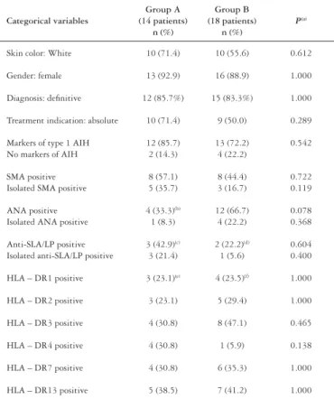
Skin color: White 10 (71.4) 10 (55.6) 0.612
Gender: female 13 (92.9) 16 (88.9) 1.000
Diagnosis: definitive 12 (85.7%) 15 (83.3%) 1.000
Treatment indication: absolute 10 (71.4) 9 (50.0) 0.289
Markers of type 1 AIH No markers of AIH
12 (85.7) 2 (14.3)
13 (72.2) 4 (22.2)
0.542
SMA positive Isolated SMA positive
8 (57.1) 5 (35.7)
8 (44.4) 3 (16.7)
0.722 0.119
ANA positive Isolated ANA positive
4 (33.3)(b)
1 (8.3)
12 (66.7) 4 (22.2)
0.078 0.368
Anti-SLA/LP positive Isolated anti-SLA/LP positive
3 (42.9)(c)
3 (21.4)
2 (22.2)(d)
1 (5.6)
0.604 0.400
HLA – DR1 positive 3 (23.1)(e) 4 (23.5)(f) 1.000
HLA – DR2 positive 3 (23.1) 5 (29.4) 1.000
HLA – DR3 positive 4 (30.8) 8 (47.1) 0.465
HLA – DR4 positive 4 (30.8) 1 (5.9) 0.138
HLA – DR7 positive 4 (30.8) 6 (35.3) 1.000
HLA – DR13 positive 5 (38.5) 7 (41.2) 1.000
AIH: autoimmune hepatitis; SMA: smooth-muscle antibody; ANA: antinuclear antibody;
SLA/LP: soluble liver antigen/liver-pancreas; HLA: human leucocyte antigen. (a) Fisher’s exact test; (b) two ANA results were missing; (c) seven patients tested; (d) 9 patients tested; (e) 13 patients tested; (f) 17 patients tested
TABLE 3 – Distribution of continuous variables in groups A and B
Continuous variables
Group A (14 patients)
Mean (SD)
Group B (18 patients)
Mean (SD)
P(a)
Age at first symptoms 27.29 (15.23) 26.00 (13.59) 0.834 Age at suspension of PA 34.29 (15.64) 31.17 (12.89) 0.543 Time of PA (years) 5.85 (3.94) 4.88 (2.52) 0.595
Time on maintenance treatment (months)(b)
44.05 (53.56) 36.40 (28.63) 0.725
Time to normalize ALT and AST (months)
30.21 (37.06) 17.26 (20.96) 0.262
Maintenance dose of azathioprine(c) 85.71 (25.41) 73.43 (19.30) 0.169
Maintenance dose of prednisone(c) 7.14 (2.161) 7.86 (2.16) 0.380
Diagnostic score (d) 18.21 (2.49) 18.39 (2.70) 0.758
Mean ALT before PA (e) 9.61 (7.64) 10.17 (7.21) 0.634
Mean AST before PA (e) 9.21 (9.55) 9.80 (7.42) 0.606
Mean γGB before PA (e) 1.75 (0.65) 1.72 (1.05) 0.776
Mean γGB before suspension of PA (e) 0.74 (0.3) 0.76 (0.49) 0.493
SD: - standard deviation; ALT: alanine aminotransferase; AST: Aspartate aminotransferase;
γGB: gamma-globulin; PA: treatment with prednisone and azathioprine; (a) Mann-Whitney test;
(b) After the last reduction of azathioprine and prednisone doses; (c) mg/dL;
(d) According to the International AIH Group (1); (e) Number of times above the upper normal limit
(Tables 2, 3), except in the number of previous relapses, due to the four patients mentioned above (28.6% x 0.0%; P = 0.028; Fisher’s exact test).
The probability of relapse was 6.49 (1.38–30.30) times greater in the historical controls as compared with the patients treated with chloroquine, during the time they received that medication. Relapses occurred in four patients while on chloroquine, and in 13 controls (23.5% x 72.2%, P = 0.031; Fisher’s exact test). None of the variables controlled in the regression analysis lowered the significance level below 0.05 (Table 4).
TABLE 4 – Binary logistic regression analysis for the prediction of relapses of autoimmune hepatitis
Independent variables P(a) OR
Study group (b) 0.018 (c) 6.39
HLA-DR4 0.029 6.67
HLA-DR3 0.020 7.58
γGB levels before suspension of PA (d) 0.017 6.44
AIH type 0.011 8.57
Maintenance doses of azathioprine 0.015 8.62 Treatment indication (e) 0.013 8.69
Total treatment time 0.008 9.53
Diagnostic score (f) 0.007 9.54
Time to normalize aminotransferases 0.011 11.11
Age at first symptoms of AIH 0.007 11.43 Age at suspension of PA 0.006 13.18 Maintenaince doses of prednisone 0.006 14.29
Time on maintenance treatment 0.009 14.45
γGB levels before PA 0.005 16.35
OR: odds ratio; PA: treatment with prednisone and azathioprine;
(a) the P values represent the significance level of the study group as a predictor of the relapse frequency, paired with
each variable; (b) group A (during chloroquine use) or B; (c) crude value obtained without pairing the “study group”
with any other variable; (d) γGB: gamma-globulin levels, number of times upper of the normal value; (e) absolute or
relative according to the AASLD3; (f) According to the revised criteria of the International AIH group1
The 10 patients who did not relapse during chloroquine intake received this medication for a mean of 14.1 (4.0 to 21.3) months. Four patients received chloroquine for more than 14 months; two of them for reasons of minor elevations of aminotransferases (less than twice the normal value), and treatment was extended in order to better ascertain the possibility that a relapse might be occurring during the use of chloroquine. After biochemical normalization, administration of the drug was stopped in these two cases. The other two patients requested to be kept on chloroquine for a longer period, because they had had a previous relapse after PA withdrawal.
The follow-up period was 20.8 months (14.2–32.9) for group A and 51.6 (14.0–80.4) months for Group B. These periods were not compared, due to the different time limits. Mean remission time was 5.0 (1.5–9.5) months for the four patients that relapsed while on chloroquine. During the follow-up, after the chloroquine withdrawal, 3 of the remaining 10 patients in group A had a relapse. In these three cases, the relapse occurred after a mean of 5.3 (2.6–8.4) months after chloroquine withdrawal.
Gamma-globulin levels before PA suspension were above 1.6 times the upper normal limit in nine patients, three of them in group A and six of them in group B. There was no relapse in the three patients from the former group, whereas, four of the six patients in the latter group relapsed. The increase in mean gamma-globulin levels was similar in both groups.
There was no difference between groups A and B in regard to alanine aminotransferase, aspartate aminotransferase, alkaline phosphatase, gamma-glutamil transpeptidase and bilirubin levels, considering the values measured before the start of PA, before the suspension of PA, at follow-up and at relapses.
Analysis for the entire follow-up period
Patients with mild to moderate increases of AST levels had more relapses than those with severe increase of AST levels at disease onset (Table 5).
Due to the small number of patients tested for anti-SLA/LP
TABLE 5 – Aspartate aminotransferase, alanine aminotransferase and gamma-globulin levels compared to total relapse rate
Relapse Mean (SD) (a) P(b)
Pre-treatment ALT No 12.37 (6.93) 0.054 Yes 8.37 (7.25)
Pre-treatment AST No 12.31 (9.26) 0.045 Yes 7.79 (7.36)
Pre-treatment γGB No 3.37 (1.52) 0.282 Yes 2.72 (1.04)
SD: standard deviation; AST: aspartate aminotransferase; ALT: alanine aminotransferase;
γGB: gamma-globulin;
(a) number of times above the upper limit; (b) Mann-Whitney test
(14 out of 28 patients), they were not included in the regression analysis. All four anti-SLA/LP positive patients relapsed, while 5 of the 10 anti-SLA/LP negative patients did (100% x 50.0%; P = 0.221; Fisher’s exact test).
Eight out of 12 HLA DR3 positive patients (66.6%) relapsed, while only 1 out of 5 HLA DR4 positive patients did (20.0%; P = 0.264; Fisher’s exact test). HLA haplotypes, when analyzed individually, had no predictive value for relapse risk and had no effect in the regression analysis. One patient in each group was not tested for HLA.
Side effects of chloroquine
Untoward symptoms were usually reported at the beginning of chloroquine treatment. The symptoms reported were nausea (five patients), headache (three patients), epigastric pain and blurred vision (two patients), sore eyes, dizziness, tremor, abdominal discomfort, fatigue and insomnia (one patient each). Only one patient had to be withdrawn from the study before completion, because of untoward side effects while on chloroquine (insomnia, nausea and anorexia). She received chloroquine for 4 months. With the exception of this patient, all untoward symptoms lasted 1 to 4 weeks and subsided spontaneously without treatment withdrawal or any need for medications or subsidiary exams. There were no cases of ophtalmopathy.
Particularities in some cases
Follow-up
Before CD1m of CD2m of CD3m of CD6m of CD9m of CD12m of CD15m of CD1.5 after SUS 2.5m after SUS
0.5m of CD1m of CD1.5m of CD3m of CD6m of CD 3
2
1
Number of times above limit
ALT AST
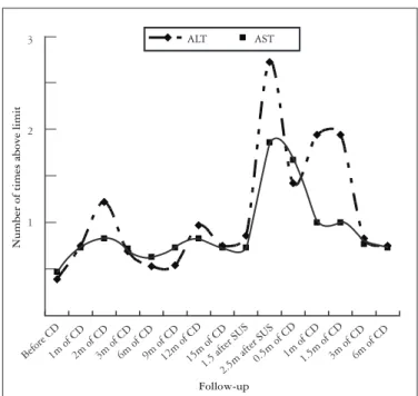
FIGURE1 – AST and ALT levels during follow-up for patient SAS
CD: chloroquine diphophate; SUS: suspension of chlo-roquine; m: months.
A second patient (ALO) relapsed after 3 months and PA was prescribed. On her return visit, she reported that, at first, she had taken chloroquine only 2 or 3 times a week and, after AIH relapse, she started taking chloroquine on a regular basis instead of PA. She was kept on chloroquine until the suspension of this drug around 14 months later. Once again, she relapsed and was also retreated with chloroquine, entering again into biochemical remission (Figure 2). Both patients had anti-SLA/LP as the sole autoantibody marker.
Before CDRelapse1m of CD3m of CD6m of CD9m of CD12m of CD15m of CD
1m after SUS2m after SUS 0.5m of CD 1m of CD3m of CD 3m after SUS
4
3
2
1
Number of times above limit
Follow-up
ALT AST
FIGURE 2 – AST and ALT levels during follow-up for patient ALO
CD: chloroquine diphophate; SUS: suspension of chloro-quine; m: months
Liver biopsies
Thirty-one of the 32 (96.9%) liver biopsies done before treatment withdrawal were reviewed. There were no significant differences between the original and revised conclusions. Spearman correlation test values were 0.876 (P <0.001) for fibrosis scores, 0.357 (P = 0.049) for periportal activity scores, 0.411 (P = 0.027) for portal activity scores and 0.447 (P = 0.015) for lobular activity scores.
DISCUSSION
The results obtained with chloroquine for the prevention of AIH relapses are promising. It was shown to be safe and effective in this pilot study, in which almost half of the cases had hepatic cirrhosis.
The follow-up period was appropriate, since more than 80% of relapses of AIH occur within 12 months of treatment withdrawal(8, 10, 17, 19). All relapses that occurred in the control group happened within 14 months, whereas 38.8% of the relapses in Group A occurred after this period (including follow-up). Relapses occurred on average 5 months after chloroquine or PA withdrawal, which suggests that both helped to prevent relapses. The two cases mentioned in which chloroquine seemed to induce a remission after the occurrence of a relapse favor this conclusion, although spontaneous biochemical fluctuations are well known in AIH.
It may be argued that small elevations in ALT or AST could not be true relapses. However, this is the criteria adopted by the International AIH Group. It is supported by a previous report in which AST elevations greater than twice the upper normal limit were invariably related to the presence of interface hepatitis, whereas AST elevations less than twice the upper limit did so only 28% of the time(12). In fact, three of the four patients that relapsed during the use of chloroquine in our study had aminotransferase levels greater than 3 times the upper normal limit, which happened also in 13 of the 18 controls. Gammaglobulin values did not help to identify which patients had a greater relapse risk.
On the other hand, the absence of laboratory abnormalities does not necessarily mean histological remission. Most of our patients on remission were not submitted to a liver biopsy on follow-up, although a patient in the group of historical controls with high gammaglobulin levels (around 2.55 g%) and normal aminotransferases was submitted to a liver biopsy that showed no periportal activity. In the report mentioned above, 11 out of 69 patients without biochemical abnormalities were shown to have moderate periportal activity, but their prognosis was relatively benign, because only 5 of them suffered clinical and biochemical deterioration in the following six to twelve months(12).
responses to antigens that have been already presented to T helper cells(14).
The four patients that have been included in both groups make it more difficult to accurately interpret the statistical tests applied, but their inclusion was necessary due to the small sample studied. However, the number of previous relapses did not influence relapse probability in the study of CZAJA et al.(10). Results obtained from crossover patients can also be interesting. Two of the patients that were included in both groups had had relapses 4.8 and 7.0 months after PA withdrawal (historical controls), and 17.2 and 22.1 months when treated with chloroquine for about 14 months.
We conducted an analysis for the entire follow-up period in order to detect variables that could help to predict the probability of relapse. During the use of chloroquine in Group A (roughly in the first year after PA stopping), the only significant prognostic variable was the study group. Including the follow-up period after chloroquine withdrawal, the only significant prognostic variable was the initial increase in aminotransferase levels. Thus patients with lesser AST elevations at onset relapsed more often in a total follow-up period of greater than 20 months after PA ceasing. One could argue that patients with more pronounced hepatitis at onset could represent cases of acute hepatitis of an etiology other than AIH. Most of our patients, however, had significant autoantibody titers and histological findings of chronic liver disease. Only one patient in the control group had had acute hepatitis at the onset without autoantibody reactivity, and she did not relapse. Another hypothesis to consider is that patients with an initially more severe disease have a greater tendency to experience a remission after treatment withdrawal. This has not been described before.
BAERES et al.(3) have found a greater relapse probability in anti-SLA/LP positive patients (74% x 54%; P = 0.02). Our study showed a similar tendency, but the significance level was not achieved, perhaps due to the small sample size. CZAJA et al.(11) found a correlation between antiasialoglicoprotein receptor antibody reactivity, immunoglobulin levels and relapse rate. There was no mention in their report of a regression analysis; therefore, it is unclear whether or not the antibody reactivity or the immunoglobulin levels were more significantly associated to relapses. Unfortunately, we did not test this type of antibody in this cohort of patients.
The adverse effects of chloroquine were mostly well tolerated and short-term. There were no cases of hepatic decompensation, though there was a cirrhosis prevalence of nearly 50% in this series of patients.
The use of historical controls was necessary because of the small number of patients treated prospectively. Biases can arise when comparing treatment results at different periods of time. There could be differences in laboratory methods, loss of data and undetected or uncontrolled prognostic variables. We partially compensated these problems with the regression analysis.
Chloroquine use was safe and the results obtained for the prevention of AIH relapses are promising. This was the first time that chloroquine was tested in patients with AIH, and one of the few reports of chloroquine for the treatment of hepatic diseases. This is a pilot study, therefore chloroquine can not be recommended for the maintenance of remission of AIH before the conduction of controlled trials. A randomized trial of chloroquine versus placebo for the prevention of relapses is currently underway in our institution.
Mucenic M, Mello ES, Cançado ELR. Cloroquina para manutenção da remissão da hepatite auto-imune: resultado de estudo piloto. ArqCloroquina para manutenção da remissão da hepatite auto-imune: resultado de estudo piloto. Arq Gastroenterol 2005;42(4):249-55.
RESUMO - Racional - Em razão dos riscos relacionados ao tratamento prolongado com prednisona e azatioprina, tenta-se a retirada dessas
drogas em pacientes com hepatite auto-imune em remissão. Como há alta taxa de recidiva, a maioria dos pacientes recebe tratamento por tempo indefinido. Objetivo - Avaliar a segurança e a eficácia do tratamento de manutenção com cloroquina na prevenção de recidiva da hepatite auto-imune. Métodos - O tratamento convencional foi suspenso após obtenção de remissão bioquímica e histológica. Difosfato de cloroquina foi administrado, 250 mg diariamente, por pelo menos 12 meses ou até a ocorrência de recidiva, definida pela elevação dos níveis de aminotransferases em, pelo menos, duas vezes acima dos valores normais. Resultados - Quatorze pacientes foram consecutivamente tratados e comparados com 18 controles históricos. Houve chance de 6,49 vezes maior de recidiva nos pacientes do grupo de controles históricos, quando comparados com os pacientes do grupo tratado com o difosfato de cloroquina. (72,2% versus 23,5%; P = 0.031). Conclusões – O grupo tratado com cloroquina teve menor freqüência de recidivas da hepatite auto-imune. Difosfato de cloroquina foi seguro em pacientes com hepatite auto-imune e cirrose hepática sem descompensação clínica, na dose de 250 mg diariamente e até 2 anos de uso. Esses resultados preliminares estimulam a realização de estudos controlados, comparando cloroquina com placebo ou com o tratamento de manutenção.
REFERENCES
1. Alvarez F, Berg PA, Bianchi FB Bianchi L, Burroughs AK, Cançado EL, Chapman RW, Cooksley WG, Czaja AJ, Desmet VJ, Donaldson PT, Eddleston AL, Fainboim L, Heathcote J, Homberg JC, Hoofnagle JH, Kakumu S, Krawitt EL, Mackay IR, MacSween RNM, Maddrey WC, Manns MP, McFarlane IG, Meyer zum Büschenfelde KH, Mieli-Vergani G, Nakanuma Y, Nishioka M, Penner E, Porta G, Portmann BC, Reed WR, Rodes J, Schalm SW, Scheuer PJ, Schrumpf E, Seki T, Toda G, Tsuji T, Tygstrup N, Vergani D, Zeniya M. International Autoimmune Hepatitis Group Report: reviewInternational Autoimmune Hepatitis Group Report: review of criteria for diagnosis of autoimmune hepatitis. J Hepatol 1999;31:929-38. 2. Arnold W, Meyer zum Büschenfelde KH. [Therapy of chronic hepatitis]. Leber
Magen Darm 1981;11:73-80.
3. Baeres M, Herkel J, Czaja AJ, Wies I, Kanzler S, Cançado EL, Porta G, Nishioka M, Simon T, Daehnrich C, Schlumberger W, Galle PR, Lohse AW. Establishment ofEstablishment of standardised SLA/LP immunoassays: specificity for autoimmune hepatitis, worldwide occurrence, and clinical characteristics. Gut 2002;51:259-64.
4. Ballot E, Homberg JC, Johanet C. Antibodies to soluble liver antigen: an additional marker in type 1 auto-immune hepatitis. J Hepatol 2000;33:208-15.
5. Bittencourt PL, Goldberg AC, Cançado ELR, Porta G, Carrilho FJ, Farias AQ, Palacios SA, Chiarella JM, Abrantes-Lemos, Baggio VL, Laudanna AA, Kalil J. Genetic heterogeneity in susceptibility to autoimmune hepatitis types 1 and 2. Am J Gastroenterol 1999;94:1906-13.
6. Brodsky FM, Lem L, Bresnahan PA. Antigen processing and presentation. Tissue Antigens 1996;47:464-71.
7. Charmot G, Goujon C. Mild hepatitis probably caused by amodiaquine. Bull Soc Pathol Exot Filiales 1987;80:266-70.
8. Czaja AJ, Wolf AM, Baggenstoss AH. Laboratory assessment of severe chronicLaboratory assessment of severe chronic active liver disease during and after corticosteroid therapy: correlation of serum transaminase and gamma globulin levels with histologic features. Gastroenterology 1981;80:687-92.
9. Czaja AJ, Pfeifer KD, Decker RH, Vallari AS. Frequency and significance of antibodies to asialoglycoprotein receptor in type 1 autoimmune hepatitis. Dig Dis Sci 1996;41:1733-40.
10. Czaja AJ. Drug therapy in the management of type 1 autoimmune hepatitis. Drugs 1999;57:49-68.
11. Czaja AJ, Freese DK, American Association for the study of liver disease. Diagnosis and treatment of autoimmune hepatitis. Hepatology 2002;36:479-97.
12. Czaja AJ, Menon KV, Carpenter HA. Sustained remission after corticosteroid therapy for type 1 autoimmune hepatitis: a retrospective analysis. Hepatology 2002;35:890-7.
13. Doxiadis TA, Doukas E, Yotsas Z, Pontidas E. Therapy of chronic active hepatitis andTherapy of chronic active hepatitis and chloroquine compounds [abstract]. Gastroenterology 1971;60:658.
14. Fox R. Anti-malarial drugs: possible mechanisms of action in autoimmune disease and prospects for drug development. Lupus 1996;5:s4-s10.
15. Freedman A. Chloroquine and rheumatoid arthritis: a short-term controlled trial. Ann Rheum Dis 1956;15:251-7.
16. Freedman A, Steinberg VL. Chloroquine in rheumatoid arthritis: a double blindfold trial of treatment for one-year. Ann Rheum Dis 1960;19:243-50.
17. Hegarty JE, Nouti Aria KT, Portmann B, Eddleston AL, Williams R. Relapse following treatment withdrawal in patients with autoimmune chronic active hepatitis. Hepatology 1983;3:685-9.
18. Helbling B, Reichen J. Reactivation of hepatitis B following withdrawal of chloroquine. Schweiz Med Wochenschr 1994;124:759-62.
19. Johnson PJ, McFarlane IG, Williams R. Azathioprine for long-term maintenance of remission in autoimmune hepatitis. N Engl J Med 1995;333:958-63.
20. Kouroumalis EA, Koskinas J. Treatment of chronic active hepatitis B (CAH B) withTreatment of chronic active hepatitis B (CAH B) with chloroquine: a preliminary report. Ann Acad Med Singapore 1986;15:149-52. 21. Mackay IR, Davies JM, Rowley MJ. Towards the pathogenesis of autoimmune liver
disease. J Autoimmun 1999;13:163-9.
22. Manns MP, Strassburg CP. Autoimmune hepatitis: clinical challenges. Gastroenterology 2001;120:1502-17.
23. Marks JS, Power BJ. Is chloroquine obsolete in treatment of rheumatic disease? Lancet 1979;1:371-3.
24. Neefjes JJ, Ploegh HL. Intracellular transport of MHC class II molecules. Immunol Today 1992;13:179-84.
25. O’Dell JR. Rheumatoid arthritis. In: Goldman L, Ausiello D, editors. Cecil textbook of medicine. 22nd ed. Philadelphia: WB Saunders; 2004. p.1644-9.
26. Olansky AJ. Antimalarials and ophthalmologic safety. J Am Acad Dermatol 1982;6:19-23.
27. O’Neill PM, Bray PG, Hawley SR, Ward SA, Park BK. 4-Aminoquinolines - past, present and future: a chemical perspective. Pharmacol Ther 1998;77:29-58. 28. Popert AJ, Meijers KA, Sharp J, Bier F. Chloroquine diphosphate in rheumatoid
arthritis: a controlled trial. Ann Rheum Dis 1961;20:18-35.
29. Reasor MJ. A review of the biology and toxicologic implications of the induction of lysosomal lamellar bodies by drugs. Toxicol Appl Pharmacol 1989;97:47-56. 30. Salako LA. Toxicity and side effects of antimalarials in Africa: a critical review. Bull
World Health Organ 1984;62(Suppl):63-8.
31. Schur PH. Systemic lupus erythematosus. In: Goldman L, Ausiello D, editors. Cecil textbook of medicine. 22nd ed. Philadelphia: WB Saunders; 2004. p.1660-9.
32. Schultz KR, Gilman AL. The lysosomotropic amines, chloroquine and hydroxychloroquine: a potentially new therapy for graft-versus-host disease. Leuk Lymphoma 1997;24:201-10.
33. Siede W. Therapy of chronic hepatitis. Dtsch Z Verdau Stoffwechselkr 1979;39:57-69. 34. Sifre RB, Gomez AO, Masso JT, Navarro RB. Favorable efecto de la cloroquina sobre
una hepatitis crónica subagresiva rebelde. Rev Esp Enf Ap Dig 1975;45:123-8. 35. Soloway RD, Summerskill WH, Baggentoss AH, Geall MG, Gitnick GL, Elveback IR,
Schoenfield IJ. Clinical, biochemical, and histological remission of severe chronic active liver disease: a controlled study of treatments and early prognosis. Gastroenterology 1972;63:820-33.
36. Wang KK, Czaja AJ, Beaver SJ, Go VL. Extrahepatic malignancy following long-term immunosupressive therapy of severe hepatitis B surface antigen-negative chronic active hepatitis. Hepatology 1989;10:39-43.
37. Wolff C, Armas R, Krause P, Parraguez A, Ramón Soto J. Treatment of porphyriaTreatment of porphyria cutanea tarda with chloroquine and its effect on associated liver disease: retrospective analysis. Rev Med Chil 1996;124:456-60.