473
A new
Aspidoras
(Siluriformes: Callichthyidae) from rio Paraguaçu basin,
Chapada Diamantina, Bahia, Brazil
Marcelo R. Britto*, Flávio C. T. Lima**, and Alexandre C. A. Santos***
During a recent ichthyological survey in Chapada Diamantina, Estado da Bahia, Brazil, a new, very distinctive Aspidoras was discovered in tributaries of the upper rio Paraguaçu. The new taxon differs from its congeners mainly in having: a poorly-developed pigmentation pattern, restricted to minute scattered blotches on dorsal region of head and body, but grouped in small, irregular blotches along the lateral body plate junction; four or five caudal vertebra, anterior to compound caudal centrum, with neural and haemal spines placed posteriorly, close to post-zygapophyses; and post-zygapophyses of the precaudal vertebrae without dorsal expansions connected with their respective neural spines. The new species shares with Aspidoras velites dorsolateral body plates not touching their counterparts dorsally, and infraorbital bones with reduced flanges that are restricted to the latero-sensory canal. Both of these are considered reductive character states, probably indicating a paedomorphic condition to both species. The new species is also compared to Aspidoras maculosus, a congener which bears the most similar color pattern and is geographically closest to the new species.
Durante um estudo recente sobre a ictiofauna da Chapada Diamantina, foi descoberta uma nova espécie de Aspidoras, bastante distinta morfologicamente de suas congêneres, em afluentes do alto rio Paraguaçu, Estado da Bahia, Brasil. O novo táxon difere das demais espécies de Aspidoras principalmente pela pigmentação pouco desenvolvida, restrita a minúsculas máculas espalhadas na região dorsal da cabeça e corpo, concentradas em pequenas manchas irregulares ao longo da junção das placas laterais; por apresentar as quatro a cinco últimas vértebras caudais anteriores ao centro caudal composto com os espinhos neurais e hemais situados posteriormente junto às pós-zigapófises; e pós-zigapófises das vértebras pré-caudais sem expansões dorsais unidas ao respectivo espinho neural. Além disso, a nova espécie compartilha com Aspidoras velites a condição em que as placas dorso-laterais não contatam suas contrapartes dorsalmente, e os ossos infra-orbitais reduzidos a pouco mais que o canal látero-sensorial, características redutivas, provavelmente indicando um estado de caráter pedomórfico para estas espécies. A nova espécie é ainda comparada a Aspidoras maculosus, congênere mais semelhante no padrão de colorido e também aquela mais próxima geograficamente.
Key words: Systematics,Aspidoras maculosus, Sand-dwelling, Paedomorphic characters.
* Museu Nacional, Universidade Federal do Rio de Janeiro, Quinta da Boa Vista, 20940-040 Rio de Janeiro, RJ, Brazil. e-mail: mrbritto2002@yahoo.com.br
** Museu de Zoologia da Universidade de São Paulo, Caixa Postal 42594, 04299-970 São Paulo, SP, Brazil. e-mail: fctlima@usp.br *** Departamento de Ciências Biológicas, Universidade Estadual de Feira de Santana, Km 03, BR-116, 44031-460 Feira de Santana, BA, Brazil. e-mail: clister@ig.com.br
Introduction
The genus AspidorasIhering includes 19 nominal species of catfishes that are generally found in shallow creeks of several river basins draining mainly the Brazilian Shield (Britto et al., 2002). Although the validity of some characters tradi-tionally used to define Aspidoras, such as the size of the frontal fontanel and the presence of a parieto-supraoccipital fontanel had been questioned previously (Nijssen & Isbrücker, 1976; Weitzman & Balph, 1979), the monophyly of the genus
was recently demonstrated through phylogenetic studies (Reis, 1998; Britto, 2003).
of the family. We describe below this new species, and we suggest that some of those features might be related to a specialized lifestyle.
Material and Methods
Morphometric and meristic data were taken following Reis (1997), with the addition of the length of the ossified portion of pectoral spine, which was measured from the articulation between the spine and pectoral girdle to distal tip of spine. Measurements were obtained with calipers to 0.1 mm. Counts of teeth and vertebrae were taken only from cleared-and-stained (cs) specimens, which were prepared according to Taylor & Van Dyke (1985). Vertebral counts include only free centra, with the compound caudal centra (preural 1 + ural 1) counted as a single element. Lateral plate counts include all dorsolateral and ventrolateral plates, except for small, irregu-lar plates on caudal peduncle. In the description, numbers in parentheses following each count represent the number of specimens with that value and counts indicated with an aster-isk represent counts from the holotype. Nomenclature of latero-sensory canals follows Schaefer & Aquino (2000), and that of preopercular pores follows Schaefer (1988). Osteologi-cal terminology follows Reis (1998), except for the use of parieto-supraoccipital instead of supraoccipital (Arratia & Gayet, 1995), compound pterotic instead of pterotic-supracleithrum (Aquino & Schaefer, 2002), and scapulocoracoid instead of coracoid (Lundberg, 1970), as adopted in Britto & Lima (2003). Homologies of barbels follow Britto & Lima (2003). Institutional abbreviations are: FMNH, Field Museum of Natural History, Chicago; MCP, Museu de Ciência e Tecnologia da Pontifícia Universidade Católica do Rio Grande do Sul, Porto Alegre; MNRJ, Museu Nacional da Universidade Federal do Rio de Janeiro; and MZUSP, Museu de Zoologia da Universidade de São Paulo, São Paulo.
Comparative material is listed in Lima & Britto (2001) and Britto (2003). In addition, the following material was studied: Aspidoras maculosus Nijssen & Isbrücker (measurements taken by P. Willink) FMNH 54808 (3), paratypes; FMNH 54809 (1), paratype; FMNH 54810, holotype; FMNH 78361 (2), paratypes;Corydoras lacerdai Hieronimus MNRJ 26017 (17, 4 cs); andScleromystax sp. MCP 28729 (20, 3 cs); MCP 29299 (5, 1 cs).
21709 (50, 2 cs, 15.0-28.4 mm SL) rio Caldeirão, 12º39’33"S 41º22’12"W; A. C. A. Santos, 13 Mar 1999. MNRJ 21710 (18, 1 cs, 16.7-24.9 mm SL) rio Roncador, 12º42’01"S 41º21’26"W; A. C. A. Santos, 6 Nov 1999. MNRJ 21711 (47, 16.5-27.9 mm SL) rio Capivara, 12º37’19"S 41º22’35"W; A. C. A. Santos, 6 Nov 1999. MNRJ 21712 (1, 23.2 mm SL) rio Ribeirão, 12º35’10"S 41º22’57"W; A. C. A. Santos, 6 Nov 1999.
Diagnosis.Aspidoras psammatides differs from its conge-ners by the following exclusive features: poorly-developed pigmentation, restricted to minute scattered blotches on dor-sal region of head and body, grouped in small, irregular blotches along lateral body plate junction (vs. dense pigmen-tation, in marbled or striped pattern); four or five caudal ver-tebra (vs. only three vertebra) anterior to compound caudal centrum, with neural and haemal spines placed posteriorly, close to post-zygapophyses; minor least interorbital distance (19.9 - 27.8% in HL vs. greater than 28% in HL); and post-zygapophyses of precaudal vertebrae not showing dorsal ex-pansions connected with its respective neural spine (vs. post-zygapophyses connected with their respective neural spine).
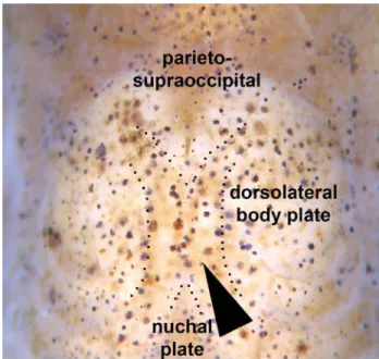
Description.Morphometric data presented in Table 1. Head compressed with slightly convex dorsal profile; roughly tri-angular in dorsal view (Fig. 2). Snout rounded. Head profile convex from upper lip to vertical through middle of parieto-supraoccipital; body slightly convex from that point to base of last dorsal-fin ray. Postdorsal-fin body profile slightly con-cave to adipose-fin spine; markedly concon-cave from this point to caudal-fin base. Ventral profile of body slightly convex from isthmus to anal-fin origin; slightly pronounced between pectoral fins. Profile markedly concave from first anal-fin ray to caudal-fin base. Body elongated; roughly cylindrical in cross section at pectoral girdle, gradually becoming more compressed toward caudal fin.
Maxillary barbel slightly longer than outer mental barbel. In-ner mental barbel fleshy. Small rounded papillae covering entire surface of all barbels, upper and lower lips, and isthmus. Gill membranes united to isthmus. Four branchiostegal rays cov-ered by thick layer of skin; distal two rays united at their tips by branchiostegal cartilage. Teeth on upper pharyngeal tooth plate 28 (2) or 34 (1), and on fifth ceratobranchial 22 (1) or 24 (2). Nasal, frontal, sphenotic, compound pterotic, and parieto-supraoccipital visible externally, all covered by thin layer of skin and bearing minute scattered odontodes. Frontal fon-tanel elongate, ellipsoid, covered by thin layer of skin, and reaching anterior border of parieto-supraoccipital. Parieto-supraoccipital fontanel round, located in the middle of bone Nasal slender, slightly curved laterally, mesial border con-tacting frontal and mesethmoid. Frontal roughly rectangular; anterior expansion in contact with nasal bone and mesethmoid, posterior portion contacting sphenotic and parieto-supraoccipital. Sphenotic trapezoid in shape,
contact-ing parieto-supraoccipital dorsally, compound pterotic pos-teriorly, and second infraorbital ventrally. Compound pterotic roughly pipe-shaped, with slender posterior expansion con-tacting first dorsal body plate dorsally, and first lateral-line ossicle posteriorly. Ventral margin of compound pterotic con-tacting opercle and cleithrum. Parieto-supraoccipital quadran-gular with posterior expansion trianquadran-gular and short, not reach-ing nuchal plate (Fig. 2).
Two narrow infraorbital bones nearly restricted to laterosensory canal, externally visible, covered by thin layer of skin. Both bearing few minute odontodes. First infraorbital with very short anterior expansion (flange; Fig. 3). Minute odontodes-bearing platelets dorsally on orbit. Opercle ex-posed, ovoid in shape and roughly elongate, with angular free border. Preopercle externally visible, slender and cov-ered by thin layer of skin. Interopercle triangular, covcov-ered by thin layer of skin.
Fig. 1. Aspidoras psammatides, holotype, MNRJ 28407, 25.7 mm SL, Brazil, Estado da Bahia, município de Lençóis, rio Caldeirão.
Paratypes (n = 51)
Holotype mean range
Standard length (mm) 25.7 25.3 20.4 - 31.0
Percents of Standard length
Depth of body 21.5 19.8 16.9 - 23.5 Predorsal distance 45.6 44.6 41.4 - 47.6 Prepelvic distance 46.0 44.7 41.7 - 48.6 Preanal distance 77.7 76.2 70.7 - 82.6 Preadipose distance 78.3 78.3 71.0 - 82.0 Length of dorsal spine 13.2 12.7 10.5 - 15.0 Length of pectoral spine 14.3 13.4 10.5 - 17.5 Length of adipose-fin spine 9.4 10.6 6.9 - 13.5 Depth of caudal peduncle 8.7 9.0 7.5 - 10.1 Dorsal to adipose distance 21.7 23.2 14.8 - 28.8 Length of dorsal-fin base 16.0 16.0 12.7 - 21.9 Maximum cleithral width 9.1 11.6 8.7 - 22.3 Head length 35.4 37.0 34.7 - 40.5 Length of maxillary barbel 13.8 13.8 10.6 - 20.0
Percents of Head length
Head depth 60.0 57.3 34.2 - 63.2 Least interorbital distance 22.4 22.5 19.9 - 27.8 Horizontal orbit diameter 21.1 21.9 18.8 - 24.6 Snout length 47.0 47.2 40.0 - 54.4 Least internareal distance 17.8 18.4 10.9 - 24.9 Table 1. Morphometric data of holotype and paratypes of Aspidoras psammatides.
Trunk lateral-line composed of one perforated dorsolateral-body plate and two laterosensory canals, reduced to small os-sicles. Lateral-line canal entering neurocranium through com-pound pterotic, splitting posterior of sphenotic into pterotic and preoperculomandibular branches, each with single pore. Sensory canal continuing through compound pterotic, enter-ing sphenotic as temporal canal, which splits into two branches: one branch giving rise to infraorbital canal, other branch enter-ing frontal through supraorbital canal. Supraorbital canal with two branches: epiphyseal branch opening in frontal bone, and anterior branch running through nasal bone. Nasal canal with single opening at each end. Infraorbital canal running through entire second infraorbital, extending to infraorbital 1 and open-ing into two pores. Preoperculomandibular branch not con-nected to preoperculomandibular canal, which runs through entire preopercle with three openings, leading to pores 3, 4, and 5, respectively.
Body plates with minute odontodes restricted to poste-rior margins. Nuchal plate exposed. Cleithrum exposed. Dor-solateral body plates between parieto-supraoccipital process and nuchal plate not touching counterparts, leaving narrow naked area; specimens less than 26.4 mm SL also shows na-ked area between last dorsal-fin ray and first preadipose plate-let. Dorsolateral body plates 26 (2), 27* (24), 28 (24), or 29 (2); ventrolateral body plates 24 (19), 25* (25), or 26 (8); dorsolat-eral body plates along dorsal-fin base 6 (12) or 7* (40); dorso-lateral body plates from adipose fin to caudal-fin base 8 (2), 9 (8), 10* (23), 11 (18), or 12 (1); preadipose platelets 2 (1), 3 (11), 4* (35), 5 (4), or 6 (1). Precaudal vertebrae 7 (2) or 8 (3); caudal vertebrae 18 (5). Four to five caudal vertebra, anterior to com-pound caudal centrum, with neural and haemal spines placed posteriorly, close to post-zygapophyses (Fig. 4). Post-zyga-pophyses of precaudal vertebrae reduced, not showing dor-sal expansions connected with respective neural spine (Fig. 5). Five (4) or seven (1) pairs of ribs, first pair conspicuously larger than others.
Dorsal fin roughly triangular; its origin just posterior to third dorsolateral body plate. Dorsal spine shorter than first five branched rays. Distal tip of spine with minute-segmented unossified portion. Anterior and posterior border of dorsal spine smooth. Dorsal-fin rays I,8 in all specimens examined. Adipose fin roughly triangular; its origin separated from base of last dorsal-fin ray by seven to eight dorsolateral body plates. Anal fin roughly ovoid; its origin located just poste-rior to 13th to16th ventrolateral body plates, at vertical through anterior margin of adipose-fin spine. Anal-fin rays ii,5,i *; one specimen (MNRJ 21710, 24.6 mm SL) ii,4,i. Pectoral fin roughly rounded; its origin located just posterior to gill opening. Os-sified portion of pectoral spine shorter than first five branched rays. Distal tip of spine with minute-segmented unossified portion. Pectoral spine with well-developed serrations along entire posterior border. Pectoral-fin rays I,9* (27) or I,10 (25). Pelvic fin ellipsoid; its origin just below second ventrolateral body plate, at vertical through base of third branched dorsal-fin ray. Pelvic-dorsal-fin rays i, 5. Caudal dorsal-fin bilobed; both lobes equal in size. Principal caudal-fin rays i,6/6,i *, one specimen (MNRJ 21711, 27.4 mm SL) i,6/5,i; upper procurrent caudal-fin rays iv; lower procurrent caudal-fin rays iv. All fins with minute odontodes scattered over all rays.
Color in alcohol. Ground coloration of head light brown to yellowish white. Several chromatophores clustered in minute brown dots scattered over dorsal and lateral surface of head. Dots more concentrated on posterodorsal portion of opercle and on side of snout forming discrete stripe from anteroventral region of orbit to snout tip. Larger individuals (up to 26.4 mm SL) with dots grouped in small blotches. Small specimens with scattered minute brown blotches on anterior portion of snout. All barbels yellowish white; few chromatophores present on barbels in some individuals.
Ground color of trunk light brown to yellowish white.
Dif-Fig. 3. Orbital region of Aspidoras psammatides, paratype, MNRJ 21709, 22.6 mm SL, right side. Arrow: latero-sensory canal.
fuse brown blotch on middle region of cleithrum. Minute dark brown dots scattered over entire surface of trunk; more concentrated on dorsal region. Dots clustered around bases of dorsal spine, last dorsal-fin ray and adipose-fin spine, re-spectively. Small diffuse brown blotches along lateral junc-tion of body plates, from cleithrum to caudal peduncle. Blotches varying in number from five to (more commonly) eight blotches. Ventral surfaces of body yellowish white.
Interradial membrane of all fins hyaline. Dorsal fin with few chromatophores scattered on dorsal spine and first two to three adjacent rays, scarcely present on their tips. Ground color of anal-fin rays yellowish white. Some individuals with sparce chromatophores on base of rays. Few chromatophores scat-tered over adipose-fin spine; some individuals with chromato-phores also on adipose-fin membrane adjacent to spine and on its apex. All pectoral-fin rays and spine yellowish white. Larger specimens with sparse chromatophores on first two branched rays. Pelvic-fin rays hyaline. Caudal-fin rays yellowish white with several chromatophores clustered in three-four patches restricted to the three outermost rays of both lobes. Color in life very similar to that of preserved specimens.
Sexual dimorphism. As usual for corydoradines (e.g. Nijssen & Isbrücker, 1980: 130; Britto, 2003: 142, fig. 23), males of Aspidoras psammatides possess a lanceolated papilla. How-ever, relatively few males show a well-developed papilla, which made the determination of sex in most specimens more diffi-cult than with other Aspidoras species (e.g. Britto, 1998, 2000, Lima & Britto, 2001, Britto et al., 2002).
Habitat and ecological notes. The species is known from af-fluents of the rio São José, one of the main tributaries of the upper course of rio Paraguaçu, which has several relatively small tributaries entering its right margin. The species was
collected in four of these tributaries, rio Roncador, rio Capivara, rio Ribeirão and rio Caldeirão. The rio São José and its tributar-ies are all blackwater rivers. The type locality of Aspidoras psammatides,rio Caldeirão, has sandy and rocky bottom, with little amount of riparian and submerged vegetation.
Distribution.Aspidoras psammatides is known only from tributaries of the upper rio Paraguaçu, a coastal river basin from eastern Brazil (Fig. 6)
Etymology. Psammatides, after “Psammatos psammatides”, “the sand sorcerer”, a character of J.R.R. Tolkien’s book “Roverandom”, from the Greek psammos, sand, and ides, son of. In allusion to the sand-dwelling behavior of the species. A noun in apposition.
Discussion
Aspidoras psammatides is remarkable among its conge-ners by displaying a suite of characters apparently related to paedomorphosis. Some of them are shared with Aspidoras velites, such as dorsolateral body plates not touching their counterparts dorsally and infraorbital bones with reduced flanges, nearly restricted to the laterosensory canal. Despite possessing the predorsal area naked as in A. velites, the con-dition found in A. psammatides is not as extreme as that found in the latter species, where not only the anterior dorsolateral plates but also the parieto-supraoccipital process and the
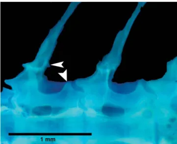
Fig. 5. Detail of the dorsal region of precaudal vertebrae of Aspidoras psammatides, paratype, MNRJ 21709, 22.6 mm SL, showing their post-zygapophyses not connected with respec-tive neural spine (arrows).
specimens. During the ontogeny the post-zygapophyses de-velop a laminar outgrowth that fuses to the respective neural spine. An additional, although not reductive, autapomorphy for the species is the presence of four or five caudal vertebra, anterior to compound caudal centrum, with neural and haemal spines placed posteriorly, close to post-zygapophyses, whereas in the remaining callichthyids invariably only the three latter vertebrae show the neural spines placed posteriorly.
AmongAspidoras species, A. psammatides shares a nar-row interorbital distance and poorly-developed color pattern withA. maculosus, a species described from the headwaters of the rio Itapicuru, an eastern coastal drainage immediately north to the rio Paraguaçu basin in Bahia, Brazil. Aspidoras maculosus is only known from its poorly preserved and faded type series, which were not examined in the present study. However, examination of Ellis’(1913: pl. XXVI, fig. 3) illustra-tion, which depicts the pigmentation of the (then) recently collectedA. maculosus holotype, plus recent pictures of the same specimen, allows a comparison between both species. Aspidoras psammatides is clearly distinct from A. maculosus in at least two characters, viz., the color pattern, which con-sists, respectively, in minute scattered blotches on dorsal re-gion of head and body, but grouped in small, irregular blotches along lateral body plate junction (vs. three longitudinal series of small rounded blotches, the more conspicuous one along lateral body plate junction), and dorsolateral body plates not touching their counterparts dorsally, leaving a small predorsal naked area (vs. dorsolateral body plates touching their coun-terparts dorsally, leaving no naked predorsal area). In addi-tion,Aspidoras maculosus shows an interorbital distance (28.6 - 33.1% HL) somewhat larger than in A. psammatides (19.9 -27.8% HL).
The low degree of development of the pigmentation found inAspidoras psammatides is unique among Aspidoras spe-cies and recalls the color pattern observed in several unre-lated catfish groups such as some African Amphiliidae (e.g. Leptoglanis spp., Psamphyletria, Dolicamphilius, Tetracamphilius, and Zaireichthys; Skelton, 1993: 219-220; Roberts, 2003), South American Trichomycteridae (e.g. Microcambeva spp.; Costa & Bockmann, 1994; Costa et al., 2004), and also some Asiatic Cypriniformes (e.g., Acanthopsoides, Cobitidae; Siebert, 1991) and North Ameri-can Perciformes (“sand darters”, Ammocrypta spp.; Page &
provided measurements from holotype and paratypes of Aspidoras maculosus deposited at that institution. A. Netto-Ferreira helped with the photographs of bones, suggesting their use in “high-contrast negative”. MRB received finan-cial support from CNPq (grant 300189/03-6); FCTL receives financial support from FAPESP (grant 01/14449-2). ACAS re-ceived financial support from CAPES/ PICDT-UEFS and from Projeto Nordeste de Pesquisa, CNPq. MRB and FCTL are participants of ACSI (All Catfish Species Inventory Project).
Literature cited
Aquino, A. E. & S. A. Schaefer. 2002. The temporal region of the cranium of loricarioid catfishes (Teleostei: Siluriformes): Morphological diversity and phylogenetic significance. Zoologischer Anzeiger, 241: 223-244. Arratia, G. & M. Gayet. 1995. Sensory canals and related bones
of tertiary siluriform crania from Bolivia and North America and comparison with recent forms. Journal of Vertebrate Paleontology, 15: 482-505.
Britto, M. R. 1998. Two new species of the genus Aspidoras (Siluriformes: Callichthyidae) from Central Brazil. Ichthyo-logical Exploration of Freshwaters, 8: 359-368.
Britto, M. R. 2000. Aspidoras depinnai (Siluriformes: Callichthyidae), a new species from northeastern Brazil. Copeia, 2000: 1048-1055.
Britto, M. R. 2003. Phylogeny of the subfamily Corydoradinae Hoedeman, 1952 (Siluriformes: Callichthyidae), with a defi-nition of its genera. Proceedings of the Academy of Natu-ral Sciences of Philadelphia, 153: 119-154.
Britto, M. R. & F. C. T. Lima. 2003. Corydoras tukano, a new species of corydoradine catfish from the rio Tiquié, upper rio Negro basin, Brazil (Ostariophysi: Siluriformes: Callichthyidae). Neotropical Ichthyology, 1: 83-91. Britto, M. R., F. C. T. Lima & C. R. Moreira. 2002. Aspidoras
velites, a new catfish from the upper rio Araguaia basin, Brazil (Teleostei: Siluriformes: Callichthyidae). Proceedings of the Biological Society of Washington, 115: 727-736. Campanario, C. M. & M. C. C. de Pinna. 2000. A new species
Costa, W. J. E. M. & F. A. Bockmann. 1994. A new genus and species of Sarcoglanidinae (Siluriformes: Trichomycteridae) from southeastern Brazil, with a re-examination of subfamilial phylogeny. Journal of Natural History, 28: 715-730. Costa, W. J. E. M., S. M. Q. Lima & C. R. S. F. Bizerril. 2004.
Microcambeva ribeirae sp. n. (Teleostei: Siluriformes: Trichomycteridae): a new sarcoglanidine catfish from the Rio Ribeira do Iguape basin, southeastern Brazil. Zootaxa, 563: 1-10.
Ellis, M. D. 1913. The plated nematognaths. Annals of the Carnegie Museum, 8: 384-413.
Lima, F. C. T. & M. R. Britto. 2001. New catfish of the genus Aspidoras (Siluriformes: Callichthyidae) from the upper Rio Paraguai system in Brazil. Copeia, 2001: 1010-1016. Lima, F.C.T. & P. Gerhard. 2001. A new Hyphessobrycon
(Characiformes: Characidae) from Chapada Diamantina, Bahia, Brazil, with notes on its natural history. Ichthyo-logical Exploration of Freshwaters, 12: 105-114.
Lundberg, J. G. 1970. The evolutionary history of North Ameri-can catfishes, Family Ictaluridae. Unpublished Ph.D. Disser-tation, The University of Michigan, Ann Arbor. xiii + 524p. Malabarba, L. R., F. C. T. Lima & S. H. Weitzman. 2004. A new
species of Kolpotocheirodon (Teleostei: Characidae: Cheirodontinae: Compsurini) from Bahia, northeastern Brazil, with a new diagnosis of the genus. Proceedings of the Biological Society of Washington, 117: 317-329. Nijssen, H. & I. J. H. Isbrücker. 1976. The South American
plated catfish genus Aspidoras R. von Ihering, 1907, with descriptions of nine new species from Brazil (Pisces, Siluriformes, Callichthyidae). Bijdragen tot de Dierkunde, 46: 107-131.
Nijssen, H. & I. J. H. Isbrücker. 1980. Aspidoras virgulatus n. sp., a plated catfish from Espírito Santo, Brazil (Pisces, Siluriformes, Callichthyidae). Bulletin Zoologisch Museum, Universiteit van Amsterdam, 7: 133-138.
Page, L. M. & B. M. Burr. 1991. A field guide to Freshwater Fishes. North America north of Mexico. Boston, Houghton Mifflin Co., 432 pp.
de Pinna, M. C. C. 1992. A new subfamily of Trichomycteridae (Teleostei, Siluriformes), lower loricarioid relationships and a discussion on the impact of additional taxa for phyloge-netic analysis. Zoological Journal of the Linnean Society, 106: 175-229.
Reis, R. E. 1997. Revision of the neotropical catfish genus Hoplosternum (Ostariophysi, Siluriformes, Callichthy-idae), with the description of two new genera. Ichthyo-logical Exploration of Freshwaters, 7: 299-326.
Reis, R. E. 1998. Anatomy and phylogenetic analysis of the neotropical callichthyid catfishes (Ostariophysi, Siluriformes). Zoological Journal of the Linnaean Society, 124: 105-168.
Reis, R. E., S. O. Kullander & C. J. Ferraris. 2003. Check List of the Freshwater Fishes of South and Central America. Edipucrs, Porto Alegre. 729p.
Roberts, T. R. 2003. Systematics and osteology of Leptoglaninae a new subfamily of the African catfish fam-ily Amphiliidae, with descriptions of three new genera and six new species. Proceedings of the California Academy of Sciences, 54: 81-132.
Schaefer, S. A. 1988. Homology and evolution of the opercu-lar series in the loricarioid catfishes (Pisces: Siluroidei). Journal of Zoology, 214: 81-93.
Schaefer, S. A. 1998. Conflict and resolution: impact of new taxa on phylogenetic studies of the Neotropical cascudinhos (Siluroidei: Loricariidae). Pp. 375-400. In: Malabarba, L. R., R. E. Reis, R. P. Vari, Z. M. S. Lucena & C. A. S. Lucena (Eds.). Phylogeny and classification of Neo-tropical fishes. Edipucrs, Porto Alegre. 603p.
Schaefer, S. A. & A. Aquino. 2000. Postotic laterosensory canal and pterotic branch homology in catfishes. Journal of Morphology, 246: 212-227.
Siebert, D. J. 1991. Revision of Acanthopsoides Fowler, 1934 (Cypriniformes: Cobitidae), with the description of new species. Japanese Journal of Ichthyology, 38: 97-114. Skelton, P. 1993. A complete guide to the freshwater fishes of
southern Africa. Southern Books Publishers, Harare. 388p. Taylor, W. R. & E. C. Van Dyke. 1985. Revised procedures for staining and clearing small fishes and other vertebrates for bone and cartilage study. Cybium, 9: 107-119. Vari, R. P. & L. R. Malabarba. 1998. Neotropical Ichthyology:
an overview. Pp. 1-11. In: Malabarba, L. R., R. E. Reis, R. P. Vari, Z. M. S. Lucena & C. A. S. Lucena (Eds.). Phylogeny and classification of Neotropical fishes. Edipucrs, Porto Alegre. 603p.
Weitzman, S. H. & M. H. Balph. 1979. Some phylogenetic implications of a discovery of Aspidoras pauciradiatus (Pisces: Siluriformes: Callichthyidae) from the rio Negro in Brazil. Proceedings of the Biological Society of Wash-ington, 92: 10-22.
Zanata, A.M. & A. Akama. 2004. Myxiops aphos, new characid genus and species (Characiformes: Characidae) from the rio Lençóis, Bahia, Brazil. Neotropical Ichthyology, 2: 45-54.