107
Taxonomic revision and phylogenetic relationships of Dasyloricaria
Isbrücker & Nijssen, 1979 (Siluriformes: Loricariidae), with
description of a new species
Alejandro Londoño-Burbano and Roberto E. Reis
1A taxonomic revision and phylogenetic analysis were completed for Dasyloricaria. The genus includes three valid species: D. filamentosa and D. latiura previously included in the genus, and a new species described herein. Dasyloricaria have a restricted trans-Andean distribution, with D. filamentosa occurring at the lower and middle Magdalena, lower Cauca, and Sinu in Colombia, and lago Maracaibo basin in Colombia and Venezuela; D. latiura in the Atrato and the Tuyra basins in Colombia and Panama, respectively; and the new species in the upper and middle Magdalena basin in Colombia. New synonyms for D. filamentosa and D. latiura are proposed, and a lectotype is designated for the latter. Dasyloricaria is herein recognized as monophyletic, with D. filamentosa as the sister group of D. latiura, and the new speciesas sister to that clade.
Spatuloricaria is hypothesized to be the sister group of Dasyloricaria based on synapomorphies of the neurocranium, branchial arches and external morphology features. The subtribe Rineloricariina was partially corroborated through the phylogenetic analysis. An identification key for the species of Dasyloricaria is provided.
Una revisión taxonómica y análisis filogenético fueron realizados para Dasyloricaria. El género incluye tres especies válidas: D. filamentosa y D. latiura previamente incluidas en el género, y una especie nueva descrita en este estudio. Dasyloricaria presenta una distribución estrictamente Transandina, con D. filamentosa ocurriendo en las porciones baja y media del rio Magdalena, bajo Cauca, y en el rio Sinú en Colombia, y en el lago Maracaibo en Colombia y Venezuela; D. latiura en la cuenca de los ríos Atrato y Tuyra en Colombia y Panamá, respectivamente; y la especie nueva en las porciones alta y media del rio Magdalena en Colombia. Nuevas sinonimias para D. filamentosa y D. latiura son propuestas, y el lectotipo es designado para esta última. Dasyloricaria es aquí reconocido como monofilético, con D. filamentosa como el grupo hermano de D. latiura, y la especie nueva como el grupo hermano de ese clado. Spatuloricaria es propuesto como el grupo hermano de Dasyloricaria, este clado está soportado por sinapomorfías del neurocráneo, arcos branquiales y características de morfología externa. La sub-tribu Rineloricariina fue parcialmente corroborada a partir del análisis filogenético. Una clave de identificación para las especies de Dasyloricaria es presentada.
Keywords: Armored catfishes, Identification key, Loricariinae, Northwestern South America, Phylogenetic analysis.
PUCRS, Faculdade de Biociências, Laboratório de Sistemática de Vertebrados, P.O. Box 1429, 90619-900 Porto Alegre, RS, Brazil. (AL-B) alondonoburbano@gmail.com (corresponding author), (RER) reis@pucsr.br
Introduction
The Loricariinae is composed by approximately 39 genera and 239 species (Eschmeyer & Fong, 2015) distributed from the río de La Plata in Argentina to the coastal basins of the Pacific Ocean and Caribbean Sea in southern Central America (Ferraris, 2003). Species belonging to this subfamily are characterized by a long depressed caudal peduncle and the absence of an adipose fin. They are bottom dwellers and show marked variations of body shape due to the different habitats, occupying lotic to lentic systems over organic or inorganic substrates (Covain et al., 2008).
Dasyloricaria Isbrücker & Nijssen, 1979 has a trans-Andean distribution in northern South America and eastern Panama. Species are currently known from the
Magdalena, Cauca, Sinú, and Atrato basins in Colombia, lago Maracaibo in Colombia and Venezuela, and in the Capeti and Tuyra basins in Panama; thus, being the only genus of the Loricariinae with a strictly trans-Andean distribution (Covain & Fisch-Muller, 2007; Ferraris, 2003, 2007). Dasyloricaria has a relatively brief taxonomic history and has not been taxonomically revised. The genus was described by Isbrücker & Nijssen (in Isbrücker, 1979) to include Loricaria filamentosa Steindachner, 1878, from the río Magdalena as its type species. In their description, those authors indicated that the genus was related to
Spatuloricaria Schultz, 1944, “Dasyloricaria is similar to
series of developed abdominal plates that cover the entire abdomen, and the presence of short, thin odontodes along the snout margin (Isbrücker, 1979: 90). In the original description of Dasyloricaria, Isbrücker & Nijssen (1979) also transferred L. capetensis Meek & Hildebrand, 1913,
L. filamentosa latiura Eigenmann & Vance, 1912, L.
filamentosa seminuda Eigenmann & Vance, 1912, and L.
tuyrensis Meek & Hildebrand, 1913, to the new genus. The first proposal of a classification for Dasyloricaria within the Loricariinae was that of Isbrücker (1980). He included Dasyloricaria in the tribe Loricariini, subtribe Rineloricariina, along with Rineloricaria Bleeker, 1862,
Ixinandria Isbrücker & Nijssen, 1979, and Spatuloricaria, implying a possible close relationship among them. Covain & Fisch-Muller (2007) offered an identification key for the Loricariinae in which only the tribes Harttiini and Loricariini were recognized and retained the relationships proposed by Isbrücker for Dasyloricaria. The authors characterized the genus by the presence of “…a secondary structure on abdominal cover consisting in double median row of plates organized in chevrons; predorsal keels strong; species of large size (generally ≥ 25 cm)” (Covain & Fisch-Muller, 2007: 20). Rapp Py-Daniel (1997) conducted a morphology-based phylogenetic analysis to test the monophyly of the Loricariinae, but did not include Dasyloricaria in her osteological analysis due to scarcity of samples. Based on the examination of a single alcohol preserved specimen, she suggested that Dasyloricaria should be included within the
Rhadinoloricaria-Spatuloricaria clade, along with Spatuloricaria and
Paraloricaria Isbrücker, 1979. In a subsequent study of the Venezuelan Loricariinae, Provenzano (2011) indicated that Dasyloricaria belongs in the Loricariini, as part of a polytomy along with Pseudoloricaria Bleeker, 1862,
Limatulichthys Isbrücker & Nijssen, 1979, Loricariichthys Bleeker, 1862, Hemiodontichthys Bleeker, 1862, and
Dentectus Martín Salazar, Isbrücker & Nijssen, 1982. Alternatively, two analyses using molecular evidence that included Dasyloricaria tuyrensis (Covain et al., 2008; Rodriguez et al., 2011) and three genetic markers (one nuclear and two mitochondrial genes) indicated that the genus is sister to all Loricariini. Finally, a recent phylogenetic study by Covain et al. (2016) of the Loricariinae, including two species of Dasyloricaria (D. latiura and D. tuyrensis), found it to be the sister group of Fonchiiloricaria Rodriguez, Ortega & Covain, 2011, this clade appearing as related to
Metaloricaria Isbrücker, 1975.
More than 30 years have passed since Dasyloricaria was described, and a century since the description of the last species included in the genus. Despite those lengthy time periods, no taxonomic or phylogenetic assessments of its five species exist. Thus, the diversity and relationships within the genus and its position within the Loricariinae were unknown. The aim of this study is a taxonomic revision of the species of Dasyloricaria and a phylogenetic analysis to test its monophyly and species-level intrarrelationships.
Material and Methods
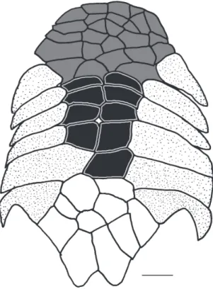
A total of 142 specimens of Dasyloricaria were analyzed. Measurements and counts follow Reis & Pereira (2000) and Thomas & Rapp Py-Daniel (2008), with the addition of the caudal-peduncle width which is measured at anal-fin base level, and the nares length which is measured from the anterior to posterior margins of the narial aperture. Measurements were taken point to point with digital calipers (0.1 mm precision). Counts and measurements were taken from the left side of the specimens except when the structure being measured or counted was damaged, in which case the data was acquired from the right side. Counts and nomenclature of body plates follow Schaefer (1997) and Rodriguez & Reis (2008), and tooth cusp names follow Müller & Weber (1992). Plates covering the abdomen were separated in groups (Fig. 1) that were used for the descriptions and for the characters in the phylogenetic analysis. The lateral abdominal plates are located on the lateral region of the abdomen from the pectoral fin to the pelvic-fin origins (= thoracic plates of early authors). The median abdominal plates are located between the lateral plates from the pectoral girdle to the pre-anal plate (Schaefer, 1997). The medial abdominal plates are further divided into three sections: the anterior abdominal plates, which are usually smaller, irregularly shaped plates in the cleithral region; the central abdominal plates which are usually larger, more symmetrical, and located between the lateral abdominal plates; and the posterior abdominal plates which form a somewhat inflexible complex of plates between the pelvic fin bases and anteriorly border the pre-anal plate.
In the Comparative Material Examined section, lots are grouped as follows: catalog number, type status if appropriate, number of specimens in alcohol and cleared and stained (indicated by c&s), country, department or state, city, river basin, collection locality and geographic coordinates. Collection dates and collector’s names are provided only for types of the new species. Institutional abbreviations follow Sabaj Pérez (2012). Osteological nomenclature follows Schaefer (1987), Rapp Py-Daniel (1997) and Paixão & Toledo-Piza (2009). Specimens for osteological observations were cleared and counterstained for bone and cartilage using the method of Taylor & Van Dyke (1985). Specimens with excess of adipose tissue which impairs the observation of anatomical structures were soaked in Xylene to dissolve the fat (Lehmann A., 2006). Observations of the anatomical structures were made via a stereomicroscope and drawings prepared via a camera lucida.
Taxon selection for the phylogenetic analysis was based on the phylogenetic hypothesis of the Loricariinae (Rapp Py-Daniel, 1997) and taxa putatively related to
Dasyloricaria (Isbrücker 1979, 1980). The outgroup was composed of Rineloricaria strigilata (Hensel, 1868), R.
microlepidogaster (Steindachner, 1907), Spatuloricaria sp.,
Spatuloricaria tuira Fichberg et al., 2014, and Ixinandria
cataphracta Linnaeus, 1758, L. clavipinna Fowler, 1940,
Pseudoloricaria laeviuscula (Valenciennes, 1840),
Pseudohemiodon sp., Loricariichthys anus (Valenciennes, 1836), and Hemiodontichthys acipenserinus (Kner, 1853) (Loricariini), Harttia loricariformis Steindachner, 1877,
Sturisoma rostratum (Spix & Agassiz, 1829), and S. robustum (Regan, 1904) (Harttiini). The outgroup was chosen in order to test both the relationships in Dasyloricaria and among the genera previously included in Rineloricariina sensu Isbrücker. Characters described mainly by Rapp Py-Daniel (1997), Fichberg (2008), and Paixão & Toledo-Piza (2009) for different members of the Loricariinae were included in the analysis with additional characters proposed for the first time herein.
Fig. 1. Abdominal plate pattern of Dasyloricaria species. Anterior abdominal plates in gray; central abdominal plates in black; lateral abdominal plates dotted; posterior abdominal plates unpigmented.
The monophyly of Dasyloricaria, the phylogenetic relationships among its species, and the monophyly of the Rineloricariina were tested using the cladistic methodology proposed by Hennig (1966). Parsimony analysis was employed to generate hypothesis of phylogenetic relationships and character state transformations. The matrix (Appendix I) was constructed in Mesquite (Maddison & Maddison, 2011). The phylogenetic analyses were performed using NONA (Goloboff, 1999) associated with Winclada 1.00.08 (Nixon, 2002). The heuristic search was performed with 1000 replications of Random Addition Sequence and branch swapping through the Tree Bisection Reconnection (TBR) algorithm, with additional TBR swapping to completion,
on the unweighted and unordered data matrix. Cladograms were rooted on Harttia loricariformis according to previous phylogenies (Rapp Py-Daniel, 1997; Fichberg, 2008; Paixão & Toledo-Piza, 2009).Tree support was calculated in NONA as decay indices or Bremer support (Bremer, 1994).
Results
Dasyloricaria Isbrücker & Nijssen, 1979
Dasyloricaria Isbrücker & Nijssen, in Isbrücker, 1979: 90 (type
species: Loricaria filamentosa Steindachner, 1878, by original designation; gender: female).
Diagnosis. Dasyloricaria is diagnosed by the following uniquely derived synapomorphies: (1) the metapterygoid is approximately rectangular (character 24.1); (2) the symplectic cartilage is one-half or more the length of the quadrate (character 26.2); (3) the ventrolateral process of epibranchial 4 is large and in the form of a curved shelf (character 37.2); (4) the lower pharyngeal tooth-plate is expanded, very thin and translucent (character 40.1); (5) the upper pharyngeal tooth-plate is triangular, with the anterior portion much narrower than the posterior region (character 44.2); (6) the anterior border of the cleithrum is strongly expanded anteriorly (character 54.2); (7) thick, fleshy filaments are present on upper lip anterior to the premaxillary teeth (character 66.1); and (8) the presence of a transverse bar of dark pigmentation extending over the eyes (character 73.1). Dasyloricaria can also be distinguished from other loricariines by having two rows of central abdominal plates with these sometimes separated laterally in juveniles from the lateral abdominal plates (vs. a single row of plates or plates without a clear arrangement or abdomen naked except in Loricariichthys), and the poorly papillated lips (vs. lips with filaments or papillae absent or with prominent rounded papillae).
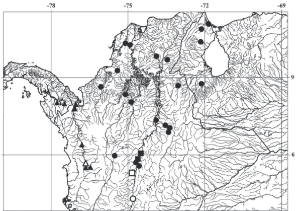
Distribution. Dasyloricaria species inhabit the trans-Andean portion of northern South America and southern Central America in the Atrato, Sinú, Cauca, and Magdalena, in Colombia, lago Maracaíbo basin in Colombia and Venezuela, and the río Tuyra basin in Panama (Fig. 2).
Dasyloricaria filamentosa (Steindachner, 1878)
Figs. 3-4, Tables 1-2
Loricaria filamentosa Steindachner, 1878: 45, pl. 9, fig. 1 (type locality: río Magdalena, Colombia, Lectotype NMW 44874).
Eigenmann, 1920b: 30 (lower río Magdalena); 1922: 90 (description). Schultz, 1944: 328 (description; tables of measurements and counts; distributed in lago Maracaibo basin).
Miles, 1947: 112; fig. 61 (brief description; lower Cauca and Magdalena; identification keys). Dahl & Medem, 1964: 46 (brief description; río Sinú). Dahl, 1971: 92, fig. 101 (description).
Galvis et al., 1997: 90 (repeat of Dahl, 1971 synonymy; brief description; Atrato, Sinú, Cauca, Magdalena, Cesar and Catatumbo basins; ventral and dorsal view photos).
Dasyloricaria filamentosa.—Isbrücker & Nijssen, in Isbrücker
1979: 87 (description of Dasyloricaria with D. filamentosa
as type species; lectotype designation). Isbrücker, 1980: 111
(composition of Dasyloricaria; species distribution; type
series location); 1981: 10 (identification key for Loricariidae).
Rapp Py-Daniel, 1997: 142 (brief description; assignment to Spatuloricaria-Rhadinoloricaria clade). Ferraris, 2003: 333 (genus distribution; synonymy; type material location; maximum body size). Maldonado-Ocampo et al., 2005: 134, fig. 130 (description; distribution). Villa-Navarro et al., 2006: 13 (río
Magdalena). Covain & Fisch-Muller, 2007: 4 (identification key
for Loricariinae; characters of Dasyloricaria). Ferraris, 2007: 233 (synonymy; type-material depositories).
Loricaria filamentosa seminuda Eigenmann & Vance, in Eigenmann,
1912: 13 (type locality: Girardot, Colombia, holotype FMNH 55116, NEW SYNONYM). Eigenmann, 1920b: 30 (upper río Magdalena in Colombia). Ibarra & Stewart, 1987: 53 (holotype at FMNH; previous CM number).
Loricaria seminuda.—Eigenmann, 1922: 91, pl. XIV, figs. 1 and
2 (brief description of 182 mm SL specimen from Girardot, Colombia). Dahl, 1971: 92 (junior synonym of Loricaria filamentosa; distribution; maximum and minimum body size; ontogeny; common names in Colombia).
Dasyloricaria seminuda.—Isbrücker & Nijssen, in Isbrücker, 1979:
87 (description of Dasyloricaria with D. filamentosa as type
species). Isbrücker, 1980: 111 (composition of Dasyloricaria;
distribution; type series depositories); 1981: 10 (identification key for Loricariidae). Ferraris, 2003: 333 (genus distribution; synonymy; types location, including “FMNH 55117” as holotype
of D. seminuda [that lot holotype of Loricaria fimbriata]; maximum body size; common names). Maldonado-Ocampo et al., 2005: 135 (brief description; distribution; types at FMNH and
USNM). Villa-Navarro et al., 2006: 13 (río Magdalena). Covain
& Fisch-Muller, 2007: 4 (identification key for the Loricariinae;
characters of Dasyloricaria). Ferraris, 2007: 234 (synonymy;
location of type material, including “FMNH 55117” as holotype
of D. seminuda [that lot holotype of Loricaria fimbriata]). Fig. 2. Map of northwestern South America (Colombia, Panama and Venezuela) showing the distributions of species of Dasyloricaria. Black dots: Dasyloricaria filamentosa; black squares: D. paucisquama; black triangles: D. latiura; white circle: holotype of Loricaria filamentosa seminuda (= Dasyloricaria filamentosa); white square: holotype of D. paucisquama; white triangle: lectotype of Loricaria filamentosa latiura; and white star: holotype of Loricaria capetensis (= Dasyloricaria
Diagnosis. Dasyloricaria filamentosa is diagnosed by having six to eight dark transverse bars on dorsum (vs. none to four bars in D. latiura and eight in D. paucisquama). It is further distinguished from D. paucisquama by having seven to 12 central abdominal plates in each row (vs. four to six); and the pelvic fin always surpassing the anal-fin origin (vs. pelvic fins never reaching the anal-fin origin).
Dasyloricaria filamentosa is further distinguished from
D. latiura by having the anterior abdominal plates smaller than central plates (vs. same size); the anal fin with well-defined spots without forming a distal band (vs. anal fin with black band on the distal portion of the rays); the black band covering the eyes never extending onto the snout (vs. the black band sometimes extending onto the snout); and the abdominal plate next to the pectoral-fin origin usually absent (vs. always present).
Description. Dorsal profile of head slightly convex to straight from snout to supraoccipital process. Dorsal profile of body slightly convex to straight from end of supraoccipital process to caudal-fin base. Plates at dorsal-fin base forming slight depression. Dorsal margin of orbit elevated; orbital notch well developed. Snout triangular in dorsal view. Odontodes poorly developed and not readily visible.
Upper lip with broad filaments that sometimes cover premaxillary teeth; short and thin filaments laterally on rictal barbel. Lower lip with filaments on posterior border similar to those on rictal barbel. Teeth slender and long in both premaxilla and dentary; number of teeth increases
with ontogeny. Teeth bifid, main cusp longer and broader, almost twice as long as lateral.
Central abdominal plates always arranged in two rows in adults, without intervening spaces between them and lateral abdominal plates. Smaller plates in juveniles always arranged in two rows and with naked intervening spaces between them, lateral abdominal plates and posterior abdominal plates. Space between central and lateral abdominal plates frequently broader than single plate row of central abdominal plates. Anterior abdominal plates rounded, without regular arrangement and having naked space between anterior most plates and filaments of lower lip border.
Plates in mid-ventral and median lateral series with well-developed keels. One pair of predorsal plates between supraoccipital and nuchal plate with two well-developed keels.
Posterior margin of dorsal fin concave; its distal tip when adpressed reaching sixth to ninth plate posterior to dorsal-fin base. Posterior pectoral-dorsal-fin margin straight or slightly convex; spine long, reaching to or barely surpassing pelvic-fin origin. Posterior pelvic-pelvic-fin margin straight or slightly convex; third and fourth branched rays longest and barely reaching anal-fin origin. Distal anal-fin margin straight to rounded; second branched ray longest. Tip of anal-fin spine reaching seventh or eighth plate posterior to its base; three plates along anal-fin base. Posterior caudal-fin margin concave, upper ray extended into long filament usually as long as or longer than SL.
Fig. 4. Lectotype of Loricaria filamentosa. NMW 44874, 233.2 mm SL, Colombia, río Magdalena basin (exact locality unknown; photo provided by W. Helmut).
Color in alcohol. Ground color of head and trunk greyish brown dorsally; pale yellow or light brown ventrally. Upper lip filaments grey or pale yellow; upper and lower lips pale yellow. Six to eight transverse, dark, dorsal bars; first bar crossing eyes, second bar on predorsal paired plates (Fig. 3). Small and irregular black spots present on dorsal-, pectoral- and pelvic-fin rays and membranes. Anal fin with scattered black spots not forming bands. Caudal-fin rays with black spots at base and complete black vertical band distally.
Sexual dimorphism. Poorly developed hyperthophied
odontodes on the lateral portions of head, pectoral- and pelvic-fin spines and sometimes on the supraoccipital in males. Thickening of pectoral-fin spine was observed, associated with sexual maturity in some males. Females lack these sexually dimorphic features.
Distribution. Dasyloricaria filamentosa occurs in the lower and middle Magdalena basin, lower Cauca, and the río Sinú in Colombia, and the lago Maracaibo basin in Colombia and Venezuela (Fig. 2).
Remarks. Steindachner (1878) described Loricaria
filamentosa from specimens collected in the río Magdalena, Colombia, without a precise locality. Eigenmann (1912) indicated that the specimens used by Steindachner were collected in the lower portion of the río Magdalena, but this information was not found in the NMW field records (W. Helmut, pers. com.). According to the observations herein, Eigenmann was correct concerning the distribution of D.
filamentosa in the lower and middle portions of the río Magdalena basin (see material examined; Fig. 2).
Dahl & Medem (1964) and Galvis et al. (1997) recorded this species in the río Sinú, Colombia, an occurrence corroborated herein. Galvis et al. (1997) and Maldonado-Ocampo et al. (2005, 2006) reported that D. filamentosa is widely distributed in northern South America, in the Catatumbo, Magdalena, Cauca, Sinú, San Jorge, and Atrato basins, what is not confirmed here, since D. filamentosa is only found in the Maracaibo, Magdalena and Sinú basins.
and D. filamentosa was a naked space between the central and the lateral abdominal plates. This difference was observed in this study to reflect ontogenetic variation with the plate arrangement described for D. f. seminuda being the juvenile condition of the plates in adults of D. filamentosa. This was also noted by Steindachner (1878: 47), who commented “…the central rows are rudimentary in young specimens and are not in contact; in adult specimens it is not strange for them to be divided…” (our translation).
Dahl (1971), in a study of the fishes of northern Colombia, proposed D. filamentosa as a senior synonym of Loricaria
fimbriata Eigenmann & Vance, 1912, Dasyloricaria
seminuda, D. latiura and D. tuyrensis. The author discussed about the few specimens used by Steindachner in the original description of D. filamentosa, and suggested that the five species belonged to an ontogenetic series of a single species, having collected himself the five species at the same locality. In this study we found that D. filamentosa is the senior synonym of D. seminuda, but we rejected the remaining synonyms proposed by Dahl, since D. latiura is a valid species with D. tuyrensis as its junior synonym and
L. fimbriata was transferred to Spatuloricaria.
The description of Loricaria filamentosa seminuda (in Eigenmann, 1912) was based on five specimens collected at Soplaviento (CM 3804a-b and IUM 12694a-c) and a single specimen from Calamar (CM 3805); both localities
in the lower Magdalena basin (Bolivar, Colombia). Those specimens were later mistakenly cataloged at FMNH as the type series of L. f. seminuda (FMNH 55113, 55114, 55116). The specimens in FMNH 55113 (CM 3804) and FMNH 55114 (CM 3805) were collected in the lower río Magdalena and identified as D. filamentosa. These were not among the specimens included in the original description of D. seminuda, and thus not types of the species. In addition, FMNH 55116 (CM 3807) is not a syntype, but the holotype of Loricaria filamentosa seminuda according to Eigenmann (1912: 13; 1922: 91), who indicated it as “type” of that species. The collection data of this specimen reads only “Girardot”, a city in Cundinamarca Department, Colombia, in the upper río Magdalena. The holotype of
Loricaria filamentosa seminuda is the only record of this species in the upper Magdalena, and a confirmation of its exact locality is needed.
Steindachner (1878) reported the presence of hypertrophied lower lip on mature males of D. filamentosa, a feature suggesting that the species is a lip brooder. That characteristic was not, however, observed on the specimens in this study. It is uncertain whether this is a function of the collection period during the year, or if the samples are composed only of females and/or non-nuptial males. Ecological studies on the species and the genus in general are lacking.
Table 1. Morphometric data for Dasyloricaria filamentosa and holotype of Loricaria filamentosa seminuda. Standard length in millimeters; holotype is included in range. HL: Head length; n: number of specimens analyzed; SD: standard deviation.
Holotype N Min Max Mean SD
Standard length (mm) 175.3 101.7 306.7 204.2
Percent of SL
Pre-dorsal length 29.4 54 28.9 34.0 30.9 1.3
Dorsal-fin spine length 22.5 54 19.8 28.2 23.3 1.6
Anal-fin spine length 17.1 54 15.2 21.1 17.2 1.3
Pectoral-fin spine length 16.7 54 15.1 19.0 16.9 0.9
Pelvic-fin spine length 15.1 54 13.0 20.4 15.9 1.8
Lower caudal-fin ray length 14.5 54 6.2 18.9 14.1 2.3
Head length 20.0 54 19.5 23.7 21.7 1.0
Trunk length 13.7 54 11.7 16.1 14.1 1.1
Abdominal length 15.3 54 12.9 17.4 15.4 1.1
Cleithral length 12.0 48 12.4 17.8 14.9 1.6
Body height 9.1 54 8.2 11.6 9.6 0.9
Caudal peduncle height 1.9 54 1.4 2.4 1.8 0.2
Post-anal length 59.8 54 50.3 61.9 56.2 2.1
Caudal peduncle width at anal-fin base 12.2 36 7.8 14.4 11.7 1.5
Percent of HL
Snout length 54.9 53 48.3 59.9 53.8 2.6
Head height 41.1 53 30.4 46.8 41.3 3.2
Interorbital distance 21.1 54 17.8 26.0 21.5 1.7
Eye diameter 14.3 54 9.6 16.4 13.3 1.6
Orbit diameter (including notch) 23.4 54 20.2 27.6 23.1 1.8
Rostral length 11.7 54 6.2 14.6 10.0 2.0
Table 2. Meristic counts for Dasyloricaria filamentosa and holotype of Loricaria filamentosa seminuda. n: number of specimens analyzed.
Counts Holotype n Min Max Mode
Premaxillary teeth 7 54 5 15 7
Dentary teeth 10 52 8 16 11
Lateral abdominal plates 9 54 5 11 9
Plates around pre-anal plate 3 54 3 5 3
Plates at median lateral series 19 54 16 21 18
Plates at mid-ventral series 18 53 15 20 18
Coalescent plates 12 53 10 15 13
Pre-dorsal plates 3 52 3 3 3
Plates at dorsal-fin base 5 54 4 6 5
Plates at anal-fin base 3 54 2 3 3
Pectoral-fin rays i-6 54 i-6 i-6 i-6
Pelvic-fin rays i-5 54 i-5 i-5 i-5
Dorsal-fin rays i-6 54 i-6 i-6 i-6
Anal-fin rays i-5 54 i-5 i-5 i-5
Caudal-fin rays i-10-i 54 i-10-i i-10-i i-10-i
Dasyloricaria latiura (Eigenmann & Vance, 1912)
Figs. 5-6; Tables 3-4
Loricaria filamentosa latiura Eigenmann & Vance, in Eigenmann, 1912: 13 (type locality: Boca de Certegai, Colombia; lectotype
FMNH 124472, BY PRESENT DESIGNATION). Meek & Hildebrand, 1916: 257 (senior synonym of L. tuyrensis; description, counts and measurements; río Atrato basin, Colombia and río Tuyra, Panama). Eigenmann, 1920b: 14 (río Atrato, Colombia and río Tuyra, Panama).
Loricaria latiura. —Eigenmann, 1920a: 10 (río Atrato basin); 1922:
91, pl. XV, fig. 3 (description; as senior synonym of Loricaria tuyrense). Miles, 1947: 112; fig. 62d, f, g (brief description; as
senior synonym of Loricaria tuyrense; lower río Magdalena
and río Atrato, Colombia, and río Tuyra, Panama; identification key). Dahl, 1971: 92 (junior synonym of Loricaria filamentosa; distribution, ontogeny; common names in Colombia).
Dasyloricaria latiura.—Isbrücker & Nijssen, in Isbrücker, 1979:
87 (description of Dasyloricaria with D. filamentosa as type
species). Isbrücker, 1980: 111 (composition of Dasyloricaria;
distribution; type series location); 1981: 10 (in identification key
for Loricariidae). Ferraris, 2003: 333 (distribution; synonymy; type material depositories; maximum size; common names). Maldonado-Ocampo et al., 2006: 150 (río Atrato). Covain &
Fisch-Muller, 2007: 4 (in identification key for Loricariinae;
Dasyloricaria characters). Ferraris, 2007: 233 (synonymy; type material depositories). Maldonado-Ocampo et al., 2012: 234 (lateral view photo; synonymy; description; color in alcohol; río Atrato basin; location of syntypes). Covain et al., 2016: 5 (in molecular phylogenetic analysis of the Loricariinae).
Loricaria capetensis Meek & Hildebrand, 1913: 80 (type locality: río Capeti, río Tuyra basin, holotype FMNH 7582; NEW SYNONYM). Meek & Hildebrand, 1916: 259, pl. XII
(description; possible synonym of Loricaria filamentosa
seminuda; paratype illustration in ventral view; río Capeti, Panama). Eigenmann, 1920b: 14 (río Tuyra, Panama, between
Canal Zone and río Atrato). Ibarra & Stewart, 1987: 53
(holotype number; type locality).
Dasyloricaria capetensis.—Isbrücker & Nijssen, in Isbrücker,
1979: 87 (description of Dasyloricaria with D. filamentosa
as type species). Isbrücker, 1980: 111 (composition of
Dasyloricaria; distribution; type series location); 1981: 10
(in identification key for Loricariidae). Ferraris, 2003: 333
(distribution; synonymy; type material location; maximum body size; common names). Maldonado-Ocampo et al., 2006: 150 (río Atrato). Ferraris, 2007: 233 (synonymy; type material location). Maldonado-Ocampo et al., 2012: 232 (lateral view photo; synonymy; description; color in alcohol; río Atrato basin).
Loricaria tuyrensis Meek & Hildebrand, 1913: 81 (type locality: río Tuyra basin, Panama; holotype FMNH 7583; NEW SYNONYM). Meek & Hildebrand, 1916: 257 (as junior
synonym of Loricaria filamentosa latiura). Eigenmann, 1922: 91 (as junior synonym of Loricaria latiura). Miles, 1947: 112;
fig. 61 (as junior synonym of Loricaria latiura; in identification key). Dahl, 1971: 92, fig. 101 (as junior synonym of Loricaria latiura; collection localities; minimum and maximum body
size; ontogeny). Ibarra & Stewart, 1987: 54 (holotype number;
number of specimens catalogued as paratypes).
Dasyloricaria tuyrensis. —Isbrücker & Nijssen, in Isbrücker, 1979:
87 (description of Dasyloricaria with D. filamentosa as type
species). Isbrücker, 1980: 112 (composition of Dasyloricaria;
distribution; type series location); 1981: 10 (in identification key
for Loricariidae). Ferraris, 2003: 333 (distribution; synonymy; type material location; maximum body size; common names). Ferraris, 2007: 234 (synonymy; type material depositories). Covain et al., 2008: 988 (in molecular phylogenetic analysis of the Loricariinae). Rodriguez et al., 2011: 3 (in molecular phylogenetic analysis of the Loricariinae). Covain et al., 2016: 4 (in molecular phylogenetic analysis of the Loricariinae). Dasyloricaria filamentosa, non-Steindachner, 1878.
Maldonado-Ocampo et al., 2006: 150 (río Atrato; specimens actually D. latiura).
Diagnosis. Dasyloricaria latiura is diagnosed by the following autapomorphies: (1) the two maxillary condyles widely separated (21.2); and (2) the posterior abdominal plates larger than the central abdominal plates (character 72.1). Dasyloricaria latiura is also differentiated from congeners by the following external characteristics: none to four dorsal, transverse dark bars on the body (vs. six to eight dorsal transverse, dark bars); the presence of a black band on the distal portions of the anal-fin rays (vs. scattered black spots on anal fin not forming bands); the presence of a black bar extending over the eyes and, sometimes onto the snout (vs. black bar only extending over the eyes and never onto the snout). The species can be further differentiated from
Description. Dorsal profile of head convex from tip of snout to supraoccipital process. Dorsal profile of body slightly convex from posterior of head to posterior of dorsal-fin base; and straight from that point to caudal-fin base. Plates along dorsal-fin base forming slight depression. Dorsal margin of orbit elevated; postorbital notch well developed. Snout triangular in dorsal view. Odontodes not well developed.
Upper lip with broad filaments that sometimes cover premaxillary teeth. Posterior border of lower lip and rictal barbel with few thin filaments. Teeth slender and long in both premaxilla and dentary; tooth number increasing ontogenetically. Teeth bifid; main cusp almost twice as long as lateral cusp.
Central abdominal plates always arranged in two rows and contacting lateral abdominal plates. Anterior abdominal plates without regular arrangement and with naked area between anterior most plates and lower lip filaments. One
well developed isolated plate next to pectoral-fin origin (Fig. 5); plate not in contact with other plates.
Plates in mid-ventral and median lateral series with developed keels. One pair of predorsal plates with two well-developed ridges between supraoccipital and nuchal plate.
Posterior dorsal-fin margin concave; distal tip of adpressed fin reaching ninth or tenth plate posterior to dorsal-fin base. Posterior pectoral-fin margin straight or slightly convex; spine long, reaching to or surpassing pelvic-fin origin. Posterior pelvic-fin margin straight to somewhat convex; third and fourth branched rays longest and reaching anal-fin origin. Distal anal-fin margin straight or rounded; second and third branched rays longest. Tip of anal-fin spine reaching seventh or eighth plate posterior of its base. Posterior caudal-fin concave, upper ray extended into long filament, sometimes equal, but never greater than SL.
Fig. 6. Lectotype of Loricaria filamentosa latiura. FMNH 124472, 220.0 mm SL, Colombia, Boca de Certegui, río Atrato basin, Atlantic versant of Colombia (photo reproduced from FMNH website with permission of Kevin Swagel).
Table 3. Morphometric data for Dasyloricaria latiura. Standard length in millimeters; Lectotype of D. latiura included in range. A: lectotype of Loricaria latiura FMNH 124472; B: holotype of L. capetensis FMNH 7582; C: holotype of L.
tuyrensis FMNH 7583; HL: Head length; n: number of specimens analyzed; SD: standard deviation.
A B C n Min Max Mean SD
Standard length (mm) 220.0 141.4 246.1 141.4 287.4 216.6
Percent of SL
Pre-dorsal length 35.2 30.2 33.0 53 29.0 35.2 31.8 1.3
Dorsal-fin spine length 24.2 25.9 23.2 53 20.5 26.6 24.1 1.6
Anal-fin spine length 18.6 17.9 18.4 53 16.6 19.6 18.1 0.9
Pectoral-fin spine length 17.0 17.5 16.0 53 15.7 18.6 17.0 0.8
Pelvic-fin spine length 19.1 14.9 17.5 53 14.3 19.6 17.4 1.3
Lower caudal-fin ray length 13.4 11.4 15.7 50 11.5 17.4 14.5 1.3
Head length 24.2 21.2 22.9 53 20.5 23.9 22.1 0.9
Trunk length 14.4 13.9 14.9 53 11.6 17.1 14.1 1.2
Abdominal length 16.6 15.5 16.9 53 13.1 17.5 15.6 1.0
Cleithral length 14.6 11.2 13.2 52 13.2 14.9 13.9 0.5
Body height 11.1 8.3 10.1 53 8.7 12.4 10.6 1.0
Caudal peduncle height 1.8 1.7 2.1 53 1.5 3.0 1.8 0.3
Post-anal length 54.0 56.7 54.6 53 51.9 58.8 56.0 1.9
Caudal peduncle width at anal-fin base 13.2 10.9 13.2 45 10.0 16.0 12.8 1.1
Percent of HL
Snout length 55.5 53.0 56.6 53 43.5 56.5 54.0 2.3
Head height 42.7 35.3 40.5 53 37.2 49.6 42.8 3.9
Interorbital distance 22.0 17.3 22.1 53 19.0 23.0 21.3 1.0
Eye diameter 14.7 14.7 11.3 53 11.1 15.7 13.5 1.3
Orbit diameter (including notch) 22.1 25.3 20.2 53 20.1 24.9 22.5 1.2
Rostral length 10.5 10.3 12.9 53 7.4 12.5 10.4 1.3
Table 4. Meristic counts for Dasyloricaria latiura. n: number of specimens analyzed.
Counts Lectotype n Min Max Mode
Premaxillary teeth 11 53 5 15 9
Dentary teeth 12 52 8 18 9
Lateral abdominal plates 8 52 6 9 8
Plates around pre-anal plate 3 53 1 5 3
Plates at median lateral series 21 53 18 22 20
Plates at mid-ventral series 20 53 18 22 20
Coalescent plates 9 53 9 12 9
Pre-dorsal plates 3 53 3 3 3
Plates at dorsal-fin base 5 53 5 6 5 Plates at anal-fin base 3 53 2 3 3
Pectoral-fin rays i-6 53 i-6 i-6 i-6
Pelvic-fin rays i-5 53 i-5 i-5 i-5
Dorsal-fin rays i-6 53 i-6 i-6 i-6
Anal-fin rays i-5 53 i-5 i-5 i-5
Caudal-fin rays i-10-i 53 i-10-i i-10-i i-10-i
Color in alcohol. Ground color of head and trunk greyish brown dorsally; pale yellow or light brown ventrally. Upper lip filaments grey or pale yellow; upper and lower lips pale yellow. Up to four transverse dorsal dark bars typically present but sometimes absent; first crossing eyes, second on predorsal paired plates (Fig. 5). Dorsal-, pectoral- and pelvic-fin rays and membranes with small, irregular black spots. Distal most portion of anal-fin rays with black band. Caudal fin with vertical black band along distal margin and dark basal spot.
Sexual dimorphism: Odontodes slightly hypertrophied in males along lateral portions of the head and pectoral- and pelvic-fin spines and sometimes the supraoccipital.
Distribution. Dasyloricaria latiura occurs in the río Atrato basin, on the Caribbean slope of Colombia, and in the río Tuyra basin of the Pacific versant of Panama (Fig. 2). Presence of D. latiura in both the Atrato and Tuyra basins can be explained by the Atrato River emptying into the Tuyra Gulf on the Pacific slope of Panama before the uplift of the Darien mountain range (Rodríguez-Olarte et al., 2011).
Remarks. Eigenmann & Vance’s (in Eigenmann, 1912) description of Loricaria filamentosa latiura is based on 12 syntypes from Boca de Certegui (originally CM 3806 and IU 12695). Seven specimens in CM 3806 were later recataloged as FMNH 55115 and the remaining five in IU 12695 were recataloged as CAS 13187. According to the CAS online catalog, CAS 13187 contains six specimens not indicated as types, and is composed of two original lots (IU 12695 and IU 12694), whose locality is “Soplaviento and Boca de Certegui”. Boca de Certegui is in the río Atrato basin, a location within the known distribution of D. latiura.
Soplaviento is, however, in the lower río Magdalena, where only D. filamentosa is known to occur. In addition, Ferraris (2007) reports one syntype of Loricaria filamentosa latiura in USNM 79219 and mentions that the location of four syntypes is unknown. The history of the original syntypes is confusing and for this reason we herein designate a lectotype (FMNH 124472, 220 mm SL, transferred from lot FMNH 55115), in accordance with the article 74 of the International Commission on Zoological Nomenclature (ICZN, 1999).
Dasyloricaria latiura was suggested to be the senior synonym of D. tuyrensis by several authors (e.g. Meek & Hildebrand, 1916; Eigenmann, 1922; Miles, 1947). Meek & Hildebrand (1916), subsequent to their description of D. tuyrensis, examined the syntypes of D. latiura, and concluded they belonged to the same species. Those authors did not elaborate as to the basis for the synonymy, presenting only a brief description of D. latiura based on specimens collected in Panama. No diagnostic features for D. tuyrensis were found in the present study to warrant the separation of that nominal species from D. latiura. Eigenmann (1920b) suggested that D. latiura is present in both the Atrato and the Tuyra basins. In that same year, Eigenmann (1920a) indicated the presence of D. latiura in the Atrato, but not San Juan, a conclusion in agreement with our findings.
The holotype of Loricaria capetensis (FMNH 7582) is a juvenile that differs from adults in various features that led Meek & Hildebrand (1913) to describe juvenile and adults as separate species (juvenile - D. capetensis; adult- D.
tuyrensis). Both species were described from the río Tuyra, Panama. Meek & Hildebrand (1916) redescribed their Loricaria capetensis, when they noticed its resemblance with D. seminuda in terms of the separation of the central and lateral abdominal plates (diagnostic character proposed for D. seminuda; see Remarks under D. filamentosa). That synonymy was not formally proposed because they did not have access to specimens of D. seminuda from the Magdalena basin to permit a proper comparison between populations of the Tuyra and Magdalena. This proposed synonymy is rejected in the present study.
Dasyloricaria paucisquama, new species
urn:lsid:zoobank.org:act:9D689322-8BCC-4CAB-9C3E-62081835CCDC
Fig. 7, Tables 5-6
Loricaria seminuda.—Miles, 1947: 112; fig. 63 (brief description; upper río Magdalena near Honda, Colombia; identification key).
Holotype. MPUJ 6019, 203.2 mm SL, Colombia,
Paratypes. Colombia: MCP 46920 (1, 179.7 mm SL), collected with holotype. CP-UCO 143 (1 c&s, 174.3 mm SL) Antioquia, río Magdalena basin, southern río Samaná, tributary to río La Miel in Butantan, 5°41’N 74°46’W, 189 masl, 31 Jan 2006, U. Jaramillo. IAvH-P 7683 (1, 157.7 mm SL) Boyacá, Puerto Boyacá, río Magdalena basin, Palagua and Velasquez creeks, 1 Jun 1995. MPUJ 5189 (1, 187.0 mm SL) Caldas, La Victoria, La Española farm at Zona El Gigante, río Magdalena basin, río Purrio, 5°22’N 74°47’W, 226 masl, 25 Feb 2010, S. Prada et al. MCP 48238 (1, 157.5 mm SL) Caldas, La Dorada, Purrio, río Magdalena basin, quebrada La Rica, 5°21’N 74°48’W, 259 masl, 23 Feb 2010, S. Prada et al.
Diagnosis. Dasyloricaria paucisquama is diagnosed by a single autapomorphy: the posterolateral border of the lateral ethmoid is slightly extended but does not contribute to ventral portion of the orbital rim (character 7.1). Additionally, this species can be differentiated from its congeners by having four to six central abdominal
plates on each row (vs. seven to 12 plates) and a pelvic fin never reaching to the anal-fin origin (vs. pelvic fin always reaching to the anal-fin origin).
Description. Dorsal profile of head straight from tip of snout to supraoccipital process. Dorsal profile of body convex from posterior of head to beginning of dorsal-fin base, then straight from that point to caudal-fin origin. Plates at dorsal-fin base forming slight depression. Dorsal margin of orbit elevated; postorbital notch present. Head broad, its width larger than its length. Snout triangular in dorsal view, lateral borders of head broad, with well-developed, thin hypertrophied odontodes in adults.
Upper lip with broad filaments never covering premaxillary teeth and laterally, with short and thin filaments on rictal barbel. Posterior border of lower lip with filaments not as broad as but longer than those on upper lip. Teeth slender and long in premaxilla and dentary. Teeth bifid with, main cusp broader than and almost twice as long as lateral cusp.
Central abdominal plates generally larger than in congeners, always arranged in two rows of 4-6 plates, with or without intervening naked spaces between them and lateral abdominal plates, but always in contact with plates bordering preanal plate; naked space never wider than one row of plates. Anterior abdominal plates irregular in size; with naked area between anterior most plates and posterior border of lower lip. Area next to pectoral-fin origin without plates. Plate in mid-ventral and median lateral series with well-developed keels. One pair of predorsal plates between supraoccipital and nuchal plate with two well-developed keels.
Posterior dorsal-fin margin straight; distal tip of adpressed fin reaching sixth plate posterior to dorsal-fin base. Posterior pectoral-dorsal-fin margin straight, first and second branched ray longest and reaching to or barely surpassing pelvic-fin origin. Posterior pelvic-fin margin straight to slightly convex; second and third branched rays longest but not reaching anal-fin origin. Distal anal-fin margin straight to rounded; second branched ray longest. Tip of adpressed anal fin reaching sixth plate posterior to its base. Posterior caudal-fin margin concave; filament on upper ray absent (possibly due to damage).
Table 5. Morphometric data for Dasyloricaria paucisquama. Standard length in millimeters; holotype included in range. HL: head length; n: number of specimens analyzed; SD: standard deviation.
Holotype n Min Max Mean SD Standard length (SL) 203.2 157.7 203.2 176.6
Percent of SL
Pre-dorsal length 62.3 6 30.3 31.5 30.7 0.5
Dorsal-fin spine length 50.4 6 19.4 24.8 21.9 2.4 Anal-fin spine length 33.6 6 15.3 16.5 16.0 0.6 Pectoral-fin spine length 33.5 6 15.2 16.9 16.0 0.9 Pelvic-fin spine length 28.8 6 14.1 14.8 14.3 0.4 Lower caudal-fin ray length 27.8 6 12.8 14.4 13.5 0.7
Head length 41.8 6 20.6 21.2 20.8 0.3
Trunk length 30.9 6 13.2 15.2 14.3 0.9
Abdominal length 32.6 6 15.3 16.9 16.1 0.7
Cleithral length 24.6 6 12.1 15.5 13.6 1.4
Body height 22.7 6 9.5 11.2 10.5 0.7
Caudal peduncle height 3.9 6 1.6 1.9 1.7 0.2
Post-anal length 113.3 6 55.5 56.3 55.8 0.3
Caudal peduncle width at
anal-fin base 25.9 6 12.0 12.9 12.6 0.4 Percent of HL
Snout length 22.4 6 52.7 53.7 53.4 0.4
Head height 19.3 5 40.8 46.3 43.9 2.8
Interorbital distance 8.9 6 20.7 21.8 21.3 0.4
Eye diameter 6.0 6 12.2 14.4 13.8 1.0
Orbit diameter (including
notch) 9.5 6 22.7 23.9 23.5 0.6
Rostral length 3.9 6 8.7 10.9 9.9 1.1
Nares length 7.5 6 14.8 17.9 15.9 1.4
Table 6. Meristic counts for Dasyloricaria paucisquama. n: number of specimens analyzed.
Counts Holotype n Min Max Mode
Premaxillary teeth 11 6 8 11 9
Dentary teeth 12 6 9 13 11
Lateral abdominal plates 5 6 4 7 5
Plates around pre-anal plate 2 6 1 4 2
Plates at median lateral series 18 6 18 19 18
Plates at mid-ventral series 18 6 17 19 18
Coalescent plates 12 6 11 13 12
Predorsal plates 3 6 3 3 3
Plates at dorsal-fin base 5 6 5 5 5
Plates at anal-fin base 2 6 2 4 3
Pectoral-fin rays i-6 6 i-6 i-6 i-6
Pelvic-fin rays i-5 6 i-5 i-5 i-5
Dorsal-fin rays i-6 6 i-6 i-6 i-6
Anal-fin rays i-5 6 i-5 i-5 i-5
Caudal-fin rays i-10-i 6 i-10-i i-10-i i-10-i
Color in alcohol. Ground color of head and trunk dark greyish brown to light brown dorsally; pale yellow or light brown ventrally. Upper lip filaments grey to pale yellow; upper and lower lips pale yellow. Eight transverse, dark, dorsal bars; first crossing eyes, second on supraoccipital and predorsal plates (Fig. 7). Dorsal-, pectoral- and pelvic-fin rays and membranes with small black spots. Distal most portions of anal-fin rays with diffuse dark spots. Caudal fin with longitudinal black band along tip of rays.
Sexual dimorphism. Males with slightly hypertrophied odontodes on lateral portions of head and on pectoral and pelvic fins.
Distribution. Dasyloricaria paucisquama is distributed in the upper and middle río Magdalena basin, Colombia (Fig. 2).
Etymology. Dasyloricaria paucisquama is named from the Latin paucis, meaning few, little and squama, meaning scale or plate, in allusion to the smaller number of central abdominal plates typical of this species. A noun in apposition.
Conservation status. Although with scarce lots, current plausible threats to the species were not detected in its distribution area, therefore Dasyloricaria paucisquama could be classified as Least Concern (LC), according to the International Union for Conservation of Nature (IUCN) categories and criteria (IUCN Standards and Petitions Subcommittee, 2014).
Remarks. Dasyloricaria paucisquama and D. filamentosa are sympatric in the middle río Magdalena, but not in the upper portions of that basin. The only record of D.
specimens of D. filamentosa were collected in the upper Magdalena. In addition, the record of Miles (1947) of D.
seminuda near Honda and Huila Department, both part of the upper Magdalena basin, is here regarded as Dasyloricaria
paucisquama based on the locality and the drawings in that publication.
Identification key for the species of Dasyloricaria
1. Four to six central abdominal plates in each row; plates well developed and usually lacking intervening naked spaces except in juveniles; pelvic fin not reaching anal-fin origin ...Dasyloricaria paucisquama
(Upper and middle río Magdalena basin)
1’. Seven to 12 central abdominal plates in each row, sometimes with intervening naked spaces between themselves and between them and the lateral abdominal plates; pelvic fin always surpassing the anal-fin origin ...2
2. None to four dark, transverse bars on dorsum; anterior abdominal plates similar in size to central plates; anal fin with vertical black band on distal portion of rays; black band covering eyes sometimes extended onto snout; conspicuous plate present ventrally next to pectoral-fin origin ...Dasyloricaria latiura
(Río Atrato and río Tuyra basins)
2’. Six to eight dark, transverse bars on dorsum; anterior abdominal plates smaller than central plates; anal fin with well-defined spots without forming distal band; black band covering eyes never extending onto snout; plate usually lacking next to pectoral-fin origin ... ...Dasyloricaria filamentosa (Río Magdalena and río Sinu, and lago Maracaibo basin)
Phylogenetic relationships
The characters descriptions are grouped by anatomical units.
Neurocranium. 1. Shape of the mesethmoid disk (Schaefer, 1987). The ventral mesethmoid disk of loricariids is circular in lateral view, robust and with edges thicker than central portion (Schaefer, 1987; state 0). In some species the mesethmoid disk is laminar but conspicuous and disk-like in lateral view (state 1), or laminar but rectangular or triangular in lateral view (state 2).
2. Development of the lateral expansion of the
mesethmoid. The variably developed lateral expansion of the mesethmoid articulates synchrondrally with the lateral ethmoid. It can be inconspicuous, approximately one-half the width of the vomer (state 0), or approximately the width of vomer (state 1; Fig. 8).
3. Ventral concavity on anterior tip of mesethmoid (Schaefer, 1990). Schaefer (1990) first described a ventral concavity on the anterior tip of the mesethmoid in scoloplacids. The ventral concavity of the mesethmoid is present in a few species (state 1) but absent in most (state 0; Fig. 8).
Fig. 8. Ventral view of neurocranium of Dasyloricaria
paucisquama, CP-UCO 143. MESD: mesethmoid disc; VOM: vomer; MESCR: mesethmoid crest; MES: mesethmoid; LAT-ET: lateral ethmoid; PSPH: parasphenoid; OSPH: orbitosphenoid; PROT: prootic; CMP-PTE: compound pterotic; EXO: exoccipital; BAS: basioccipital; BA-LIG: Baudelot’s ligament; TPWEB: transverse process of Weberian apparatus; AOR-CHA: aortic cannel; 6V: sixth vertebra. Scale bar = 5 mm.
4. Extension of the naked area on the snout tip. Among the examined groups, the Harttiini have an elliptical, naked area on the snout tip that does not reach the last sensory pore (state 0). In other species, the naked area on the snout tip can be elliptical or elongate and reaches the last pore of the infraorbital sensory canal (state 1). Dasyloricaria and most of the Loricariini possess a roundish naked area that does not reach the last sensory pore (state 2). Finally, Loricariichthys and Pseudohemiodon have the snout tip completely covered with dermal plates without a naked area (state 3).
5. Shape of the parasphenoid (Schaefer, 1991). The parasphenoid of loricariids is generally narrow along its entire length (Schaefer, 1987, 1991; Rapp Py-Daniel, 1997; Armbruster, 2004; Lehmann A., 2006; Pereira, 2008), as observed in Dasyloricaria paucisquama (state 0). Alternatively, the parasphenoid of Sturisoma, D. latiura and
6. Dorsolateral process of the lateral ethmoid (Schaefer, 1987). Dorsolateral processes of the lateral ethmoid are bar-like projections in the Harttiini and some other taxa (state 0). The projections are shaped as large wings, which contribute to the orbital rim and can extend a little beyond the orbital rim in Dasyloricaria paucisquama and some other taxa (state 1; Fig. 8). Finally, in some species the dorsolateral process of the lateral ethmoid is reduced (state 2).
7. Posteroventral border of the lateral ethmoid (Howes, 1983). The posteroventral border of the lateral ethmoid can be simple and match the anterior orbital rim (state 0). Alternatively, it can be slightly extended but still not contribute to the ventral portion of the orbital rim (state 1) or distinctly extended and contribute to the ventral portion of the orbital rim (state 2).
8. Lateral process of the sphenotic (Schaefer, 1987). The sphenotic process has been subject of discussions regarding its homology with the lateral spine of other Siluriformes. Schaefer (1987) proposed that homology contra the hypothesis of Howes (1983) who hypothesized that the lateral spine is absent in most siluriforms. The lateral process of the sphenotic can be short, never reaching one-half of the sphenotic height (state 0), about one-half the height of the bone (state 1), or still very long process, similar to the height of the sphenotic (state 2; Fig. 9) as in Dasyloricaria and Spatuloricaria. Conversely, in some species the lateral process is much reduced or absent (state 3).
9. Postorbital notch (Fichberg, 2008, character 163, modified). The postorbital notch is a feature found only in some Loricariini among loricariids. We followed Fichberg (2008) and considered the notch as short when it was less than one-half the orbital length (state 1), and long when it is at one-least half the orbital length (state 2; Fig. 9). The postorbital notch is absent in other loricariids (state 0).
10. Ornamentation of the parieto-supraoccipital and predorsal plates. Loricariines commonly have a marked development of odontodes crests on the parieto-supraoccipital and predorsal plates. Such crests can be absent (state 0) and when present their degree of development is quite variable among loricariids. Crests can be low and poorly developed (state 1), or well developed and conspicuous (state 2; Figs. 3,5,7), as in Dasyloricaria and Hemiodontichthys.
11. Lateral processes of the basioccipital (Schaefer, 1990). The basioccipital of loricariids has lateral processes with a wide variation in orientation and size. These lateral processes (described as dorsal processes by Schaefer, 1990) synchondrally articulate to the prootic. Such processes can be expanded not only laterally but also ventrally (state 0), or laterally expanded and as, or almost as, long as the occipital (state 1), or highly laterally expanded passing the exoccipital (state 2; Fig. 8). Conversely, the processes can be short and not reaching beyond one-half of the exoccipital (state 3).
Cephalic sensorial canals. 12. Ventral process of
preopercle (Rapp Py-Daniel, 1997, Character 53). The ventral process of the preopercle along the posterior margin of that bone was described by Rapp Py-Daniel (1997) and contacts a bony plate (the subpreopercle of Rapp Py-Daniel, 1997; and the canal-bearing plate of Schaefer, 1991). Such a process is present in the Harttiini (state 0) but absent in the remaining Loricariinae (state 1; Figs. 10a-c).
13. Shape of the nasal (Rapp Py-Daniel, 1997: 171, modified). The nasal bone located anterior to the frontal and mesial to the nostril includes the terminus of the supraorbital sensory canal. All of the Loricariini except
Ixinandria have a long, slim nasal (state 1; Fig. 9); whereas the Harttiini and Ixinandria share a short, broad, more square nasal (state 0).
14. Extension of the nasal sensorial canal (Rapp Py-Daniel, 1997; Character 170, modified). The supraorbital sensory canal usually ends in the nasal (state 0). In a few loricariids, however, the canal continues as a small tube into a dermal plate immediately anterior to the nasal (state 1; Fig. 9).
Fig. 9. Dorsal view of neurocranium of Dasyloricaria
15. Terminus of the parietal branch of the supraorbital sensory canal (Schaefer, 1987). The parietal branch of the supraorbital canal of Hypostomus plecostomus has its terminal pore in the sphenotic (Schaefer, 1987), but varies signifi cantly in position among examined loricariids. In Harttia, the terminal pore is in the sphenotic (state 0). In other groups the terminal pore can be at the articulation between the frontal and the supraoccipital (state 1); between the frontal and the sphenotic (state 2); on the supraoccipital (state 3); or on the frontal (state 4; Fig. 9).
16. Shape of the epiphyseal pore of the supraorbital sensory canal (Schaefer, 1987). The epiphyseal pore of the supraorbital sensory canal of most loricariids (Fig. 9) is usually large and readily visible between the frontals (state 1). Alternatively, the epiphyseal pore can be reduced in size (state 0) or form a groove as in Dasyloricaria and some loricariines (state 2).
Jaws and autopalatine. 17. Shape of the autopalatine (Arratia, 1990). The autopalatine is generally a rod-shaped bone connecting the lateral ethmoid to the upper jaw; sometimes with ventrolateral or dorsolateral expansions (Arratia, 1990, Figs. 11a-c). Studied loricariids vary in
the shape of the autopalatine. In Harttia the autopalatine is simple, rod-shaped and lacks expansions (state 0, Fig. 11c). Alternatively, in Dasyloricaria and other taxa the autopalatine has expansions extending two-thirds of the bone (state 1; Fig. 11a), or the expansions can extend along the entire length of the bone (state 2). Finally, the autopalatine can be curved and bear a small lateral expansion (state 3, Fig. 11b).
Fig. 11. Lateral view of autopalatine of: a. Dasyloricaria
paucisquama, CP-UCO 143; b. Loricaria cataphracta, MCP 41395; c. Harttia loricariformis, MCP 11707. Scale bar = 5 mm.
18. Autopalatine splint (Schaefer, 1987). The autopalatine of most loricariids bears a thin, approximately straight, sesamoid ossification that extends parallel to the autopalatine from the anterior palatine cartilage to the nasal opening and the lateral ethmoid (state 0). Conversely, the splint is absent in the Loricariini (state 1).
19. Shape of the premaxilla (Schaefer, 1987). The premaxillae of loricariids are loosely attached to the mesethmoid and highly mobile (Schaefer & Lauder, 1986). The variation in shape is notable with the premaxilla being long and rectangular, thick, and tridimensional (state 0); quadrangular, thick, and strongly tridimensional (state 1); slim and elongate (state 2); or very thin and reduced to a lamina (state 3).
20. Length of the premaxilla. The length of the premaxilla is variable among examined loricariids. The premaxilla can be long and approximately equal to or slightly longer than the autopalatine (state 0); or short and approximately one-half the length of the autopalatine (state 1), or very short and measuring one-fourth the length of the autopalatine (state 2).
21. Shape of the maxilla (Rapp Py-Daniel, 1997; Character 29, modified). The maxilla of loricariids is connected to the autopalatine via one or two condyles (Rapp Py-Daniel, 1997). Among the examined taxa the maxilla has one condyle (state 0), two closely positioned condyles (state 1; Figs. 12b,c) or two more widely separated condyles (state 2; Fig. 12a).
22. Size of the dentigerous area of the dentary (Schaefer, 1987). As is the case with the premaxilla, the dentary has a cup-shaped concavity where the teeth are implanted. In some loricariids the dentigerous area is almost as long as the dentary itself (state 0). Conversely, in most of the Loricariini the dentigerous area is shorter and never occupies the entire length of the bone (state 1). Finally, in Hemiodontichthys and Pseudohemiodon the tooth-bearing concavity is much reduced or absent (state 2).
Fig. 12. Dorsal view of maxillary bone of: a. Dasyloricaria
paucisquama, CP-UCO 143; b. D. latiura, USNM 293296; and lateral view: c. Dasyloricaria paucisquama, CP-UCO 143. MAX-COND: maxillary condyles. Scale bar = 5 mm.
Suspensorium. The suspensorium of Loricariidae is
composed by the hyomandibula, metapterygoid, quadrate and preopercle (Schaefer, 1987; Arratia, 1990; Figs. 10a-c), with the ectopterygoid and endopterygoid being absent in the family (Rapp Py-Daniel, 1997). Arratia (1990) discusses the possible homologies of the metapterygoid and the absence of the endopterygoid and ectopterygoid. Despite the absence of the latter bones in loricarioids and the several studies across the Siluriformes (Howes, 1983; Schaefer & Lauder, 1986; Schaefer, 1987; Arratia, 1987, 1990; Mo, 1991) there is no consensus on the homology of the pterygoid bones of loricarioids. Diogo et al. (2001) suggest that the real metapterygoid is fused to the hyomandibula, and that what is usually called metapterygoid is a dermal bone homologous to the ectopterygoid and endopterygoid of other fishes. We follow Schaefer (1987) and Arratia (1990) in interpreting a metapterygoid separate from the hyomandibula.
23. Canal of the metapterygoid (Howes, 1983). The metapterygoid of loricariids contacts the hyomandibula posteriorly and the quadrate ventrally (Figs. 10a-c). A feature first noted by Howes (1983) and later by Schaefer (1987) is the presence of a metapterygoid canal. This canal is formed by a lateral, laminar wall of the metapterygoid, which contacts the mesethmoid dorsally thereby forming a closed or partially closed canal. In some species the canal is deep and completely covered by the lateral laminar wall (state 0). In other species, the canal is deep but not completely covered by the lateral wall (state 1). Alternatively, a shallow but noticeable canal occurs in some members of the Loricariini (state 2) or the canal is absent or inconspicuous as in Dasyloricaria (state 3).
24. Shape of the metapterygoid. The metapterygoid of
Dasyloricaria has a characteristic shape, being roughly rectangular because of a posterior expansion that articulates by means of an interdigitated suture to the hyomandibula. Consequently, the dorsal portions of these bones are widely separated and lack further contact (state 1; Fig. 10a). In other examined loricariines the hyomandibula is approximately quadrangular and although also articulated to the hyomandibula, is not expanded posteriorly and the dorsal portions of the bones are not widely separated (state 0; Figs. 10b,c).
25. Crest for attachment of the levator arcus palatini onto the hyomandibula (Schaefer, 1987). The hyomandibula of loricariids usually has a conspicuous crest for the insertion of the levator arcus palatini, which usually occupies more than one-half the length of the bone (state 0; Fig. 10a). In a few taxa the crest is reduced and shorter than one-half the length of the bone (Schaefer, 1991; state 1).
hyomandibula and quadrate. The cartilage varies, ranging from reduced, almost one-eighth of the length of the quadrate (state 0), of approximately one-fourth the length of the quadrate (state 1, Fig. 10c), or one-half or more of the length of the quadrate (state 2; Figs. 10a,b).
27. Depth of the quadrate (Arratia, 1990). The quadrate of the loricariids ranges in form. In some taxa, its depth reaches at least one-half the depth of the hyomandibula (state 0), or alternatively, its depth is distinctly less than one-half the depth of the hyomandibula (state 1; Fig. 10).
28. Shape of the preopercle (Rapp Py-Daniel, 1997, Character 50; modified). The generalized shape of the preopercle of loricariids was described by Rapp Py-Daniel (1997) as thin and slender (state 1), but in some members of the group is wide and robust (state 0). In the present study, we corroborate those observations and found that a thin, slender preopercle is a non-exclusive synapomorphy of the Loricariini.
29. Shape of the sensory canal of the preopercle (Rapp Py-Daniel, 1997, Character 54; modified). The sensory canal of the preopercle of Harttia and Pseudohemiodon has a curved form (state 0). A straight tube was alternatively observed in most examined taxa (state 1).
Hyoid and branchial arches. The hyoid arch is composed of the paired anterohyals, posterohyals, branchiostegal rays, hypohyals, and the unpaired urohyal. The dorsal hypohyals are absent in loricariids but present in callichthyids (Reis, 1998) among loricarioids. The branchial arches are composed of two unpaired hypobranchials and the paired elements: three basibranchials, five ceratobranchials, four epibranchials, and two pharyngobranchials.
30. Relative length of laminar expansion along the ventral border of anterohyal (Paixão & Toledo-Piza, 2009; Character 10, modified). Paixão & Toledo-Piza (2009) described three states for this character. In the plesiomorphic state the laminar expansion along the ventral border of the anterohyal flares abruptly and is restricted to the lateral portion of the bone (state 0). In other taxa the laminar expansion begins gradually in the middle of the anterohyal and expands towards the lateral portion of the bone, but is only slightly developed (state 1). Finally the laminar expansion can commence gradually in the middle of the anterohyal and expand towards the lateral portion and is wide and well developed (state 2).
31. Projection of urohyal processes through the hypohyal foramina. The urohyal possesses two anterior projections with different degrees of development among the taxa studied. These processes can be medium-sized, just crossing the foramina (state 0), or they can be short and not cross the hypohyal foramina (state 1).
32. Shape of basibranchial 3 (Rapp Py-Daniel, 1997; Character 67, modified). Basibranchial 1 is absent in loricariids while basibranchial 2 (thus, the first in the series) is always ossified. Basibranchial 3 is ossified in loricariines and some hypostomines, and cartilaginous in most other loricariids, while the basibranchial 4 is variably present and always cartilaginous (Pereira, 2008). Among the taxa examined, basibranchials 3 (the second basibranchials of Rapp Py-Daniel, 1997) were always ossified while basibranchial 4 was always present and cartilaginous. Basibranchial 3 can have the shape of a short rod (state 0), a small nodule or biconcave drum (state 1), or can be an elongate rod (state 2).
33. Degree of ossification of the epibranchial gill filaments. Gill filaments on the epibranchials display variable degrees of ossification in loricariines. They can solely be ossified basally (state 0), up to their midlength (state 1), or for more than one half of the total length (state 2).
34. Extend of dorsolateral process of epibranchial 1 (Rapp Py-Daniel, 1997; Character 73, modified). Rapp Py-Daniel (1997) suggested that epibranchial 1 is devoid of processes in loricariines, but we found processes to be present and variably developed in the group. The dorsolateral process can be small (state 0, Fig. 13a), relatively elongate and triangular in shape (state 1; Fig. 13b), or elongate and rounded in shape (state 2; Fig. 13c).
35. Dorsolateral process of epibranchial 2 (Rapp Py-Daniel, 1997; Character 74, modified). The dorsolateral process of epibranchial 2 is highly variable in shape and position among the examined loricariids. This process can be large, well-developed and posteriorly directed (state 0; Figs. 13f,g), short and posteriorly directed (state 1; Fig. 13e), short and anteriorly directed in a mode similar to that of epibranchial 1 (state 2; Fig. 13b), or absent (state 3; Fig. 13d).
36. Dorsolateral process on epibranchial 3. Schaefer (1987) described a small dorsolateral process on epibranchial 3 of Hypostomus plecostomus, which was observed to be quite variable in shape among examined loricariids. The dorsolateral process can be elongate (state 0; Fig. 13d), or uncinate thereby forming a hook (state 1; Fig. 13b). Alternatively, the process on epibranchial 3 can be absent (state 2; Fig. 13a).