OCCURRENCE AND CHARACTERISTICS OF VIRULENCE GENES OF ESCHERICHIA COLI STRAINS ISOLATED FROM HEALTHY DAIRY COWS IN INNER MONGOLIA, CHINA
Simujide Huasai 1, Aorigele Chen 1*, Chun-jie Wang 2, Yu Li 1, Bai Tongrige 1
1
College of Animal Science, Inner Mongolia Agricultural University, Hohhot 010018, P. R. China; 2College of Veterinary Medicine, Inner Mongolia Agricultural University, Hohhot 010018, P.R. China.
Submitted: October 15, 2010; Returned to authors for corrections: July 13, 2011; Approved: January 16, 2012.
ABSTRACT
Virulence genes of Escherichia coli (E. coli) isolates from healthy dairy cows were identified and
characterized by a multiplex PCR assay and serogrouping test. The results showed that among the target
genes, eaeA was most frequently detected, accounting for 22.11% (67/303) in all strains from 101 cows. For
categorization of E. coli, aEPEC was the category with widest distribution detected in 55 (18.15%) strains
from 22 cattle. All of 84 PCR-positive strains belonged to 14 O serogroups, and O149 (25.00%) was most
common identified, followed by O2 (17.86%), O8 (11.90%) and O103 (9.52%) with relatively high
prevalence.
Key words: E. coli; Virulence gene; Multiplex PCR; Serogroup; Healthy dairy cow
INTRODUCTION
E. coli is an opportunistic species comprising the normal
flora in the gastrointestinal tracts of the warm-blooded animals.
It is usually non-pathogenic kept in host intestines. However,
in the immunocompromised or intestinal flora disturbed hosts,
or when the E. coli in the host intestines become translocated,
normal E. coli strains can cause relevant diseases as a
pathogenic one (1).
Pathogenic E. coli is increasingly isolated from cases of
human and animal diseases (2, 3). Moreover, it has also been
detected from healthy humans and animals. In 2001, Orden et
al. (2001) (4) reported some causative agents (cytotoxic
necrotizing factors, verotoxins and eae gene) of pathogenic E.
coli in healthy cows, sheep and goats. Amar et al. (2007) (5)
reported existence of enteroaggregative E. coli in
asymptomatic individuals in England. Some researches in other
countries also isolated pathogenic E. coli from healthy animals
(6, 7). However, very few studies have examined the
pathotypes of E. coli in healthy animals in China. In present
study, the virulence genes of E. coli were investigated and
characterized to elucidate the prevalence of pathogenic E. coli
in healthy cattle.
PCR (Polymerase chain reaction) method, particularly
multiplex PCR, being a rapid and effective gene detection
assay, is increasingly popular in identification of various
bacteria (8, 9). Some multiplex PCRs have been reported
elsewhere to detect virulence genes of E. coli. Bai et al. (2010)
(10) reported a multiplex PCR procedure that detected fliC,
stx1, stx2, eae, rfbE, and hlyA genes, which are the major
virulence genes of E. coli O157:H7. In the study conducted by
Kwai and Ke (2009) (11), a multiplex PCR assay was applied
to a panel of 87 E. coli isolates from different sources to
simultaneously detect virulence genes, such as st1, lt1, lt2, vt1,
vt2 and eaeA. Moreover, some researches focused on the
detection of virulence genes in combination with
serogroup-specific genes to test serogroups of E. coli using multiplex
PCR methods (12, 13). We can effectively save time and effort
involved in the determination of various virulence genes
through multiplex PCR technique.
MATERIALS AND METHODS
E. coli strains and control strains
Forty unrelated family dairy farms in Hulunbeier area
(Inner Mongolia, Northern China), were randomly selected for
test. The population in this area is predominantly engaged in
animal husbandry. Stool specimens were collected from the
rectums of 212 apparently healthy dairy cows (20 % of the
herd or a minimum of 5 cows from each farm). The specimens
were transported on ice to the laboratory. Swabs of stool were
inoculated onto the surface of eosin–methylene blue agars
(EMB) and streaked for isolated colonies. After incubation for
24 h at 37 °C, three or four colonies with typical E. coli
morphology and one of another morphological type were
streaked on fresh plates. Each colony was independently
subjected to Gram staining, microscopic examination,
biochemical test and serogrouping. Eventually, 303 E. coli
strains from 101 cows (three strains for one cow), were
randomly selected for characterizing virulence genes. E. coli
strains used as controls were C83922 (O101:K99, F41; st+), C83902 (O8:K87, K88ac; st+ lt+), E. coli O157 (stx1+ stx2+), E. coli O139 (eae+) and E. coli O127 (eaeA+ bfpA+). All strains were stored at -20 °C in LB broth with 10% glycerol.
PCR protocols
Bacterial DNA used for PCR analyses was prepared with
a genomic DNA purification kit (Wizard® Genomic DNA Purification kit, Promega Corporation, USA) used according to
the manufacturer’s recommendations. Alternatively, bacterial
DNA was extracted by a boiling procedure. Single colonies
were cultured overnight at 37℃ in 1.5 ml tubes containing 1 ml
LB broth. Each bacterial suspension was centrifuged for 15
min at 12,000 rpm, and the pellet was resuspended in 200μl
double distilled water. After 10 min of boiling in a water bath
and centrifugation for 15 min at 12,000 rpm, the supernatant
was used as template DNA for PCR assays.
All strains were evaluated by multiplex PCR for
identification of six virulence genes. The PCR primers were
presented in Table 1. We consulted the references about the
conditions for DNA template amplification and further
optimized for present multiplex PCR procedures.
Table 1. PCR markers for detection of virulence gene of E. coli
Target gene Nucleotide sequence (5’→3’) Size of amplified product (bp) References
eaeA F: GTGGCGAATACTGGCGAGACT
R: CCCCATTCTTTTTCACCGTCG 891 14
bfpA F: AATGGTGCTTGCGCTTGCTGC
R: GCCGCTTTATCCAACCTGGTA 326 14
stx1 F: AAATCGCCATTCGTTGACTACTTCT
R: CAGTCGTCACTCACTGGTTTCATCA 370 14
stx2 F: TGCCATTCTGGCAACTCGCGATGCA
R: GGATCTTCTCCCCACTCTGACACC 283 14
st F: ATTTTTCTTTCTGTATTGTCTT
R: CACCCGGTACAAGCAGGATT 190 15
lt F: GGC GAC AGA TTA TAC CGT GC
Bacterial DNA was amplified in the PCR reaction mixtures
as follows. Each reaction system had a final volume of 50μl and
contained 2.0μl of 10×EX Taq buffer, 4μl of 25 mM MgCl2, 4μl of dNTPs (each 2.5 mM), 1.25U of EX Taq DNA polymerase,
20-40μM of each primer, and 2μl of the DNA template. All reagents
were the products of TaKaRa Biotechnology (Dalian) Co., Ltd,
China). The mixtures were preheated at 94℃ for 1 min before
submitted to recycling step. The amplification conditions for the
multiplex PCR assays were 30 cycles at 94℃ for 30 sec, 55℃ for
30 sec, and 72℃ for 1 min. A final extension step was performed
at 72℃ for 10 min. The PCR products were kept at 4℃ until
removed and were separated in agarose gel (2% w/v)
electrophoresis and visualized under UV light after staining with a
nucleic acid stain (GoldViewTM, SBS Genetech, Beijing, China).
Serogrouping
Serological typing of all PCR-positive strains was carried out
by slide agglutination with E. coli O antisera (O1-O160). The
antisera were obtained from the China Institute of Veterinary Drug
Control (Beijing, China).
RESULTS
Of 101 healthy dairy cows, 34 (33.66%) carried at least one
virulence gene of E. coli, and of 303 E. coli strains from all
animals (three strains were tested for one animal), 84 (27.72%)
were positive for the virulence genes (Table 2, seen in page 16).
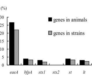
Among the six virulence genes identified, eaeA was the most
common gene harbored by 67 (22.11%) strains from 27 cattle. The
prevalence of the remaining genes was that bfpA and st were both
recovered in 10 (3.30%) strains from four different cattle, lt and
stx1 were in seven (2.31%) strains from three cattle respectively
and six (0.94%) strains from three cattle, and none of the strains
carried the genefor stx2 (Fig. 1).
Table 2. Distribution of virulence genes of E. coli isolates
No. of Virulence gene of E. coli
Serogroup
Animals Strains* eaeA bfpA stx1 stx2 st lt
O2 6 15 + - - - - -
O8 3 7 + - - -
O8 1 3 - - - +
O10 2 5 + - - -
O44 1 2 - + - - - -
O55 1 3 + + - - - -
O78 1 3 - - - - + -
O78 1 3 - - - - + +
O86 1 2 + - + - - -
O86 1 3 + - - -
O103 2 5 + + - - - -
O103 1 3 + - - -
O125 1 2 - - + - - -
O136 1 3 - - - - + -
O145 1 2 + - + - - -
O149 8 21 + - - -
O152 1 1 + - - -
O159 1 1 - - - - + +
0 5 10 15 20 25 30
eaeA bfpA stx1 stx2 st lt
(%)
genes in animals
genes in strains
Figure 1. Frequency of virulence genes of E. coli isolates
E. coli isolates were categorized into enteropathogenic E.
coli (EPEC), Shiga-toxin producing E. coli (STEC) and
enterotoxigenic E. coli (ETEC), and EPEC was further divided
into typical EPEC (tEPEC) and atypical EPEC (aEPEC)
according to the related virulence genes detected by the
multiplex PCR assays. The distribution of the categories
according to priority was as follows: aEPEC detected in 55
(18.15%) strains from 22 cattle, ETEC in 13 (4.29%) strains
from five cattle, tEPEC in eight (2.64%) strains from three
cattle, and STEC in six (1.98%) strains from three cattle (Fig.
2). An example of the multiplex PCR products for
representative isolates is shown in Fig. 3.
0
5
10
15
20
25
tEPEC
aEPEC
STEC
ETEC
(%)
categories in animals
categories in strains
Figure 2. Frequency of categories of E. coli isolates
Figure 3. Multiplex PCR products for representative strains from aEPEC, tEPEC, STEC and ETEC. Lanes: M, DNA
markers; 1, an aEPEC-strain only detected eaeA; 2, a
ETEC-strain only detected lt; 3, a STEC-ETEC-strain only detected stx1; 4,
an aEPEC-strain only detected bfpA; 5, a ETEC-strain only
detected st; 6, an tEPEC-strain detected eaeA and bfpA; 7, a
STEC-strain detected eaeA and stx1; 8, a ETEC-strain detected
lt and st.
Based on the serogrouping test, all of 84 PCR-positive
strains covered 14 O serogroups (Table 2, seen in page 16). As
shown in the table, O149 (25.00%) was the most common
serogroup, followed by O2 (17.86%), O8 (11.90%), O103
(9.52%), O78 (7.14%), O10 (5.95%), O86 (5.95%), O55
(3.57%), O136 (3.57%), O44 (2.38%), O125 (2.38%), O145
(2.38%), O152 (1.19%), and O159 (1.19%).
DISCUSSION
Cattle have been considered as important asymptomatic
carriers of potential risk factors for diseases of humans and
animals. The present study has shown that key virulence genes
of pathogenic E. coli could be identified in fecal E. coli isolates
from healthy cattle in northern China. The virulence genes
examined in this article were chosen because of their
association with E. coli strains causing animal and human
infections. Our findings showed that eaeA is the most common
gene identified in 22.11% (67/303) strains, whereas the
(Table 2). Moreover, in the 67 eaeA-positive strains, only 12
(17.91%) were found the combination of eaeA with other
genes; the rest 55 strains were recovered eaeA alone (Table 2,
seen in page 16). The above results were the main reason for
why aEPEC was the most frequent one among the four
categories tested. In our study, E. coli strains possessed A/E
genotype (eaeA+) or EAF plasmid (bfpA+) were classified as
atypical EPEC (aEPEC), while the strains with both of them
were as typical EPEC (tEPEC) regardless of their serogroups.
For the other categories, the strains with and without the eaeA
genotype that harbored Shiga toxin genes (stx1 and/or stx2)
were classified as STEC; the strains harbored enterotoxins (st
and/or lt) were as ETEC. The aEPEC is increasingly isolated
from human and different animal species, and domestic
animals have been seen as important reservoir of aEPEC.
Krause et al. (2005) (16) reported prevalence of aEPEC as high
as 92.8% in attaching and effacing Escherichia coli (AEEC)
strains (equivalent to eae-positive E. coli strains in current
article) from healthy animals when testing seven different
species of domestic animals. Yuste et al. (2008) (17) described
166 of 185 AEEC strains that isolated from diarrheic calves,
lambs, and goat kids and from healthy cattle, sheep, and goats
were aEPEC. The results of these reports also showed that
aEPEC strains were more prevalent than tEPEC strains; this is
consistent with our results. In present test, the percentage of
aEPEC was significantly higher than tEPEC in AEEC when
accounting for 82.09% (55/67), whereas the latter was only
found in eight strains. In addition, we found that aEPEC was
most frequently detected in all PCR positive strains compared
to other categories. The findings of our research indicated that
healthy dairy cows were a natural reservoir of aEPEC. STEC is
an important pathogen recovered from different clinical
isolates and even associated with hemolytic uremic syndrome
(HUS) (1), and STEC strains of bovine origin have been
detected elsewhere (6, 18). Rigobelo et al. (2006) (18) reported
different countries had shown that 10-80% of cattle might carry
STEC. However, in our study, a lower rate (1.98%) of STEC
isolation was found, and among the identified six STEC
strains, four carried eaeA and stx1, two carried stx1 alone,
while none of the strains carried stx2. Similar results were
described by some other reports in which stx1 genes were more
prevalent in bovine STEC isolates than stx2 genes (7, 19).
ETEC strains are the major cause of diarrhea in human and in
some animal species, especially in neonates (20, 21). ETEC
causes diarrhea by producing enterotoxins labile (lt),
heat-stable (st) or both (18). The two toxins were frequently found
in diarrheal cases of humans, cattle and pigs, sometimes in
healthy humans and pigs from different countries (3, 18, 22,
23). However, the distribution of ETEC in healthy cattle has
been less noticed. As far as we know, Houser et al. (2008) (24)
recently reported O-groups associated with ETEC in healthy
lactating dairy cows of a Pennsylvania dairy herd when
investigating phenotypic and genotypic diversity of E. coli in
10 cows. Blanco et al. (25) reported the prevalence of ETEC in
1% calves with diarrhea and 4% healthy controls. In present
study, 6.93% (7/101) cattle were confirmed to carry ETEC and
13 strains were positive for st and/or lt. The gene for st was
detected from 10 strains and for lt was from seven strains, four
of them were positive for both, it seems that st was more
prevalent. Similar result was described by Rigobelo et al.
(2006) (18) in Brazil, for fecal samples from diarrheal dairy
calves.
A wide range of serogroups in E. coli strains of bovine
origin has already been reported either in disease cases or in
healthy cattle, when some of them were only found in cattle
and some were associated with humans (26). Among the
serogroups detected in this survey, O2, O8, O103 and O136
belong to the prevalent serogroups in healthy cattle. For the
rest serogroups, O10, O55, O125, O145, O149, and O159 were
also identified from healthy cattle whereas with low
proportions, especially, O55 was found only in one Japanese
healthy cow (27). On the contrary, in our research, O149 was
most frequent accounting for 25% (21/84) and O55 was
serogroups (O44, O78, O86, and O152) not reported in healthy
cattle strains so far. Three of them have been reported in
association with several diseases in human and cattle, namely,
O44, O78 and O86 were detected from diarrheic patients (28),
O78 was from septicemic calves (29), and O86 was from
diarrheic calves (18). The majority of PCR-positive strains in
present study belonged to serogroups previously found among
isolates of cattle cases (i.e. gastrointestinal infections and
diarrhea), and even found in patients with severe diseases, such
as sudden infant death syndrome (SIDS), HUS, hemorrhagic
colitis (HC), and urosepsis (26, 30, 31, 32). The result
indicated that healthy cattle are the reservoir of E. coli strains
associated with infections and diseases.
CONCLUSION
Our data confirm dairy cow as important asymptomatic
carrier of E. coli strains pathogenic for humans, and as a major
reservoir of atypical EPEC. To our knowledge, this is the first
survey on the occurrence of these pathogenic strains in healthy
cattle in China. Therefore, more epidemiological characteristics
and virulence properties of these strains are necessary to be
clarified in the future work.
ACKNOWLEDGEMENTS
This work was supported by the Major Special Project of
National Dairy Industry of China (2006BAD04A15), the Key
project of Natural Science Fund of Inner Mongolia, China
(200607010403), and the Talent Fund of Inner Mongolia,
China.
REFERENCES
1. Afset, J.E; Bergh, K; Bevanger, L. (2003). High prevalence of atypical enteropathogenic Escherichia coli (EPEC) in Norwegian children with diarrhoea. J. Med. Microbiol. 52, 1015-1019.
2. Aidar-Ugrinovich, L; Blanco, J; Blanco, M; Blanco, J.E; Leomil, L; Dahbi, G; Mora, A; Onuma, D.L; Silveira, W.D; Pestana de Castro, A.F.
(2007). Serotypes, virulence genes, and intimin types of Shiga toxin-producing Escherichia coli (STEC) and enteropathogenic E. coli (EPEC) isolated from calves in São Paulo, Brazil. Int. J. Food Microbiol. 115, 297-306.
3. Amar, C.F.L; East, C.L; Gray, J; Iturriza-Gomara, M; Maclure, E.A; McLauchlin, J. (2007). Detection by PCR of eight groups of enteric pathogens in 4, 627 faecal samples; re-examination of the English case-control Infectious Intestinal Disease Study (1993-1996). Eur. J. Clin. Microbiol. Infect. Dis. 26, 311-323.
4. Antikainen, J; Tarkka, E; Haukka, K; Siitonen, A; Vaara, M; Kirveskari, J. (2009). New 16-plex PCR method for rapid detection of diarrheagenic Escherichia coli directly from stool samples. Eur. J. Clin. Microbiol. Infect. Dis. 28, 899-908.
5. Bai, J.F; Shia, X.R; Nagarajaa, T.G. (2010). A multiplex PCR procedure for the detection of six major virulence genes in Escherichia coli O157:H7. J. Microbiol. Methods. 82, 85-89.
6. Beutin, L; Jahn, S; Fach, P. (2009). Evaluation of the ‘GeneDisc’ real-time PCR system for detection of enterohaemorrhagic Escherichia coli (EHEC) O26, O103, O111, O145 and O157 strains according to their virulence markers and their O- and H-antigen-associated genes. J. Appl. Microbiol. 106, 1122-1132.
7. Blanco, M; Blanco, J; Blanco, J.E; Ramos, J. (1993). Enterotoxigenic, vertoxigenic, and necrotoxigenic Escherichia coli isolated from cattle in Spain. Am. J. Vet. Res. 54, 1446-1451.
8. Blanco, M; Schumacher, S; Tasara, T; Zweifel, C; Blanco, J.E; Dahbi, G; Blanco, J; Stephan, R. (2005). Serotypes, intimin variants and other virulence factors of eae positive Escherichia coli strains isolated from healthy cattle in Switzerland. Identification of a new intimin variant gene (eae-η2). BMC Microbiol. 5, 23.
9. Costa, M.M; Drescher, G; Maboni, F; Weber, S.S; Schrank, A; Vainstein, M.H; Schrank, I.S; Vargas, A.C. (2010). Virulence factors, antimicrobial resistance, and plasmid content of Escherichia coli isolated in swine commercial farms. Arq. Bras. Med. Vet. Zootec. 62, 30-36. 10. Fratamico, P.M; Yan, X.H; Liu, Y.H; DebRoy, C; Byrne, B; Monaghan,
A; Fanning, S; Bolton, D. (2010). Escherichia coli serogroup O2 and O28ac O-antigen gene cluster sequences and detection of pathogenic E. coli O2 and O28ac by PCR. Can. J. Microbiol. 56, 308-316.
11. Ghanbarpour, R; Oswald, E. (2009). Characteristics and virulence genes of Escherichia coli isolated from septicemic calves in southeast of Iran. Trop Anim. Health. Prod. 41, 1091-1099.
12. Gillespie, B.E; Olive, S.P. (2005). Simultaneous detection of mastitis pathogens, Staphylococcus aureus, Streptococcus uberis, and Streptococcus agalactiae by multiplex real-time polymerase chain reaction. J. Dairy Sci. 88, 3510-3518.
Escherichia coli in cases of sudden infant death syndrome does not differ from that in other infant deaths and healthy infants. J. Med. Microbiol. 58, 285-289.
14. Hornitzky, M.A; Mercieca, K; Bettelheim, K.A; Djordjevic, S.P. (2005). Bovine feces from animals with gastrointestinal infections are a source of serologically diverse atypical enteropathogenic Escherichia coli and Shiga toxin-producing E. coli strains that commonly possess intimin. Appl. Environ. Microbiol. 71, 3405-3412.
15. Houser, B.A; Donaldson, S.C; Padte, R; Sawant A.A; DebRoy, C; Jayarao, B.M. (2008). Assessment of phenotypic and genotypic diversity of Escherichia coli shed by healthy lactating dairy cattle. Foodborne Pathog. Dis. 5, 41-51.
16. Hussein, H.S; Sakuma, T. (2005). Invited review: prevalence of Shiga toxin-producing Escherichia coli in dairy cattle and their products. J. Dairy Sci. 88, 450-465.
17. Johnson, J.R; Stell, A.L. (2000). Extended virulence genotypes of Escherichia coli strains from patients with urosepsis in relation to phylogeny and host compromise. J. Infect. Dis. 181, 261-272.
18. Kobayashi, H; Shimada, J; Nakazawa, M; Morozumi, T; Pohjanvirta, T; Pelkonen, S; Yamamoto, K. (2001). Prevalence and characteristics of Shiga toxin-producing Escherichia coli from healthy cattle in Japan. Appl. Environ. Microbiol. 67, 484-489.
19. Krause, G; Zimmermann, S; Beutin, L. (2005). Investigation of domestic animals and pets as a reservoir for intimin-(eae) gene positive Escherichia coli types. Vet. Microbiol. 106, 87-95.
20. Kwai, L.T; Ke, X.Y. (2009). Multiplex PCR for simultaneous detection of virulence Genes in Escherichia coli. Malaysian J. Sci. 28, 1-14. 21. López-Saucedo, C; Cerna, J.F; Villegas-Sepulveda, N; Thompson, R;
Velazquez, R. F; Torres, J; Tarr, P.I; Estrada-García, T. (2003). Single multiplex polymerase chain reaction to detect diverse loci associated with diarrheagenic Escherichia coli. Emerg. Infect. Dis. URL: http://www.cdc.gov/ncidod/EID/vol9no1/01-0507.htm
22. Müller, D; Greune, L; Heusipp, G; Karch, H; Fruth, A; Tschäpe, H; Schmidt, M.A. (2007). Identification of unconventional intestinal pathogenic Escherichia coli isolates expressing intermediate virulence factor profiles by using a novel single-step multiplex PCR. Appl. Environ. Microbiol. 73, 3380-3390.
23. Nagy, B; Fekete, P.Zs. (1999). Enterotoxigenic Escherichia coli (ETEC)
in farm animals. Vet. Res. 30, 259-284.
24. Nataro, J.P; Kaper, J.B. (1998). Diarrheagenic Escherichia coli. Cin. Microbiol. Rev. 11, 142-201
25. Orden, J.A; Ruiz-Santa-Quiteria, J.A; Cid, D; Díez, R; Martínez, S; De la Fuente, R. (2001). Quinolone resistance in potentially pathogenic and non-pathogenic Escherichia coli strains isolated from healthy ruminants. J. Antimicrob. Chemother. 48, 421-424.
26. Orden, J.A; Ruiz-Santa-Quiteria, J.A; García, S; Cid, D; De la Fuente, R. (1999). In vitro activities of Cephalosporins and Quinolones against Escherichia coli strains isolated from diarrheic dairy calves. Antimicrob. Agents. Chemother. 43, 510-513.
27. Rigobelo, E.C; Gamez, H.J; Marin, J.M; Macedo, C; Ambrosin, J.A. Ávila, F.A. (2006). Virulence factors of Escherichia coli isolated from diarrheic calves. Arq. Bras. Med. Vet. Zootec. 58, 305-310.
28. Rivera, F.P; Ochoa, T.J; Maves, R.C; Bernal, M; Medina, A.M; Meza, R; Barletta, F; Mercado, E; Ecker, L; Gil, A.I; Hall, E.R; Huicho, L; Lanata, C.F. (2010). Genotypic and phenotypic characterization of enterotoxigenic Escherichia coli (ETEC) strains isolated from Peruvian children. J. Clin. Microbiol. doi:10.1128/JCM.00644-10.
29. Rodriguez-Siek, K.E; Giddings, C.W; Doetkott, C; Johnson, T.J; Fakhr, M.K; Nolan, L.K. (2005). Comparison of Escherichia coli isolates implicated in human urinary tract infection and avian colibacillosis. Microbiology. 151, 2097-2110.
30. Walter, J; Hertel, C; Tannock, G.W; Lis, C.M; Munro, K; Hammes, W.P. (2001). Detection of Lactobacillus, Pediococcus, Leuconostoc, and Weissella species in human feces by using group-specific PCR primers and denaturing gradient gel electrophoresis. Appl. Environ. Microbiol. 67, 2578–2585.
31. Wells, J.G; Shipman, L.D; Greene, K.D; Sowers, E.G; Green, J.H; Cameron, D.N; Downes, F.P; Martin, M.L; Griffin, P.M; Ostroff, S.M; Potter, M.E; Tauxe, R.V; Wachsmuth, I.K. (1991). Isolation of Escherichia coli serotype O157:H7 and other Shiga-like-toxin-producing E. coli from dairy cattle. J. Clin. Mirobiol. 29, 985-989.
32. Yuste, M; Orden, J.A; De la Fuente, R; Ruiz-Santa-Quiteria, J.A; Cid, D; Martínez-Pulgarín, S; Domínguez-Bernal, G. (2008). Polymerase chain reaction typing of genes of the locus of enterocyte effacement of ruminant attaching and effacing Escherichia coli. Can. J. Vet. Res. 72, 444-448.