Identification and adhesion profile of
Lactobacillus spp.
strains isolated from poultry
Ticiana Silva Rocha, Ana Angelita Sampaio Baptista, Tais Cremasco Donato,
Elisane Lenita Milbradt, Adriano Sakai Okamoto, Raphael Lucio Andreatti Filho
Laboratório de Ornitopatologia, Faculdade de Medicina Veterinária e Zootecnia, Universidade Estadual Paulista, Botucatu, SP, Brazil.
Submitted: September 18, 2012; Approved: March 14, 2014.
Abstract
In the aviculture industry, the use ofLactobacillusspp. as a probiotic has been shown to be frequent and satisfactory, both in improving bird production indexes and in protecting intestine against coloni-zation by pathogenic bacteria. Adhesion is an important characteristic in selecting Lactobacillus probiotic strains since it impedes its immediate elimination to enable its beneficial action in the host. This study aimed to isolate, identify and characterize the in vitro and in vivo adhesion of Lactobacillusstrains isolated from birds. TheLactobacillusspp. was identified by PCR and sequenc-ing and the strains and its adhesion evaluatedin vitrovia BMM cell matrix andin vivoby inoculation in one-day-old birds. Duodenum, jejunum, ileum and cecum were collected one, four, 12 and 24 h af-ter inoculation. The findings demonstrate greaaf-ter adhesion of strains in the cecum and an important correlation betweenin vitroandin vivoresults. It was concluded that BMM utilization represents an important technique for triage ofLactobacillusfor subsequentin vivoevaluation, which was shown to be efficient in identifying bacterial adhesion to the enteric tract.
Key words:Lactobacillus, identification, intestinal adhesion.
Introduction
Probiotics are defined as microorganisms that, when administered in suitable quantities, confer health benefits to the host (FAO/WHO, 2002). In the aviculture industry, supplementation with probiotics, especiallyLactobacillus, has been shown efficient in augmenting weight gain, im-proving the alimentary conversion rate, and diminishing mortality in birds of production (Huanget al., 2004). Due to the growing interest in the utilization of these microor-ganisms as probiotics, their correct identification becomes necessary (Moreiraet al., 2005).
Given that manyLactobacillusspecies have similar nutritional and growth requirements, it is often difficult to apply classical microbiology methods to identify them with precision. Several research studies have focused on the ap-plication of molecular biology techniques to achieve rapid detection and differentiation of this specie. The use of prim-ers and probes that rDNA sequences coding for the 16S and 23S rRNA has been validated as a means of identification. (Dubernetet al., 2002).
The adhesion ofLactobacillusto the epithelium was defined as a characteristic of interest for selection of probiotic strains, since it represents the first step in the for-mation of a barrier to prevent colonization by undesirable microorganisms due to competition for nutrients and adher-ence sites, in addition to preventing its immediate elimina-tion by intestinal peristalsis (Collado et al., 2005); but despite this, information on its adhesion to the intestinal ep-ithelium of poultry remains scarce (Morelli, 2002).
The use ofin vitromodels,including epithelial cells, mucosal components and extracellular matrix components such as laminin, fibronectin, collagen and proteoglycans, found in the commercial product Basement Membrane Ma-trix - BMM (BD - Becton Dickinson, USA), could be very useful, once it demonstrate the adhesion capacity of Lactobacillus, both individually and collectively (Horieet al., 2002; Vellezet al., 2007).
Although none of the previously cited methodsin vi-troreflect the complex interactions that occur in the mu-cosa of the gastrointestinal tractin vivo, they represent a
Send correspondence to T.S. Rocha. Ornitopatology Laboratory, College of Veterinary Medicine and Animal Science, São Paulo State University, 18618-970 Botucatu, SP, Brazil. E-mail: ticirocha@yahoo.com.br.
rapid method for characterizing and selecting strains of Lactobacillus, and according to Muñoz-Provencio et al. (2009), in most cases there is a high correlation between re-sults foundin vitroand those obtained fromin vivotests.
The staining of Lactobacillus by the fluorescence technique is a highly accurate and reproducible technique for tracking and quantifying bacterial cells, especially via in vivomodels (Bianchiet al., 2004), and one option is to utilize carboxyfluorescein succinimidyl amino ester (CFDA SE), which is colourless and nonfluorescent until its acetate group is cleaved by intracellularesterase to yield highly fluorescent, amine-reactive carboxyfluorescein succinimidyl ester (Bouzaineet al., 2005) that allows easily distinguish form from the native bacterial population of the intestine, without having their adhesion, survival capacity or membrane properties altered by the staining agent (Fulleret al., 2000).
The present work aimed to identify, by polymerase chain reaction (PCR), strains ofLactobacillus spp.isolated from chicken, to evaluate their adhesion capacityin vitro andin vivoin one-day-old birds and posterior sequencing.
Material and Methods
Isolation ofLactobacillusspp.strains
The isolation and genus identification of Lactobacillus strains were done as described before by Barroset al.(2009) using crop and cecum of thirty Cobb breeders, aged 65 weeks collected aseptically.
Confirmation ofLactobacillusspp. identification by Polymerase Chain Reaction (PCR) technique
The bacterial strains that presented characteristics compatible with theLactobacillusgenus were submitted to confirmation by means of PCR. The following strains were utilized as positive reaction controls: Lactobacillus fermentum CCT0559-ATCC9338, L. reuteri CCT3433 -ATCC23272, L. acidophilus CCT3258-ATCC4356, L. casei ssp. caseiCCT 1465- ATCC 393,L. delbrueckii ssp. lactisCCT7520-ATCC7830 andL. helveticus CCT 3747 -ATCC 15009.
For this purpose, DNA was extracted from the strains utilizing the kit QIAamp DNA blood Mini Kit (Qiagen, USA) according to the manufacturer’s instructions. The following primers:ForwR16-1 (5’-CTT GTA CAC ACC GCC CGT CA- 3’) andRevLbLMA1- (5’-CTC AAA ACT AAA CAA AGT TTC -3’), the reaction and cycle em-ployed were according to previously described by Dubernet et al.(2002) in order to amplify a product of approximately 250 pb.
In vitroadhesion in cell matrix type BMM
EachLactobacillusstrain was submitted to an adhe-sion test, as described by Bouzaineet al.(2005) utilizing the cell matrix type Basement Membrane Matrix (BMM).
To prepare the slides with BMM matrix, the same was diluted (1:20) in PBS; and 35 uL was deposited on Falcon® (BD) slide at 4 °C. The slides were maintained in repose at 37 °C for two h and then incubated with phosphate buffer solution (PBS) with the addition of 2% bovine serum albu-min (BSA) for one hour at room temperature.
The preparation of theLactobacillusstrains consisted of incubating the bacterial cells in MRS broth for 24 h at 37 °C, centrifuging (13,000 x g for 3 min) and washing twice with one mL of PBS. Subsequently, 200mL of this solution at the concentration of 107UFC mL-1was depos-ited on slides previously prepared with BMM and incu-bated for two h at room temperature. After incubation, the slides were washed three times with PBS, submitted to Gram staining and read in an optical microscope.
Three repetitions were performed for each strain tested, with eight fields being counted in each one in a rect-angular area of 1.7 x 1.0 cm. The area for counting in each microscopic field was 3.8 x 10⁻²mm2, utilizing a 100x ob-jective. The counts were performed by double-blind study. A strain ofEscherichia coliisolated from chicken and a Ba-cillus subtilis168 were utilized as positive and negative control, respectively.
In vivo adhesion ofLactobacillus spp.in intestinal epithelium of birds
Culturing and bacterial staining with carboxyfluo-rescein succinimidyl amino ester (CFDA SE).
Based on the in vitro adhesion test, the five Lactobacillus spp.strains that had obtained the best adhe-sion results in BMM were selected for thein vivotest.
The strains ofLactobacillus spp.andBacillus subtilis were cultured in MRS broth and Luria broth (LB), respec-tively, until they reached a stationary phase (16 h). Subse-quently, they were centrifuged at 9300 xgfor five minutes, washed twice in PBS and resuspended in PBS (pH 7.5) to obtain a concentration of 1010cfu mL-1. The strains were stained with Vybrant CFDA SE Tracer (Invitrogen) accord-ing to the manufacturer’s recommendations using 50mmol L-1of CFDA SE solution at 37 °C for 30 min. The stained cells were then centrifuged, washed twice in PBS and re-suspended to its initial volume in the proper culture broth (MRS or LB) and incubated again at 37 °C for 30 min. The stained cultures were protected from light and main-tained at 4 °C until inoculation in birds.
Experimental design
Prior to thein vivoassay a pilot test was performed with a group in which the birds did not receive any type of stained microorganism, to certify that only the inoculated strains would be detected by fluorescence.
The inoculum was administered orally, with the aid of a gavage needle, directly into the crop. Alimentation was suspended 12 h before this procedure. The collection mo-ments were at one, four, 12 and 24 h after treatment.
Preparation of intestine fragments from birds
At each of the four moments after bacterial inocula-tion, six birds per group were humanely euthanized by cer-vical dislocation, and cranial one-centimeter portions of duodenum, jejunum, ileum and cecum were collected. All visible residues were carefully removed to avoid damaging the intestinal mucosa. The collected fragments were imme-diately frozen via submersion in liquid nitrogen and stored at -80 °C until the histological preparation. The fragments were collected with the aid of a micro-cryostat and pre-pared on slides containing five histological cuts (8 to 10mm each). The slides were protected from light and stored at -20 °C until analysis.
To quantify the attachedLactobacillus spp.and Ba-cillus subtilis, two random microscopic fields were ob-served in each cut, totaling ten counting fields per slide/fragment. The counting area in each microscopic field was 3.8 x 10⁻²mm2. The reading was performed in a fluo-rescence microscope and the results were expressed as the median number of stained bacteria per histological cut.
Sequencing
The PCR products of the five best adhesive strains at in vitrotest were cloned at pGem vector (Promega), accord-ingly to the manufacture’s recommendation, utilizing
vec-tor’ primers Fw-M13
5’-CACGACGTTGTAAAACGAC-3’ e rev-M13
-5’-GGATAACAATTTCACACAGG-3’. The sequences of the 16S/23S ribosomal RNA intergenic spacer region of phylogenetically related species:L. acidophilus(U32971), L. amylovorus(AF182732),L. casei subsp. casei(Z75478), L. crispatus(AF074857),L. delbrueckii subsp. bulgaricus (Z75475), L. fermentum (AF080099), L. gasseri (AF074859), L. hamsteri (AF113601), L. helveticus (Z75482), L. jensenii (AB035486), L. johnsonii (AF074860), L. plantarum (U97139), L. reuteri (AF080100), L. rhamnosus (AF121201), L. sakei (U97137), L. salivarius (AF113600), L. vaginalis (AF182731) were retrieved from GenBank (www.ncbi.nlm.nih.gov) and used to perform multiple alignments with the our strains sequences using Clustal W (Tamuraet al., 2011).
Statistical analysis
The results obtained in vitro were transformed to log10 form; the mean and standard deviation and
subse-quently the confidence interval were calculated to deter-mine the upper and lower limits, which were compared with the mean of the positive control. A 5% significance level was adopted (Sampaio, 2002).
For thein vivocounts we used a non-parametric anal-ysis of variance (Dunn test), utilizing the median values of bacterial counts, with a two-factorial scheme (Zar, 2009). A 5% significance level was adopted in all tests and the datas were submitted to the software SIGMASAT (Surhone et al., 2010).
Results
Identification ofLactobacillus spp.
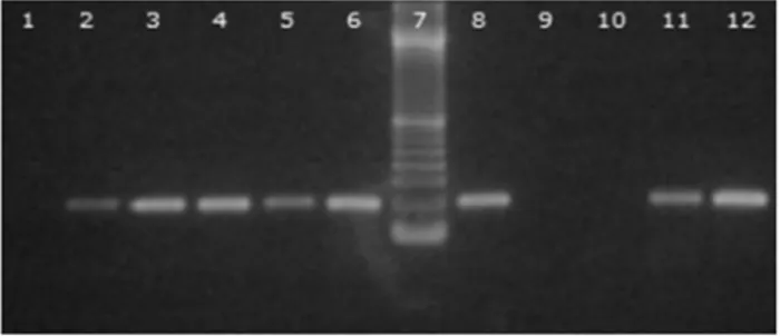
Of the strains analyzed, 123 were compatible with the genusLactobacillusin the aforementioned tests and were then submitted to PCR. Of this total, only 73 strains were positive (see Figure 1).
In vitro adhesion in cellular matrix type BMM
After identification of the genus Lactobacillus by PCR, each of the strains (73) was submitted toin vitro ad-hesion test in BMM. Table 1 displays the counts of the mi-croorganisms adhering to the BMM matrix.
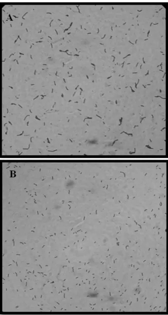
The visualization of theEscherichia colistrain uti-lized as a positive control and the Bacillus subtilis 168 strain employed as a negative control is represented in Fig-ure 2, A and B.
In the present study, it was possible to determine that the strains ofLactobacillus spp.isolated from the bird in-testines are capable of adhering to BMM (Figure 3 A and B). We can observe greater adhesion capacity in strains 206, 231, 258, 262 and 311 with mean counts of bacteria adhering in BMM closer to the positive control (Table 1). In this manner, these five strains were selected for thein vivo adhesion test.
Adhesionin vivoofLactobacillus spp. strains
The median counts of adherent cells in thein vivotest of Bacillus subtillis (negative control), Lactobacillus reuteri(positive control) and of the test of strains 206, 231, 258, 262 and 311 are shown in Table 2.
Figure 1 - Agarose gel 1.5%. 1-Negative control; 2- Lactobacillus acidophilusCCT3258; 3-L. caseiCCT 1465; 4-L. delbrueckiiCCT 7520; 5-L. fermentum CCT 0559; 6-L. helveticus CCT 3747; 8-L. reuteri
CCT3433; 9 and 10-Samples negative forLactobacillusspp.; 11 and
Table 1- Result of thein vitroadhesion test represented by mean number of bacterialLactobacillusspp cells adhering to BMM matrix.
Identification of strains Mean Identification of strains Mean
E. coli(positive control) 206.12 250 2.62
B. subtilis(negative control) 7.08 251 6.79
203 6.00 252 2.33
204 2.70 253 2.08
206 111.70 254 2.00
208 71.70 255 3.33
210 3.37 256 6.16
211 6.87 257 4.25
212 56.29 258 106.25
214 19.5 259 3.83
215 65.66 260 3.33
216 42.65 261 18.54
217 13.16 262 231.41
218 40.29 263 5.87
219 49.16 264 2.75
220 29.16 265 3.33
221 17.70 266 3.66
222 6.75 267 5.70
224 8.12 268 2.79
226 5.83 271 7.25
227 19.91 273 1.12
230 13.66 305 2.04
231 254.87 306 1.08
234 7.08 307 2.04
237 10.04 308 16.16
238 21.00 309 1.83
239 26.45 310 23.70
240 12.79 311 109.54
241 15.25 312 9.91
242 11.12 318 2.29
243 6.45 319 2.08
244 18.29 320 7.04
245 11.83 321 9.95
246 2.20 322 12.95
247 1.66 323 4.66
248 5.5 324 5.16
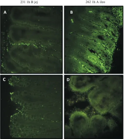
As expected, in the group in which the birds did not receive stained microorganisms, no type of fluorescence or staining was found. The visualization of theLactobacillus spp.strains stained in different intestinal segments is repre-sented in Figure 4.
The greatest bacterial count was found in strain 262 in the cecum, four hours after inoculation. However, a de-crease in the counts was observed at the moments 12 and 24 h after inoculation.
In the four different intestinal segments during the time of the experiment, it was observed that all strains pre-sented a greater quantity ofLactobacillusadhering at 1 h and 4 h than at 24 h, but this was not found in the cecum fragments of the treatments with strains 231 and 206.
The positive control presented reduced counts in all segments as time elapsed, demonstrating low adherence to intestinal epithelium. In general, the cecum was the
seg-ment that demonstrated the greatest quantity of adhering Lactobacillus, with the exception of strain 258. But this strain showed stability in values that did not differ signifi-cantly (p > 0.05) during the collections at 12 and 24 h (see Table 2), demonstrating a homogenous distribution throughout the intestine, but in a lesser quantity when com-pared to the otherLactobacillusstrains.
In all the strains, the bacterial counts observed in the duodenum at 12 and 24 h were similar of the negative con-trol, demonstrating low adherence of the Lactobacillus evaluated in this segment.
The results obtained with the sequencing alignment showed a highly conserved region between nucleotides 369-391 corroborating with Dubernet et al. (2002). The strains 206, 231, 258, 262, 311 and 316 showed an identity
Figure 2- Optical microscopic image (100x) ofEscherichia coli(A) and
Bacillus subtillis(B), stained by Gram, demonstrating high and low
adher-ence of strains in BMM matrix byin vitroadhesion tests, respectively. Figure 3- Optical microscopic image (100x) of Strains 231 (A) and 262
Table 2- Median count of bacteria adhering to intestinal epithelium, byin vivoassay,considering the strain, time after inoculation and intestinal segment.
Strain Time (h) Intestinal Segment
Duodenum Jejunum Ileum Cecum
Control -B. subtillis 1 12.2ab x* 3.8b x 49.6a x 1.3b x
4 0.7a x 0.8a x 0.4a x
0.8a x
12 0.8a x 0.3a x 0.0a y 0.5a x
24 1.9a x 1.0ab x
0.7b y 0.5b x
Control +L. reuteri 1 215.5ab x 300.7a x 301.9a x 57.3b y
4 22.9b y 3.0b y
5.2b y 104.0a xy
12 6.4b y 4.3b y
3.4b y 190.4a x
24 3.8a y 10.2a y 2.8a y 9.9a z
311 1 146.7a x 38.7ab x 14.5bc x 2.1c x
4 19.4a y 7.5a xy 3.4a x 4.3a x
12 1.3a y 1.4ab y 1.4ab x 10.1b x
24 1.4ab y 1.4ab y 0.7b x 2.6a x
262 1 251a x 328.4a x 192.2a x 14.1b z
4 21.3b y 105.3b y 125.4ab x 534.0a x
12 1.4b z 1.5b z 1.2b y 155.5a y
24 0.5b z 0.8b z 0.6b y 59.3a yz
258 1 163.0a y 102.9a y 67.4a x 0.7b y
4 63.6a y 8.9a x 5.8a y 55.8a x
12 0.5a x 0.7a x 0.4a y 5.7a y
24 0.9a x 0.1a x 0.1a y 1.7a y
231 1 17.2ab x 25.5ab x 42.7a x 0.0b y
4 65.2ab y 192.2a y 18.0b xy 150.8ab x
12 18.4ab x 16.6ab x 7.7b y 53.2a x
24 24.0ab x 12.6ab x 5.6b y 71.4a x
206 1 155.3a y 368.7a z 172.6a x 0.2b z
4 85.5a y 46.8a xy 33.8a y 125.8a xy
12 22.9b x 67.0ab y 34.2b y 252.9a x
24 18.8a x 9.1ab x 1.4b z 33.9a y
*a,b,cTwo medians followed by at least one different superscript, in the same row, do differ significantly (p
³0.05), comparing the same strain and same
time in different intestinal segments (Line).
x,y,zTwo medians followed by at least one different superscript, in the same columm, do differ significantly (p
of 100% with up to eight sequences of L. reuteri (CP002844.1; AP007281.1; CP000705.1; EU547293.1, EU547292.1, EU547290.1, EF412989.1; EF412988.1), al-lowing us to conclude that all strains analyzed belong to this specie.
Discussion
The difference found between the PCR and biochemi-cal analyses corroborates De Martinis (2002), who consid-ers the utilization of a fermentation pattern of carbohydrates unsatisfactory as a single criterion for identi-fying lactic acid bacteria, since frequent variations occur in the fermentations, enabling subjectivity in their interpreta-tions.
Adhesion to intestinal mucosa can confer an impor-tant competitive advantage for bacterial maintenance in the gastrointestinal tract and it is generally accepted that adhe-sive properties contribute to the efficacy of probiotic strains (Servin and Coconnier, 2003).
Bouzaineet al.in 2005, utilizing the same methodol-ogy employed in the present study for the evaluation ofin vitro adhesion, reported respective mean counts for the strains Lactobacillus rhamnosus TB1 and Lactobacillus reuteri LRT1 greater than 2000 and 500 cels per field,
while those of the strainsLactobacillus johnsoniiLJT2 and Lactobacillus salivariusLST1 were less than 250 cells per field. But Horieet al.in 2002 evaluated the adhesion capac-ity in BMM of Lactobacillus crispatus JCM 5810 and found an average of 70 bacterial cells per microscopic field. The counts reported in previous studies are higher than most of the values observed in our study (except for the strains selected for thein vivoadhesion test), which may be explained by the fact that the physical-chemical nature of the external membrane of the cell wall, the conformation of macromolecules of the bacterial surface and the suscep-tibility to external factors (time, medium and general cul-ture conditions) determine the propensity ofLactobacillus to adhere to a surface, factors that vary even within the same specie (Schar-Zammaretti and Ubbinik, 2003).
The CFDA SE cell tracer, utilized in thein vivoassay, is a compound capable of staining cells without compro-mising their viability or altering their adhesive characteris-tics, although during bacterial growth, when the bacterial concentration is expected to rise, the number of cells stained can decrease due to dilution of intracellular CFDA SE after cellular division (Fuller et al., 2000). Thus, to avoid discrepant results between the quantities of adhering bacteria vs. the number effectively stained, the present
study opted to evaluate adhesion for a maximum period of 24 h after bacterial inoculation in the birds.
Lee et al.(2004) in an investigational study of the growth and colonization of Lactobacillus casei shirota stained by CFDA SE in rat intestines; determined that the Lactobacillusstrain utilized presented in the first collec-tion, at 24 h, greater adhesion to the jejunum, in contrast to the data found in the present study, in which the strains, ex-cept for 258, presented greater adhesion to ceca at 24 h after inoculation of birds.
According to Bouzaine et al. (2005) Lactobacillus rhamnosus TB1presented a low bacterial count in intestinal epithelium of birds, despite having demonstrated greater affinity for adhesion in the rectum, ileum and jejunum. The cecum was the segment that presented the least adhesion by Lactobacillus rhamnosus TB1, in agreement with the re-sults of Edelmanet al.(2002), who also observed low adhe-sion of different Lactobacillus species in the cecal epithelium of birds.
In our present work it was possible to identify a ten-dency among the strains evaluated to colonize the ceca, with less capacity to establish themselves in the duodenum or jejunum, especially in the 24 h period after inoculation, corroborating the results of Fuller and Turvey (1971) who reported a massive bacterial colonization of the ceca.
We verified that a single strain (258) presented homo-geneity in intestinal colonization but also the lowest counts in the four intestinal segments evaluated at 24 h after inocu-lation. In contrast, strain 231 presented the highest adhe-sion values at 24 h, but showed variations in its adhesive capacity in the different intestinal segments, at the mo-ments analyzed.
These data confirm the strain-dependent nature of the intestinal adhesion capacity, since they may have differ-ences in the structural conformation and composition of the bacterial cell wall, with these characteristics varying within the same genus and even within the same species of Lactobacillus(Servin and Coconnier, 2003; Deepika and Charalampopoulos, 2010).
Strain 231 also presented better adherence in thein vi-trotest utilizing BMM, and satisfactory results in the in vivoadhesion tests, significantly superior to the other ones and especially to the positive control (Table 2), demonstrat-ing the relevance and correspondence of thein vitrowith thein vivoresults and suggesting that thein vitrotechnique employed may be utilized for the selection of possible probiotic strains.
Early studies on chicken microbiota found that Lactobacillus salivarius, L. reuteri, and L. acidophilus inhabited the crop and the chicken digestive tract. Our re-sults show that the five strains sequenced are compatible withLactobacillus reuteri specie, and agrees with the fact that this is the most abundantLactobacillusspecies in the chicken gastrointestinal tract (Abbas Hilmiet al., 2007).
From the results obtained in the present study it can be concluded that the PCR technique is an important tool for identifyingLactobacillusspp., and can be utilized to validate the result of biochemical identification tests.
It may also be concluded that thein vitro adhesion process of Lactobacillus, utilizing the Basement Mem-brane Matrix (BMM) permits the selection of probiotic strains with adhesion capacity; also, the staining of Lactobacillusby CFDA SE is shown to be suitable for the visualization and counting of this bacterium adhering to the intestinal epithelium of birds, with both techniques being efficient for evaluating the adherence capacity of probiotic strains.
Acknowledgments
The authors thank the Sao Paulo State Research Sup-port Foundation- FAPESP for funding the project.
References
Abbas Hilmi H, Surakka A, Apajalahti J, Saris PEJ (2007) Identi-fication of the Most Abundant Lactobacillus Species in the Crop of 1- and 5-Week-Old Broiler Chickens. Appl Environ Microbiol 73:7867-7873.
Barros MR, Andreatti Filho RL, Oliveira DE, Lima ET, Crocci AJ (2009) Comparison between biochemical and polymerase chain reaction methods for the identification of
Lactobacillusspp. isolated from chickens. Arq Bras Med Vet Zoot 61:319-325.
Bianchi MA, Del Rio D, Pellegrini N, Sansebastiano G, Neviani E, Brighenti F (2004) A fluorescence-based method for the detection of adhesive properties of lactic acid bacteria to Caco-2 cells. Lett App Microbiol 39:301-305.
Bouzaine T, Dauphin RD, Thonart P, Urdaci MC, Hamdi M (2005) Adherence and colonization properties of
Lactobacillus rhamnosusTB1, a broiler chicken isolate. Lett App Microbiol 40:391-396.
Collado MC, Gueimonde M, Hernadez M, Sanz Y, Salminen S (2005) Adhesion of selected Bifidobacterium strains to hu-man intestinal mucus and the role of adhesion in enteropathogen exclusion. J Food Protec 68:2672-2678. Deepika G, Charalampopoulos D (2010) Surface and Adhesion
Properties of Lactobacilli. Adv App Microbiol 70:127-152. De Martinis ECP (2002). Identification of meat isolated bac-teriocin-producing lactic acid bacteria using biotyping and ribotyping. Arq Bras Med Vet Zoot 54:659-661.
Dubernet S, Desmasures N, Guéguen M (2002) A PCR-based method for identication of lactobacilli at the genus level. FEMS Microbiol Lett 214:271-275.
Edelman S, Westerlund-Wikstrom B, Leskelas S, Kettunen H, Rautonen N, Apajalahti J, Korhonen TK (2002) In vitro ad-hesion specificity of indigenous Lactobacilli within the avian intestinal tract. Appl Environ Microbiol 68:5155-5159.
FAO/WHO (2002) Guidelines for the Evaluation of Probiotics in Food. Working Group Report.
Fuller ME, Streger SH, Rothmel RK, Mailloux BJ, Hall JA, Onstott CT, Fredrickson KJ, Balkwill LD (2000) Develop-ment of vital fluorescent staining method for monitoring bacterial transport in subsurface environments. Appl Envi-ron Microbiol 66:4486-4496.
Horie M, Ishiyama A, Fujihira-Ueki Y, Sillanpaa J, Korhonen TK, Toba T (2002) Inhibition of the adherence of Esche-richia coli strains to basement membrane by Lactobacillus crispatus expressingan S-layer. Appl Environ Microbiol 92:396-403.
Huang MK, Choi YJ, Houde R, Lee JW, Lee B, Zhao X (2004) Ef-fects of Lactobacilli and an Acidophilic Fungus on the Pro-duction Performance and Immune Responses in Broiler Chickens. Poul Sci 83:788-795.
Lee YK, Ho PS, Low CS, Arvilommi H, Salminen S (2004) Per-manent colonization by Lactobacillus casei is hindered by the low rate of cell division in mouse gut. App Environ Microbiol 70:670-674.
Moreira JLS, Motta RM, Horta MF, Teixeira SMR, Neumann E, Nicoli JR, Nunes AC (2005) Identification to the species level of Lactobacillus isolated in probiotic prospecting stud-ies of human, animal or food origin by 16S-23S rRNA re-striction profiling. BMC Microbiol 5:15-19.
Morelli, L (2000) In vitro selection of probiotic lactobacilli: a crit-ical appraisal. Curr Issues Intest Microbiol 1:59-67.
Munoz-Provencio D, Llopis M, Antolin M, Torres I, Guarner F, Pérez-Martinex G, Monedero V (2009) Adhesion properties of Lactobacillus casei strains to resected intestinal fragments and components of the extracellular matrix. Arch Microbiol 191:153-161.
Sampaio, IBM (2002) Estatística Aplicada à Experimentação An-imal. Editora FEPMVZ, Belo Horizonte.
Schar-Zammaretti P, Ubbinik J (2003) The cell wall of lactic acid bacteria: Surface constituents and macromolecular confor-mations. Bioph J 85:4076-4092.
Servin AL, Coconnier MH (2003) Adhesion of probiotic strains to the intestinal mucosa and interaction with pathogens. Best Pract Res Clin Gastroenterol 17:741-754.
Surhone LM, Timpledon MT, Marseken SF (2010) Probiotics. VDM Verlag AG & Co. Kg, p. 164.
Tamura K, Peterson D, Peterson N, Stecher G, Nei M, Kumar S (2011) MEGA5: Molecular Evolutionary Genetics Analysis using Maximum Likelihood, Evolutionary Distance, and Maximum Parsimony Methods. Molecular Biology and Evolution.
Velez M P, De Keersmaecker SCJ, Vanderleyeden J (2007) Ad-herence factors of Lactobacillus in the human gastrointesti-nal tract. FEMS Microbiol Lett 276:140-148.
Zar JH (2009) Bioestatistical Analysis. Pearson, Prentice Hall, Upper Saddle River, p. 960.


