LUCIANA RODRIGUES DA CUNHA
GENOTYPIC AND PHENOTYPIC CHARACTERIZATION OF
Lactobacillus gasseri ISOLATED FROM A NEWBORN INFANT
Tese apresentada à Universidade
Federal de Viçosa, como parte das
exigências do Programa de
Pós-Graduação em Ciência e Tecnologia
de Alimentos, para obtenção do
título de
Doctor Scientiae
.
VIÇOSA
LUCIANA RODRIGUES DA CUNHA
GENOTYPIC AND PHENOTYPIC CHARACTERIZATION OF
Lactobacillus gasseri ISOLATED FROM A NEWBORN INFANT
Tese apresentada à Universidade
Federal de Viçosa, como parte das
exigências do Programa de
Pós-Graduação em Ciência e Tecnologia
de Alimentos, para obtenção do
título de
Doctor Scientiae
.
APROVADA: 18 de março de 2011.
Pesq. Cláudia Lúcia de O. Pinto
(Coorientadora)
Prof. Paulo César Stringheta
(Coorientador)
Prof
aElisabeth Neumann
Pesq. Marcelo Bonnet Alvarenga
To my parents
Abel Fernandes da Cunha e Lúcia M. Rodrigues da Cunha
ACKNOWLEDGMENTS
I would like to thank GOD for everything I have in my life.
I would like to thank my chair Célia L. L. F. Ferreira for her assistance, guidance, opportunities and friendship over these years.
I would like to thank Dr. Todd R. Klaenhammer for his scientific support, friendship and enormous assistance with my Doctoral Training in USA. Thank you so much for believing me and giving me the opportunity to conduct part of my Doctoral studies in your laboratory, and be part of your wonderful team. I really appreciate all your reassurances and encouragement.
I would like to thank the Federal University of Viçosa and the Department of Food Science and Technology for the opportunity.
I would like to thank National Council for Scientific and Technological Development (CNPq) for providing me the fellowship during part of my Doctoral Training in Brazil and also to Foundation Coordination for Improvement of Higher Education Personnel (CAPES) for providing me the fellowship to conduct the research experiment in North Carolina State University (NCSU), USA (Grant number, BEX 4654-08-0).
I would like to thank my committee members, Dr. Marcelo Bonnet Alvarenga, Dra Elisabeth Neumann, the EPAMIG`s researcher Dra. Cláudia Lúcia de O. Pinto and Dr. Paulo César Stringheta for their receptivity and suggestions.
I would like to thank all the professors of the Department of Food Science and Technology and Food Microbiology for their support and contributions to my scientific background. Special thanks to Nélio José de Andrade, Nilda de Fátima Ferreira Soares, Antônio Fernandes de Carvalho and Maria Cristina Dantas Vanetti for believing me and my potential.
I would like to thank all my co-works of the Laboratory of Lactic Cultures, specially Célio, Juliana, Milene, Carolina, Tatiane, Joice, Fabiana, Eder, Mônica, Sara, Érika, for their friendship and for creating a great atmosphere in the lab. I very much appreciate Karina da Silva Chaves and Juliana Nóbrega for their friendship and always being there to listen and laugh with me.
TRK lab for their receptivity, generous help in the lab and for making my experience in USA so enjoyable. Jun Goh, Evelyn and Rosemary deserve a significant portion of my gratitude for their technical assistance and precious help in my experiment. I can not express how thankful I am to have those three angels in my life in the USA. They are outstanding scientists and taught me to love the `` world of genetics``. They were always there to help me as I needed and without them, none of this would have been possible. From the botton of my heart, thank you so much!
I would like to thank all the collaborators of the Department of Food Science and Technology, UFV specially, Vaninha, Osvaldo, Geralda, Perereca, Adão, Sr. Manuel, Dimas, Pio, Fernando, Divino, Piu, Valério, Juarez, Sr Luis and José Geraldo for their support and friendship over these years. I really would like to express my gratitude to Geralda and Gilcemir that did not measure efforts to help me with CAPES process. I also wish to extend my gratitude to all staff and Faculties at the Department of Food, Bioprocessing and Nutrition Sciences of North Carolina State University (NCSU), Raleigh, North Carolina, USA, in special Dr. Brian Farkas, Sue Strong, Carol Reilly, Shirley Lyles, and Sabrina Whitley-Ferrell, for their support, friendship and help during my training in the USA.
I deeply would like to thank Edith Ramos Neta, who is the best friend I could have. I cannot express how thankful I am to have Edith around these years. She greatly assisted me in obtaining my training at the Dr. Klaenhammer`s Lab and help me a lot during my training in USA. She has always being a great friend and I am very grateful for her being there to listen and advice me.
I would like to thank Marcelo Augusto Ferraz, who has also been a wounderful friend over these years. Marcelo is a unique person that I love very much. He has always being there to help and make me laugh.
I want to thank my friends in the Federal University of Viçosa, specially Michele Bertoldi, Danilo Pereira, Rodrigo Resende, Roberta Careli, Geruza Dias, Maria Patricia Milagres, João Paulo Rigueira, Wender Souza, Alexandre Resende, Washington da Silva, Nathalia Ramos, Joesse, Rita Superbi, Vanessa de Castro, Junia Capua, Rosineia de Paula, Maurício, José Carlos, Ana Andrea, Nathan Pimentel, for their friendship and fun times we`ve had together.
I would like to give my special thank you to my boyfriend Joel Oliveira for the support he has given me, for his unconditional love, and for all the wonderful memories that we shared together in the USA and are still sharing in Brazil.
I would like to extend my special thank you to Denilce de Fátima da Cunha (Pepete) and Chumbinho, who are not my blood, but the sister and brother I could choose. Thank you for supporting and caring me.
I want to express my gratitude to my family, my grandmother Carlita, aunts, in special Maria Arruda Rodrigues, Leonor Rodrigues da Silva, Carlita Helena Rodrigues and Lia Cristina Rodrigues Brandi, uncles, cousins for providing a loving environment for me.
Lastly, but not least, I deeply would like to express my gratitude to my parents Abel e Lúcia, my sister Mariana and, my brother Leandro, for their active participation and constant support. I am very fortunate to have such a loving family and I would not be the person I am without their love and support.
BIOGRAPHY
LIST OF CONTENTS
LIST OF FIGURES ... xi
LIST OF TABLE ... xv
RESUMO ... xix
ABSTRACT ... xvii
1 - INTRODUCTION ... 1
2 - LITERATURE REVIEW ... 3
2.1 - Human Milk and Child Health ... 3
2.2 - Human Milk Banks ... 4
2.3 - Probiotics - History and definitions ... 4
2.4 - Principal Probiotic Microorganisms ... 6
2.4.1 - Genus Lactobacillus ... 6
2.4.1.1 - Lactobacillus gasseri ... 7
2.5 - Probiotics and beneficial effects to health of children ... 9
2.5.1 - Modulation of the intestinal microbiota ... 9
2.5.1.1 - Production of antimicrobial compounds ... 9
2.5.1.2 - Competition for nutrients and adhesion sites ... 12
2.5.1.3 - Stimulation of the immune system ... 14
2.5.2 - Maturation of the Intestinal Microbiota ... 14
2.5.3 - Probiotics and Gastrointestinal Diseases ... 18
2.5.3.1 - Diarrhea ... 18
2.5.3.1.1 - Antibiotic Associated Diarrhea (DAA) ... 19
2.5.3.1.2 - Diarrhea associated with Rotavirus infection ... 20
2.5.3.2 - Necrotizing Enterocolitis ... 22
2.5.3.3 - Probiotics and Alergies ... 23
2.5.3.3.1 - Eczema and atopic dermatitis ... 24
2.5.3.4 - Probiotics and Respiratory infections ... 25
2.5.3.5 - Probiotics and obesity ... 26
2.5.4 - Selection of probiotic bacteria ... 27
2.5.4.1.2 - Antagonism to pathogens ... 29
2.5.4.1.3 - Adherence to intestinal tissues ... 29
2.5.4.2 - Technological Aspects ... 30
2.5.4.3 - Aspects of Safety ... 31
2.5.4.3.1 - Identification of probiotic strain ... 31
2.5.4.3.2 - Determination of virulence factors ... 32
2.5.4.3.3 - Resistance to antibiotics ... 32
2.5.5 - Legislation on probiotics ... 34
2.6 - REFERENCES ... 36
3 - EXPERIMENTAL HYPOTHESES ... 57
CHAPTER I CHARACTERIZATION OF Lactobacillus gasseri ISOLATES FROM A BREAST-FED INFANT ... 60
ABSTRACT ... 60
RESUMO ... 61
1 - INTRODUCTION ... 62
2 - MATERIALS AND METHODS ... 63
2.1 - Bacterial strains and growth conditions ... 63
2.2 - Bacterial identification ... 64
2.3 - Pulsed-Field Gel Electrophoresis (PFGE) ... 64
2.3.1 - Preparation of genomic DNA in agarose blocks ... 64
2.3.2 - Restriction enzyme digestion and PFGE. ... 65
2.4 - Testing of Hemolytic Activity and Bile Tolerance ... 65
2.5 - Antagonistic activity against pathogenS ... 66
2.6 - Testing of Antibiotic Susceptibility ... 67
2.7 - Plasmid analysis ... 67
2.8 - Tolerance to simulated gastric and small intestinal juices ... 68
2.9 - Adherence assay ... 68
3 - RESULTS ... 69
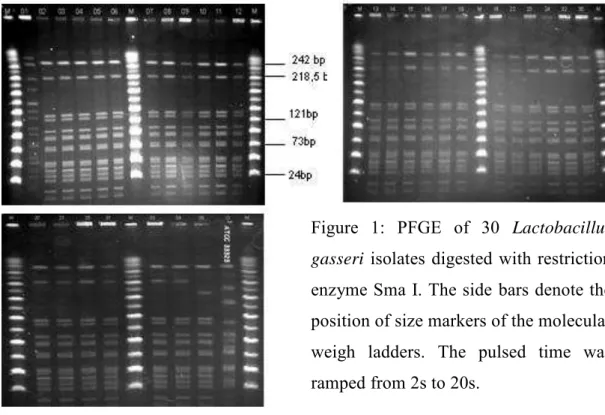
3.1 - Bacterial identification and Pulsed-Field Gel Electrophoresis (PFGE) ... 69
3.2 - Testing of Hemolytic Activity and Bile Tolerance ... 70
3.3 - Antagonistic activity against selected pathogens ... 71
3.4 - Antibiotic Susceptibility ... 72
3.6 - Tolerance to simulated gastric and small intestinal juices ... 74
3.7 - Adherence assay ... 75
4 - DISCUSSION ... 76
5 - CONCLUSION ... 82
6 - REFERENCES ... 83
CHAPTER II SEQUENCE ANALYSIS OF FOUR PLASMIDS OF Lactobacillus gasseri NCK2141 ISOLATED FROM A NEWBORN INFANT ... 90
ABSTRACT ... 90
RESUMO ... 91
1 - INTRODUCTION ... 92
2 - MATERIALS AND METHODS ... 93
2.1 - Bacterial strain, media and growth condition ... 93
2.2 - DNA manipulation, plasmid isolation and sequencing ... 94
2.3 - Sequence annotation ... 94
2.4 - Blunt cloning ... 95
2.4.1 - Restriction enzyme digestion of pTRK989 ... 95
2.4.2 - Ligation and transformation ... 97
2.5 - Phenotypic assay - Antibiotic susceptibility ... 97
3 - RESULTS AND DISCUSSION ... 99
3.1 - Plasmid DNA analysis and sequencing ... 99
3.2 - General features of the plasmids ... 99
3.3 - Putative replication functions ... 104
3.4 - Transposable elements ... 105
3.5 - Mobilization ... 106
3.6 - Partitioning ... 106
3.7 - Lantibiotic biosynthesis ... 107
3.8 - Putative transport regions ... 108
3.9 - Collagen adhesion ... 109
3.10 - Multidrug resistance transporter (lmrB) ... 110
3.10.1 - Construction of lmrB recombinants ... 111
3.10.2 - Phenotypic assay ... 112
4 - CONCLUSIONS ... 113
CHAPTER III FUNCTIONAL ANALYSIS OF FIBRONECTIN BINDING
PROTEIN IN Lactobacillus gasseri ... 120
ABSTRACT ... 120
RESUMO ... 121
1 - INTRODUCTION ... 122
MATERIALS AND METHODS ... 124
1.1 - Bacterial strains, media and growth conditions ... 124
1.2 - DNA manipulations and sequence analysis ... 124
1.3 - Construction of L. gasseri fbp mutants ... 126
1.4 - Adherence assay ... 127
1.5 - Tolerance to simulated gastric juice ... 128
2 - RESULTS ... 130
2.1 - Adherence to immobilized fibronectin and effect of the growth condition on the adhesion ... 130
2.2 - Characterization of Fibronectin-binding protein from L. gasseri NCK2141 ... 131
2.3 - Construction of fbp mutants ... 131
2.4 - Phenotypic analysis of the fbp insertion mutants ... 132
2.4.1 - Survival to simulated gastric juice ... 132
2.4.2 - Adherence ability of NCK 2147 and NCK 2148 ... 133
3 - DISCUSSION ... 134
4 - CONCLUSION ... 137
5 - REFERENCES ... 138
LIST OF FIGURES
Figure 1: Phylogenetic tree showing the relationships among the species of the Family Lactobacillaceae, including genera Lactobacillus(abbreviated with ―L” in the tree), Paralactobacillus and Pediococcus (abbreviated with ``P`` in the tree). Extracted from Felis et al 2009. ... 8
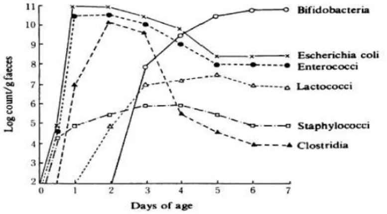
Figure 2: Changes in the intestinal microbiota in babies from birth to 7 days (Mitsuoka, 1989). ... 15
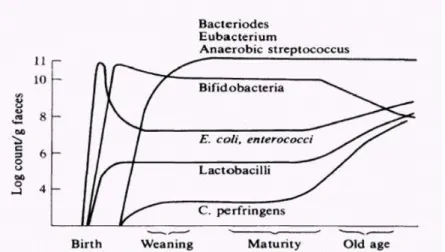
Figure 3: Changes in the intestinal microbiota with age (Mitsuoka, 1989). ... 16
CHAPTER I CHARACTERIZATION OF Lactobacillus gasseri ISOLATES FROM A BREAST-FED INFANT ... 60
Figure 1: PFGE of 30 Lactobacillus gasseri isolates digested with restriction enzyme Sma I. The side bars denote the position of size markers of the molecular weigh ladders. The pulsed time was ramped from 2s to 20s... 70
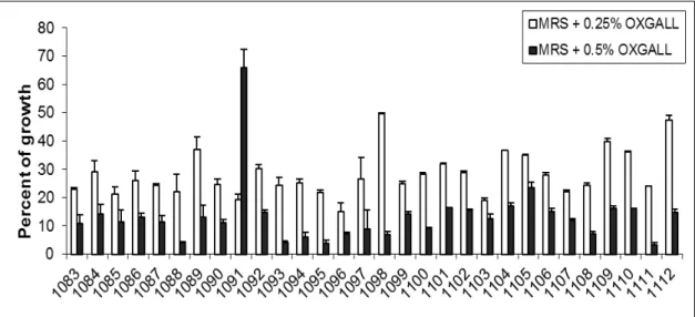
Figure 2: Percent of growth (A600nm) in the presence of oxgall compared to the control (MRS medium without bile). Each column represents the mean of two replicates. The bars represent the standard deviation. ... 71
Figure 3: (a) Inhibition of E. coli by Lactobacillus gasseri (UFVCC1091). (b) Inhibition of L. bulgaricus by NCFM (used as a control in this assay). The numbers show the proteases (1mg/ml ) dropped (5 µl) around the colony: 1 - Proteinase K; 2- Bacillus protease; 3- Bovine; 4- Papain; 5- Pronase E type XIV; 6- Chymotrypsin; 7- Trypsin; 8- Fungal; 9- Pepsin . .. 72
Figure 4: (a) Agarose gel electrophoresis plasmid profile of Lactobacillus gasseri
Figure 5: Survival of stationary phase cells of Lactobacillus gasseri in simulated small intestinal juice at pH 8.0 (Panel a) and gastric juice at pH 2.0 (Panel b). Percent survival represents viable cells (CFU/mL) remaining after exposure at the time points indicated versus pre-treatment (time 0). The data represent the means of two independent replicates. ... 74
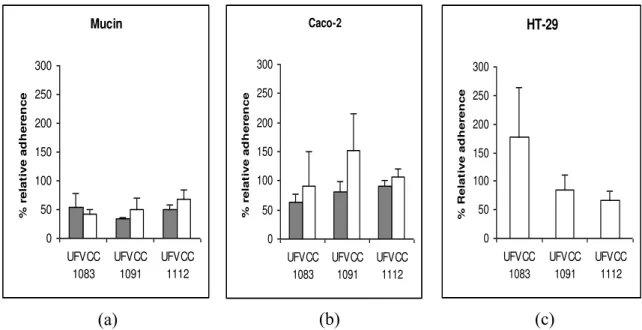
Figure 6: Adherence of Lactobacillus gasseri UFVCC1083, 1091 and 1112 to mucin (Panel a), Caco-2 cells (Panel b) and HT-29 (Panel c) in vitro. The bacteria grown in static MRS broth in ambient atmospheric (filled) and on MRS agar under anaerobic conditions (open). The data represent the means ± standard errors of the means for two independent replicates. ... 75
CHAPTER II SEQUENCE ANALYSIS OF FOUR PLASMIDS OF Lactobacillus gasseri NCK2141 ISOLATED FROM A NEWBORN INFANT ... 90
Figure 1:Construction strategy and restriction enzymes sites of pTRK1027 vector. .... 98
Figure 2: Physical and genetic map of plasmid pTRK1023 (a), pTRK1024 (b), pTRK 1025 (c) and pTRK1026 (d) of Lactobacillus gasseri NCK2141. Each ORF is numbered, and arrows indicate positions and directions of predicted ORFs. ORF numbers refer to Tables 2, 3, 4, and 5. ... 100
Figure 3: Areas of significant similarity among RepA pSK118-44 (standard) and RepA protein from pTRK1023, pTRK1025 and pTRK 1026. The green areas show high matches at the same base position. ... 104
Figure 4: Verification of recombinant plasmids by restriction enzyme digestion with XhoI. Lane M, 1 kb plus DNA ladder marker. Lane 1, undigested pTRK1027 isolated from E. coli MC1061 (4.8 kb). Lane 2, digested pTRK1027 isolated from E. coli MC1061 (control). Lanes 3 and 4, digested pTRK1027 isolated from L. gasseri NCK2144 isolates. Lanes 5 and 6, digested pTRK1027 isolated from L. gasseri NCK2145 isolates. The black and red arrows indicate the XhoI-digested plasmids. ... 112
Figure 1: Construction of the pTRK1028 integration vector. ori, origin of replication of pWV01; erm, gene encoding Em resistance; MCS, multiple cloning sites from pUC19. Only unique restriction sites are shown (Adapted from Goh et al 2009). ... 129
Figure 2: Adherence of Lactobacillus gasseri NCK2140, 2141, 2142 and the control strain NCK99 to fibronectin in vitro. The bacteria grown in static MRS broth in ambient atmosphere (black bars) and on MRS agar under anaerobic condition (gray bars) were used for adherence testing at a concentration of 1.0 x 10⁸ cells/ml. Bacterial cells were exposed to microwells coated with fibronectin for 1 h at 37ºC, followed by plating on MRS agar medium for enumeration of adhered bacterial cells. The data represent the means ± standard errors of the means for four independent replicates. A Student`s t-test indicated that the results are significantly different (*) at a value of P<0.05 for adherence of the strain grown in MRS broth compared to the same strain grown in MRS agar. ... 130
Figure 3: Colony PCR analysis of 16 selected Erm-sensitive double recombinants. Lane M, DNA size marker (1 Kb plus); lane 1 to 8, selected NCK2148
isolated Erm-sensitive recombinants; lanes
9 to 16, selected NCK2147 isolated Erm sensitive recombinants; Lane WT, parent strain NCK 1041 (control). The expected amplicon sizes generated from wild-type and ∆fbp genotypes are approximately 3.311 kb and 1.8 kb, respectively. ... 132
Figure 4: Survival of stationary phase cells of Lactobacillus gasseri NCK 2140 (a) and NCK 2141 (b) (open square) compared with their respective fbp mutants
NCK 2148 (a) and NCK 2147
(b) (dark square) in simulated gastric juice at pH 2.0. Percentage of survival represents viable cells (CFU/ml after treatment at various time points versus before treatment (time 0). The data are the means ± standard errors of the means for two independent replicates. ... 133
LIST OF TABLE
CHAPTER I CHARACTERIZATION OF Lactobacillus gasseri ISOLATES
FROM A BREAST-FED INFANT ... 60
Table 1: Primers tested in the study. ... 68
Table 2: MIC values of L. gasseri to human therapeutic antibiotics. ... 72
Table 3: Lactobacillus gasseri grouped according to their intrinsic characteristics. ... 74
CHAPTER II SEQUENCE ANALYSIS OF FOUR PLASMIDS OF Lactobacillus gasseri NCK2141 ISOLATED FROM A NEWBORN INFANT ... 90
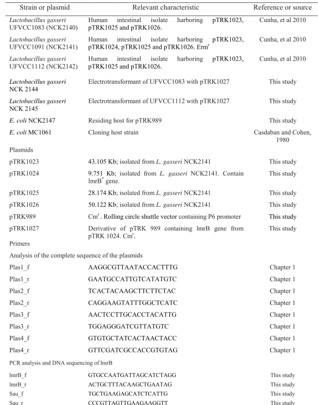
Table 1: Bacterial strains, primers and plasmids used in this study. ... 96
Table 2: ORF analysis of the native plasmid pTRK1023 from L. gasseri NCK2141 with best matches to sequences in the public databases... 101
Table 3: ORF analysis of the native plasmid pTRK1024 from L. gasseri NCK2141 with best matches to sequences in the public databases... 102
Table 4: ORF analysis of the native plasmid pTRK1025 from L. gasseri NCK2141 with best matches to sequences in the public databases... 102
Table 5: ORF analysis of the native plasmid pTRK1026 from L. gasseri NCK2141 with best matches to sequences in the public databases... 103
CHAPTER III FUNCTIONAL ANALYSIS OF FIBRONECTIN BINDING PROTEIN IN Lactobacillus gasseri ... 120
LIST OF ABBREVIATIONS
GRAS - Generally Recognized as Safe
LAB – Lactic Acid Bacteria
FAO/WHO – Food and Agriculture Organization and World Health Organization
HMB – Human Milk Bank
UFVCC – Universidade Federal de Viçosa Culture Collection
NCK – North Carolina Klaenhammer Culture Collection
MIC – Minimum Inhibitory Concentration
PFGE – Pulsed Field Gel Electrophoresis
CDS - Coding DNA Sequence
GAMOLA - Global Annotation of Multiplexed On-site bLasted DNA-sequences
BLAST - Basic Local Alignment Search Tool
TA System – Toxin-Antitoxin System
RESUMO
CUNHA, Luciana Rodrigues da, D.Sc.,Universidade Federal de Viçosa, março de 2011. Caracterização genotípica e fenotípica de Lactobacillus gasseri isolatedos de recém nascidos. Orientadora: Célia Lúcia de Luces Fortes Ferreira. Coorientadores: Cláudia Lúcia de Oliveira Pinto and Paulo César Stringheta.
O objetivo deste estudo foi caracterizar novas estirpes de Lactobacillus gasseri
mostrou maior tolerância a bile (0.5%) e resistência a eritromicina, cefalotina e oxacilina. Todas as estirpes apresentaram antagonismo frente aos patógenos avaliados sendo a inibição assossiada apenas à produção de ácidos orgânicos. Os três isolados selecionados NCK2140, 2141 e 2142 foram resistentes ao suco gástrico e ao suco pancreático e apresentaram boa capacidade de adesão in vitro a mucina, fibronectina e às linhagens de células humanas do câncer de cólon, Caco-2 e HT-29. Os quatro plasmídeos identificados, pTRK1023, 1024, 1025 e 1026 apresentaram morfologia circular, replicação por mecanismo teta e 36, 7, 20 e 46 open reading frames (ORF), respectivamente. Os conteúdos de G+C foram consistentes com aqueles encontrados em outras estirpes de Lactobacillus (37 - 40%), exceto o pTRK1025 que foi de 44,57%. Algumas propriedades funcionais como, proteínas de ligação ao colágeno (pTRK1026), biossíntese de lantibióticos (pTRK1023) e transporte de carboidratos (pTRK1023 e pTRK1026) foram identificados nos plasmídeos, as quais podem promover vantagens competitivas para as células hospedeiras. Mecanismo de manutenção celular como, Sistema de Separação e Toxina-Antitoxina também foi observado, explicando a dificuldade de cura dos plasmídeos. O gene lmrB
identificado no pTRK1024, é associado à resistência a alguns antibióticos, no entanto, o experimento de clonagem não confirmou a relação do mesmo com a maior resistencia à eritromicina, cefalotina e oxacilina apresentada pelo isolado NCK2141 carreador desse plasmideo. Por questões de segurança, essa estirpe não pode ser utilizada como um probiótico. Maior adesão in vitro foi observada à fibronectina quando as estirpes NCK2140, 2141 e 2142 foram cultivadas em ágar MRS e incubadas em condições de anaerobiose. Observou-se também que os mutantes fbp
ABSTRACT
CUNHA, Luciana Rodrigues da, D.Sc.,Universidade Federal de Viçosa, March, 2011.
Genotypic and phenotypic characterization of Lactobacillus gasseri isolated from a newborn infant. Adviser: Célia Lúcia de Luces Fortes Ferreira. Co-advisers: Cláudia Lúcia de Oliveira Pinto and Paulo César Stringheta.
showed strong resistance to small intestinal and gastric juices, and adhered in vitro to mucin, fibronectin and two intestinal epithelial cell lines, Caco-2 and HT-29. The complete nucleotide sequence of the plasmids (pTRK1023, 1024, 1025, and 1026) revealed that they are circular and contain 36, 7, 20 and 46 open reading frames, respectively. All four plasmids are predicted to replicate by the theta mechanism, and have G+C contents consistent with those found in other lactobacilli strains (37-40%), with the exception of pTRK1025 (44.57%). The plasmids appear to encode functional properties, such as collagen binding (pTRK1026), lantibiotic biosynthesis
(pTRK1023) and carbohydrate transport (pTRK1023 and pTRK1026) that may
provide competitive advantages to the plasmid-carrying cells. Cell maintenance
mechanisms (partitioning and TA system) were also observed, thus explaining the
1 -
INTRODUCTION
Breastfeeding is recognized worldwide as nurturing and protective to the developing infant. Human milk supplies the nutrients and energy needs of the newborn and protects them against infections. Despite the numerous benefits conferred by this human milk (HM), many children are deprived from its consumption because their mothers cannot breastfeed them. Therefore, human milk banks have a fundamental role as an important food source for such newborns.
Brazil is a world reference in the Human Milk Banking with more than 290 banks currently under operation. In 2010, the 152,813 thousand liters of HM collected provided protection to more than 139,000 newborns. Almost all of these children were prematurely born and were not able to breast feed. Furthermore, it is estimated that 7,000 Brazilian children would die every year if they were not breastfed properly, which emphasizes the importance of the human milk banks (HMB). Pasteurization (65oC x 30 min-1) insures the microbial safety of the milk deposited in the HMB. However, studies from Borba et al (2003) indicated that this heat treatment reduces, inactivates or eliminates some prebiotic constituents, which are essential for establishing Bifidobacterium and other desirable lactic acid bacteria (LAB) in the intestinal tract of the newborn, therefore compromising the formation of a beneficial intestinal microbiota for them.
The development of a beneficial microbiota contributes to the health of the host. The gut microbiota acts as an important intestinal immune-modulator, not only educating the naive infant immune system but also serving as an important source of non inflammatory immune stimulators throughout life in healthy individuals. In addition, the enteric microbiota can secrete molecules that inhibit host pathogens, metabolize compounds that harm the host to less toxic substances and produce a range of bioactive compounds such as conjugated linoleic acid, and short chain fatty acids that may play a role in protection from lifestyle illnesses such cancer, obesity and cardiovascular disease.
pasteurized milk provided in the milk banks. With this in mind, 30 LAB strains were isolated in previous work in our laboratory of Lactic Acid Bacteria, from the target population, healthy newborn fully breast fed.
In order to claim that a bacterial strain is a potential probiotic, in 2002, the Food and Agriculture Organization (FAO) and World Health Organization (WHO) established guidelines with some safety and functional criteria. According to this document, all probiotic strains need to be properly identified at the genus and species level using current and internationally scientific practices. In addition, they also need to be evaluated for resistance to bile, antagonism toward pathogens, ability to adhere to intestinal epithelial cells, tolerance to small intestine and gastric juices, hemolytic activity and resistance to antibiotics. The importance of assessing the antibiotic resistance profile pattern of new isolates is to limit the use of probiotic cultures harboring plasmids containing transferable antibiotic-resistance genes.
2 -
LITERATURE REVIEW
2.1 - Human Milk and Child Health
Human milk is considered the ideal food for the newborn. It has a balanced nutritional composition, containing the right amount of fatty acids, lactose, water and amino acids for newborn digestion, brain development and growth (Willians and Stehlin, 2005), and it is enough to supply all the nutritional needs of newborns during the first six month of life (Workgroup on Breastfeeding, 1997). In addition, it has more than 45 bioactive compounds, which contribute to the maturation of the newborn gastrointestinal tract (Kunz, 1999).
Besides providing optimal nutrition to infants, human milk contains a multitude of immunological components, which are transferred from the mothers to infant via breastfeeding. About 80 percent of the cells in breast milk are macrophages, cells that kill bacteria, fungi and viruses (Willians and Stehlin, 2005). In addition to macrophages, human milk contains proteins such as lactoferrin, lysozyme and casein, lipids, oligosaccharides, enzymes, prostaglandins, growth factors, hormones, and cells that work in many different ways to prevent infections and modulate the immune system. This natural immune protection is not available to artificially fed infants (Hanson, 2007). Studies have been shown that breastfed babies are less susceptible to certain diseases, such as bacterial meningitis, bacteremia, diarrhea, respiratory tract infections, necrotizing enterocolitis, otitis, urinary tract infections (Singhal et al 2002), hypercholesterolemia, and asthma (Chulada et al 2003) than not breastfed infants. These special properties of human milk also provide long-term protection from many diseases seen at higher rates in artificially fed infants, including an increased risk of obesity, type 1 and 2 diabetes, and childhood leukemia (Hamosh, 2001). In addition, the protective effect of human milk against HIV postnatal transmission in humans (Walter et al 2009) and in the reduction of the progression of the disease (Coutsoudis et al 2003; Tozzi et al 1990) also has been reported.
2.2 - Human Milk Banks
Human milk banks (HMB) are specialized centers established for encouraging breastfeeding and for collecting, screening, processing, storing and distributing donated human milk (colostrum, transition milk and mature human milk). They must be linked to a maternity or hospital and are responsible for quality control of the milk. Brazil is a world reference in the Human Milk Banking with 283 banks under current operation (Fiocruz, 2010). In 2007, 1,350 million liters of human milk were collected through the Nacional Net of Human Milk providing protection to more than 107,000 newborns. Almost all these children were prematurely born and were not able to breast feed. Furthermore, it is estimated that 7,000 Brazilian children would die every year if they were not breastfed properly (Bom dia Brazil, 2008) which emphasizes the importance of the human milk banks (HMB).
As evidenced in literature, human milk can be easily contaminated and vehicle of various infectious diseases, such as AIDS, herpes, rubella, hepatitis B and C (Goldin, 1997). Thus, for safety reasons, all milk is pasteurized (65oC x 30 min-1). However, studies from Borba et al (2003) indicated that this heat treatment reduces, inactivates or eliminates some prebiotic constituents. The prebiotic character of human milk is important to establishing Bifidobacterium and other desirable probiotic lactic acid bacteria (LAB) in the newborn´s intestinal tract. A possible way to augment any undesirable effects of heat treatment could be through the addition of well selected endogenous intestinal probiotic strains into the pasteurized milk provided in the milk banks.
2.3 - Probiotics - History and definitions
physiological and therapeutical properties of these microorganisms, the definition has been suffering alterations until today and there is still no consensus, however the definition most utilized internationally (Grupta and Garg, 2009; Petrof, 2009; Sanders, 2009; Reid, 2008; Pineiro and Stanton, 2007) is that they are live microorganisms which when administered in adequate concentrations benefit the health of the host (World Health Organization [WHO] and Food and Agriculture Organization [FAO], 2002). In Brazil, the department which regulates probiotic products is the ANVISA (National Agency for Sanitary Vigilance), and according to this Agency probiotics are live microorganisms capable of improving intestinal microbial equilibrium, producing beneficial effects to the health of the individual.
According to the last two definitions, microorganisms must be alive to exert beneficial effects. However, the scientific basis of this affirmation has been questioned by some authors, since there are studies showing that non-viable microorganisms (inactivated by heat or UV radiation), products or even bacterial DNA may exert heath benefits to the host in specific situations (Kataria et al 2009). Zhang et al (2005) reported that thermally inactivated Lactobacillus rhammnosus GG promoted a reduction in IL-8 (interleukin-8) production induced by TNFα (tumor necrosis factor alpha) in Caco-2 cells. Xiao et al (2003) administered capsules of active or thermal inactivated Lactobacillus acidophilus LB to 64 and 59 patients with chronic diarrhea, respectively. After four weeks of treatment, it was verified that the inactive probiotic form was significantly more effective in the combat of diarrhea. Children (3-24 months old) with acute diarrhea and moderate degrees of dehydration also showed clinical improvement after receiving thermally inactivated L.acidophilus
(Simakachorn et al 2000). Besides the ability to modulate the immune system and combat diarrhea, it has also been reported that inactivated probiotic bacteria have potential to adhere to intestinal cells (Chauviere et al 1992).
by Saccharomyces boulardii, isolated from lychee (Litchi chinensis Soon) in the treatment of diarrheas associated to antibiotics and Clostridium difficile (Kotowska et al 2005; Vanderhoof & Young, 2002), ulcerative colitis (Guslandi et al 2000), irritated bowel syndrome (Maupas et al 1983) and Crohn’s disease (Guslandi et al 2000).
2.4 - Principal Probiotic Microorganisms
The microorganisms most commonly used as a probiotics are producers of lactic acid such as lactobacilli and bifidobacteria (Ferreira, 2003; Kopp-Hoolihan, 2001). Enterococcus faecium and Enterococcus faecalis have also been employed as functional supplements in foods and their utilization is permitted in Brazil by the ANVISA. However, there exists a concern on the part of the scientific community in relation to utilization of these bacteria as probiotics due to the evidences relating these microorganisms to cases of bacteremia (Gasser et al 1994), septicemia (Poh et al 2006; Schaberg et al 1991), urinary infections (Poh et al 2006; Lopes et al 2005; Schaberg et al 1991) and endocarditis (Adam et al 1999). Moreover, virulence factors such as hemolysins, gelatinases and surface proteins (Jahangiri et al 2010; Mundy et al 2000), as well as conjugative plasmids encoding antibiotic resistance genes (Eaton & Gasson, 2001) have also been encountered in these bacteria.
2.4.1 - Genus Lactobacillus
Lactobacillus are Gram positive microorganisms, nonspore-forming rods or coccobacilli, deprived of flagella and are facultative anaerobic. They possess optimal pH and temperature of growth in the range of 5.5 – 6.3 and 30 – 40ºC respectively (Gomes and Malcata, 1999), and lower G+C content (Carr et al 2002). This genus is amply distributed in the environment, especially in the intestinal tract (Wall et al 2007; Reuter 2001), mouth (Smith et al 2001), vagina (Martin et al 2008) and stomach (Ryan et al 2008) of humans and other animals, as well as in plants, organic material, sewage and fermented or deteriorated foods (Bernardeau et al 2006; Axelsson, 1989).
homofermentative, facultative heterofermentative and obligatory heterofermentative (Kandller and Weiss, 1986). Obligatory homofermentative lactobacilli include those which exclusively ferment glucose in lactic acid and do not ferment pentoses or gliconate. The obrigatory heterofermentatives include lactobacilli which ferment hexoses in lactic acid, acetic acid and/or ethanol and carbon dioxide, being that the production of gas from glucose is a distinctive characteristic of these bacteria. The facultative heterofermentatives include Lactobacillus which ferment hexoses in lactic acid and may produce gas from gliconate, but not from glucose. These microorganisms also ferment pentoses by activity of an induced phosphoketolase to produce lactic and acetic acid (Vásques et al, 2005).
The genus Lactobacillus is phylogenetically diverse (Figure 1) and with development of molecular techniques and genetic characterization, this was reorganized and new groups were identified, based on the 16S rRNA region (Collins et al 1991). Currently, in accordance with data obtained from the NCBI (National Center of Biotechnology Information, USA), there are 195 different species of
Lactobacilli, including subspecies.
2.4.1.1 - Lactobacillus gasseri
Lactobacillus gasseri are obligate saccharoclastic, homofermentative organism, with optimum growth at 35 to 38ºC, and forms small rods with rounded ends from 0.6 to 0.8 by 3 to 5 µm in size. L. gasseri is considered one of the true autochthonous species of the human intestinal ―probiome,‖ defined as commensal intestinal bacteria considered to have a beneficial influence on human health (Azcarate-Peril et al 2008). Depending on consumption habits and geographic location, L. gasseri has been determined to be one of the Lactobacillus species native to the human gastrointestinal tract (GIT) of neonates (Wall et al 2007) and adults (Reuter, 2001). L. gasseri has also been described as a common member of the vaginal (Martin et al 2008) and oral Lactobacillus biota (Smith et al 2001), and it has been proposed that the oral cavity acts as a reservoir and source of intestinal lactobacilli (Dal Bello et al 2006).
L. gasseri was routinely classified as ―L. acidophilus‖ since morphologically it
gasseri was differentiated by DNA/DNA hybridization patterns from L. acidophilus
and named after Francis Gasser, who studied lactate dehydrogenases of Lactobacillus
species (Gasser and Mandel, 1968). Today, the complete genome of L. gasseri ATCC 33323 are deposited in the Bacterial Genome Database at the National Center for Biotechnology Information (NCBI) and was sequenced by the Department of Energy-Joint Genome Institute in collaboration with the Lactic Acid Bacteria Genomics Consortium (LABGC) (Klaenhammer et al 2002).
2.5 - Probiotics and beneficial effects to health of children
2.5.1 - Modulation of the intestinal microbiota
The relation between a balanced intestinal microbiota and health of the host has been documented in literature. The enteric microbiota not only functions as a barrier, but is involved in the immune response, inhibition of pathogen growth and synthesis of bioactive compounds, such as conjugated linoleic acid, -aminobutíric acid and short chain fatty acids, which exert an important role in the protection against diseases including cancer, obesity and cardiovascular diseases (Wall et al 2009). Analogously, when there is a breakdown of intestinal homeostasis, and pathogenic and/or opportunistic microorganisms are excessive, the enteric microbiota may also contribute to the development of diseases (Kelly et al 2007). Therefore, this suggests that predominance of a beneficial microbiota is associated with the promotion of metabolism, nutrition and health of the host (Mackie et al 1999).
Probiotics may promote the modulation of the intestinal microbiota by competition for epithelium adhesion sites and nutrients, production of antimicrobial compounds and stimulation of the immune system (Sobko et al 2006).
2.5.1.1 - Production of antimicrobial compounds
Organic acids
Lactobacilli and bifidobacteria produce metabolites, such as acetic and lactic acid, which possess an inhibitory effect on growth of pathogenic microorganisms (Servin, 2004). This effect is related to the non-dissociated form of the acid (Podolak et al 1996), which being lipophilic and apolar passively diffuse through the membrane (Kashket, 1987) and promote acidification of the cellular cytoplasm. Therefore, the photoelectrochemical gradient is collapsed promoting interruption of the substrate transport system to the cell (Snijders et al 1985) and impairment of the metabolic function of the microorganism. Acetic acid has a greater inhibitory effect than lactic acids for the same pH value due to its pKa (pKa=5.76) being greater than that of lactic acid (pKa=3.86), resulting in greater concentrations of the non-disassociated form in the bacterial cytoplasm (Vasseur et al 1999).
Gram-negative bacteria (Alakomi et al 2000) and as a chelation agent, capturing elements such as iron, essential for bacterial growth (Presser et al 1997).
Studies in vitro have shown organic acids to be the principal inhibiting agent to growth of pathogenic bacteria, as Escherichia coli (Maragkoudakis et al 2006; Forestier et al 2001; Ogawa et al 2001; Dembeté et al 1998), Listeria monocytogenes
(Dembeté et al 1998), Salmonella typhimurium (Maragkoudakis et al 2006; Keersmaecker et al 2006; Forestier et al 2001), Clostridium difficile (Forestier et al 2001), Helicobacter pylori (Maragkoudakis et al 2006; Midolo 1995) and
Staphylococcus aureus (Dembeté et al 1998).
Bacteriocin and/or molecules similar to bacteriocin
Bacteriocin are antimicrobial compounds of proteic origin, ribosomally synthesized and produced by bacteria, and are generally active against species intimately related with the producing strain (Klaenhammer, 1988; Tagg et al 1976). However, some bacteriocin are capable of inhibiting growth of phylogenetically distant microorganisms, as for example, Clostridium botulinum, Bacillus sp,
Enterococcus faecalis, Listeria monocytogenes and Staphylococcus aureus
(Tichaczek et al 1992). These compounds were principally characterized in gram-negative bacteria, in which colicins (Escherichia coli) are the most studied (Lazdunski, 1988). Posteriorly, it was found that Gram-positive bacteria also produce these compounds and within this class, lactic acid bacteria have been amply explored for utilization of their bacteriocin in the conservation of foods (Cleveland et al 2001).
These molecules act promoting collapse of the membrane potential by means of electrostatic bonds with the phospholipids (negatively charged). These bonds are favored since the majority of bacteriocins are amplified and cationic. After bonding, the hydrophobic portion of the bacteriocin is inserted in the membrane forming pores. Formation of these pores allow for the exit of ions, principally potassium and magnesium, promoting dissipation of the promoter force, compromising synthesis of macromolecules and production of energy, resulting in cell death (Montville et al 1995).
inhibitory activity on growth of Staphylococcus aureus and Bacillus cereus. Yildirim and Johnson (1998) identified bacteriocin (bifidocina B) produced by Bifidobacterium bifidum with inhibitory effects on growth of some species of the genera Listeria, Bacillus, Enterococcus, Lactobacillus, Leuconostoc and Pediococcus. Gassericin A, a bacteriocin produced by Lactobacillus gasseri LA39 isolated from feces of newborns, inhibited growth of Listeria monocytogenes, Bacillus cereus and Staphylococcus aureus (Itoh et al 1995). Ten Brink et al (1994) identified bacteriocin produced by
Lactobacillus salivarus M17 (salivaricin B), which present inhibitory activity on growth of Listeria monocytogenes, Bacillus cereus and Enterococcus ssp. The authors identified another bacteriocin produced by Lactobacillus acidophilus M46 (acidocin B), which has an inhibitory effect on Clostridium speorogenes.
Lactobacillus and Bifidobacterium may also antagonize growth of pathogens by the production of lipid or protein compounds of low molecular weight. Collado et al (2005) encountered inhibition of Helicobacter pylori by protein compounds, thermostable and sensitive to proteases, produced by Bifidobacterium. The growth of
Giardia intestinalis and its bonding to Caco-2 human colon adenocarcinoma cells lines was significantly reduced by antimicrobial substances of low molecular weight (La1) produced by Lactobacillus johnsonii (Perez et al 2001). Liévin et al (2000) attributed the antagonism of bifidobacteria on growth of Salmonella typhimurium to production of lipophilic molecules of low molecular weight (<3500Da).
Production of H2O2
O2 + 1e- O2- (superoxide radical)
O2 + 1e- + 2 H+ H2O2 (hydrogen peroxide) Eq. 1 H2O2 + 1e- OH- + OH. (hydroxyl radical)
OH. + 1e- + H+ H2O
In the membrane peroxidation process, the free hydroxyl radical captures one hydrogen atom from the unsaturated fatty acid of the phospholipid membrane and is transformed in energy. The fatty acid, in the presence of oxygen, generates a free radical and initiates a chain reaction which will destroy the cell membrane. The damaged membrane loses its flexibility and its barrier functions allowing the entrance of calcium into the cell and activating the phospholipases. These enzymes continue to damage the cell since they attack the lysosomal membrane and liberate its enzymes, accelerating degradation. In the DNA, the peroxide radical reacts with the iron ions present in the molecule, producing the hydroxyl radical. This principally attacks the pyrimidine in the bond with deoxyribose, rupturing the sugar-phosphate bond and liberating the free bases of the nucleotides (Silva, 2003).
Lactobacillus plays an important role in maintaining vaginal health and preventing the growth of pathogens including Escherichia coli and Gardnerella vaginalis by the production of acid and H2O2. Lactobacillus crispatus and
Lactobacillus jensenii inhibit growth of gonococci in vitro by the production of H2O2 (Amant et al 2002). Aroutcheva et al (2001), while examining the antimicrobial activity of twenty-one strains of Lactobacillus spp. of vaginal origin, observed that approximately 80% of these produced H2O2, lactic acid and bacteriocins, and were effective in inhibiting the growth of Gardnerella vaginalis, Lactobacillus brevis,
Lactobacillus salivarius and Lactobacillus gasseriin vitro.
2.5.1.2 - Competition for nutrients and adhesion sites
Collado et al (2007b) verified inhibition of adhesion of pathogenic bacteria, as
Bacteroides vulgatus DSM 1447, Clostridium histolyticum DSM 627, Clostridium difficile DSM 1296, Escherichia coli K2, Enterobacter aerogenes DSM 30053, Listeria monocytogenes ATCC 15313, Salmonella enterica serovar Typhimurium
ATCC 12028, Staphylococcus aureus DSM 20231 to human intestinal mucus by probiotic bacteria (Lactobacillus rhamnosus GG [ATCC 53103], L. rhamnosus
LC705, L. casei, Shirota [Yakult], L. fermentum ME3, L. acidophilus NCFM, L. plantarum Lp-115, L. salivarius Ls-33, Bifidobacterium longum 46, B. lactis Bb12, B. lactis 420, B. breve) isolated from commercial milk products. The authors also reported that these not only inhibited adhesion, but the probiotic bacteria were capable of removing the pathogens already adhered to the intestinal mucus.
In another study performed by the same group, the authors verified inhibition of pathogenic bacteria adhesion (Bacteroides vulgatus DSM 1447, Clostridium histolyticum DSM 627, C. difficile DSM 1296, Escherichia coli K2, Listeria monocytogenes ATCC 15313, Salmonella enterica serovar Typhimurium ATCC 12028, Staphylococcus aureus DSM 20231) to the human intestinal mucus by the combination of probiotic bacteria Lactobacillus rhamnosus GG (ATCC 53103), L. rhamnosus LC705 (DSM 7061), B. breve 99 (DSM 13692) and B. lactis Bb12 (DSM 10140). The authors also found that the probiotic bacteria in the form of a pool promote greater inhibition to adhesion that when alone (Collado et al 2006). However, the researchers, suggested the individual evaluation of each probiotic strain for later selection of the pool since adhesion level of these bacteria to the intestinal cells are dependent on genus, species and even the strain (Collado et al 2007b; Collado et al 2006).
Lee et al (2003) verified a reduction in the adhesion levels of E. coli and
Salmonella sp. to the intestinal mucus (isolated from the feces of healthy humans) and in Caco-2 human colon adenocarcinoma cell line when incubated together with
Lactobacillus rhamnosus GG and Lactobacillus casei Shirota. Lactobacillus johnsonii
LJ1 and Lactobacillus casei Shirota significantly inhibited adhesion of Salmonella typhimurium to the human intestinal mucus (Tuomola et al 1999).
degraded by the digestive enzymes of the host, making them unavailable to the pathogen (Gibson et al 2005).
2.5.1.3 - Stimulation of the immune system
The intestine represents the largest lymphoid organ of the human body, being responsible for 80% of the immunological response. This action is due to the presence of antibodies and various immunocompetent cells dispersed on the inner wall and epithelium, or organized in structures, which play fundamental role in the antigenic presentation and elaboration of the immune response to the microorganisms and proteins of the diet (Morais and Jacob, 2006).
In vitro studies and animal and human models suggest that probiotics may stimulate the immune system. This effect is attributed to the capacity of these strains to interact with the Peyer’s patches and the intestinal epithelial cells, stimulating the Ig A producing B cells and the migration of intestinal T cells (Perdigon and Holgado, 2000). It has also been shown that probiotics favor unexpected phagocytic activity of the alveolar macrophages, suggesting systemic action by secretion of mediators which stimulate the immune system (Cross, 2002). Increase in the secretory IgA serum levels and production of macrophages are extremely important in the immune response of the intestine since this is the first line of non-specific defense of the organism (Vanderhoof, 2001). Schiffrin et al (1997) in a randomized double blind study administered of supplemented or non-supplemented (control) fermented milk with Bifidobacterium bifidum Bb12 (1 x 10¹⁰ CFU/day) and Lactobacillus acidophilus
LA1 (7 x 10¹⁰ CFU/day) to 28 adults (23 – 62 years old) for 3 weeks and reported a significant increase in phagocytic activity and in the serum levels of granulocytes and monocytes, but not in lymphocytes or T cell activity. Fukushima et al (1998) administered a formula supplemented with Bifidobacterium bifidum Bb12 (109 CFU/day) to Japanese children (15-31 months). After 21 days of treatment a significant increase was verified in levels of Ig A in the feces of the infants fed with the probiotic strain. The authors concluded that this increase in Ig A may increase the resistance of mucus to intestinal infections.
2.5.2 - Maturation of the Intestinal Microbiota
microorganisms present in the environment and in the mother (Mitsuoka, 1982). Roughly 24 hours after birth, the feces of most newborns already contain bacteria, such as coliforms, Enterococcus, Clostridium and Sthaphylococcus in various proportions (Figure 2), whose growth is favored at the beginning of colonization in function of the positive oxido-reduction potential in the intestine.
Figure 2: Changes in the intestinal microbiota in babies from birth to 7 days (Mitsuoka, 1989).
Although some of these microorganisms belong to species with pathogenic potential, they do not cause harm to the host (Morelli, 2008). The first colonizers consume the oxygen and produce new metabolics, preparing the intestinal environment for the establishment of strictly anaerobic bacteria, as for example
Bifidobacterium, Clostridium and Bacteroides, which play an important role in maturation of the intestinal tract of the new-born (Adlerberth and Wold 2009; Morelli, 2008).
Three to four days after birth, bacteria of the genus Bifidobacterium begin to proliferate and predominate. In response to the increase of these bacteria, coliforms,
Enterococcus and putrefactive bacteria are restricted and diminish gradually, probably by the reduction of nutrients and low oxido-reduction potential of the environment (Mitsuoka, 1978). The bacterial microbiota of the intestinal tract stabilizes in the first week of the newborns, with predominance of bifidobacteria, believing that this predominance has an effect on prevention of intestinal infections (Mitsuoka, 1989).
understood, but it is known that they are stimulated by prebiotics (Konstantinov et al 2003) and that they produce ruminocin A, a bacteriocin effective in inhibiting growth of different species of Clostridium.
During weaning, when an adult diet is introduced to the child, a bacillary microbiota, Gram negative, similar to adults becomes predominant. The number of
Bifidobacterium diminishes, being surpassed by Bacteroides, eubacterium and
peptostreptococci. As time passes, in the third age, the concentration of
Bifidobacterium diminished even further and some undesirable groups such as clostridia, including Clostridium perfringens increase significantly, as well as lactobacilli, streptococci and enterobacteriaceae (Figure 3) (Mitsuoka, 1996).
Figure 3: Changes in the intestinal microbiota with age (Mitsuoka, 1989).
The bifidobacteria species also suffer alterations with the advancing of age, where the species of Bifidobacterium infantis and Bifidobacterium breve, which are typical of newborns, are succeeded by Bifidobacterium adolescentis. Bifidobacterium longum endures throughout the life of the host, and for this reason, is one of the most sought for integration in functional foods (Mitsuoka, 1990).
These alterations are amply influenced by the composition of the diet of the child (maternal milk x formulas), type of birth (cesarean section x normal birth), gestational age, antibiotic therapy, state of health of the mother and hospitalization (Adlerberth and Wold 2009; Morelli, 2008).
microbiota of the mother, however, in those born by cesarean section, the maternal microbiota does not have a strong influence in the initial contamination (Neu and Douglas-Escobar, 2008), where the infants are first exposed to bacteria of the hospital environment (doctors, nurses, equipment and other newborns) [Reinhardt et al, 2009, Morelli, 2008]. According to Morelli (2008), infants born by cesarean section possess a smaller concentration of bifidobacteria and Bacteroides fragilis, and greater numbers of Clostridium difficile than those of vaginal birth. Grounlund et al (1999) observed a lower concentration of Bacteroides fragilis and later colonization by
Bifidobacterium and Lactobacillus in newborns of cesarean section birth.
In relation to the effect of the diet, traditionally, the microbiota of the newborns feed with breast milk has been considered to possess a greater number of
Bifidobacterium and lower concentration of facultative anaerobes, including
Streptococci, Staphylococci, Enterococci, Lactobacilli and Enterobacteria, while the microbiota of newborns fed with formulas is more diverse and includes bacteria of the group Bacteroides, Clostridium and Enterobacteriaceae (Wall, et al 2009). Some authors attributed this difference to the presence of secretory IgA, lysozyme, lactoferrin, nucleotides and bifidogenic factors in breast milk which stimulate the growth of bifidobacteria and inhibit the pathogenic microbiota (Coppa et al 2004). Additionally, breast milk appears to be a continuous source of bacteria for the intestinal tract of the infant (Martin et al 2008). Bacteria with probiotic potential have been isolated in breast milk, for example, L. gasseri, L. rhamnosus, L. plantarum, L. fermentum, B. Breve, B. Adolescentis and B. Bifidum (Martin et al 2008). However, the effect of the diet on intestinal microbiota composition, more specifically, on the predominance of bifidobacteria in breastfeed newborns still controversial. Some authors encountered no difference between the two types of feeding, and suggested that modern formulas supplemented with prebiotics would increase the number of bifidobacteria in the intestine to values similar to those encountered in breastfeed babies (Adlerberth, et al 2009).
composition of intestinal microbiota of the infant by decreasing the number of anaerobic bacteria [bifidobacteria and Bacteroides] (Martin et al 2008).
The environment is another factor which infers on intestinal colonization of newborns. In developed countries, it is believed that the rigorous aseptic practices at birth and the immediate care, resulting in low environmental contamination, may affect intestinal colonization pattern (Martin and Walker, 2006). The hygiene hypothesis, which is based on the practices of good sanitation conditions in developed countries, diminishes exposure of newborns to microorganisms, resulting in reduced microbial immune stimulation. On the other hand, infants born in poorer countries are exposes to successive contaminations with pathogenic bacteria, thus increasing the risk of infectious diseases (Kalliomaki and Walker, 2005). One study performed in Pakistan accompanied the microbiota of infants delivered by vaginal birth at home, and a premature and intense colonization of E. coli of the environment was observed (Alderth et al 1998).
Besides the maturation and modulation of enteric microbiota, probiotics has also shown an important role in the prevention and treatment of gastrointestinal disturbances.
2.5.3 - Probiotics and Gastrointestinal Diseases
2.5.3.1 - Diarrhea
In Brazil and other regions of the world, infant mortality due to acute and persistent diarrhea, malnutrition and dehydration among children younger than 5 has decreased in the last decades. A study performed by the Brazilian Department of Heath revealed a 93.9% decrease in infant death caused by diarrhea in the last 25 years. With this reduction, the problem is no longer the second leading cause of infant mortality (24.3% in 1980) in the country and is now in fourth (4.1% in 2005), of a total of six principal causes (Brazil, 2009).
Brazil, its importance is also related to the impact of the diseases of the population, brought by its harm to human health and affecting infant development as well as the society due to the costs generated by the demand for medical care services and hospitalizations. Therefore, prevention of diarrhea is a great challenge to public health.
Therapies with probiotics have shown to be effective both in prevention as well as cure of acute diarrhea of different etiologies.
2.5.3.1.1 - Antibiotic Associated Diarrhea (DAA)
Antibiotic Associated Diarrhea (DAA) is an intestinal disturbance which affects 5-15% of patients subjected to antibiotic therapy and results from disequilibrium in the enteric microbiota causes by action of the antibiotics (Bartlett, 2002).
Treatment with probiotics has shown to reduce the incidence and severity of DAA as indicated in various studies. One of the pioneering studies performed in this area was conducted by Vanderhoof et al 1999, who administered Lactobacillus GG to 210 children between 6 months and 10 years old who had received antibiotics for treatment of respiratory infections. The use of probiotics resulted in a significant reduction (26% to 8%) in the frequency of diarrhea and in the duration of the disease. In a similar study, Arvola et al 1999 administered Lactobacillus rhamnosus GG (2 x 10¹⁰ CFU) in capsule two times a day to 167 children with respiratory infections. Again, the use of the probiotic resulted in a reduction (16% to 5%) of diarrhea occurrence. Corrêa et al (2005) verified the effect of formulas supplemented with
Bifidobacterium lactis and S. thermophilus in the prevention of diarrhea associated to antibiotics in 157 children between 6 and 36 months old. The percentage of children who developed the disease in the next 30 days after implementation of the antibiotic for respiratory disturbances was significantly lower in the group receiving the probiotic strain (16.3%) than in the control (31.2%).
However, not all studies support these results. Thomas et al 2001 administered
2.5.3.1.2 - Diarrhea associated with Rotavirus infection
Since its discovery in 1970, rotavirus has been considered one of the greatest causers of infections associated to diarrhea in children throughout the world (Maldonado et al 1990). In developing countries, this microorganism is responsible for severe cases of dehydration associated to infectious diarrheas, causing the death of more than 440 thousand people per year. In 2003, it was estimated that 1205 children die daily from diarrhea caused by the rotavirus, where 80% of these are infants in undeveloped countries (Parashar et al 2003).
Studies performed with probiotic microorganisms have shown promising results in the treatment and prevention of acute diarrhea, as well as reducing the severity and duration of infections promoted by the rotavirus in children (Isolauri, 2003; Szajewska and Mrukowict, 2001).
- Guandalini et al (2001) administered oral rehydration supplemented or not (control) with Lactobacillus GG (1x 10¹⁰ CFU/ 250 mL) to children (1-36 months old) and reported a reduction in the duration of diarrhea in the group which received the probiotic strain. The authors also verified that in children with rotaviral infection, the duration of diarrhea was 76.6 ± 41.6h in the control group and 56.2 ± 16.9 h in the group given L. rhamnosus GG.
- Guarino et al (1997) administered oral rehydration supplemented or not (control) with Lactobacillus GG for 6 days to 100 children (3 to 36 months old). The authors observed a reduction in the duration of diarrhea from 6 to 3 days in children who received the probiotic strain.
- Majamma et al (1995) administered 2.8 x 10⁹ CFU of Lactobacillus GG and 109 CFU of Lactobacillus casei in capsule to 49 children (4 – 35 months old) with acute gastroenteritis promoted by the rotavirus, showing symptoms for more than 7 days. After 5 days of treatment, a reduction in diarrhea was observed along with increase in rotavirus specific IgA, IgG, Ig M and secretory IgA.
- Saaverdra et al (1994) reported significant reduction in diarrhea promoted by rotavirus in children (5 – 24 months old) administered formula supplemented with
Bifidobacterium lactis (1.9 x 10⁸ UFC/g) and Streptococcus thermophilus (0.14 x 10⁸ UFC/g).
children (4 to 45 months old) for 5 days. The authors reported a reduction in the duration of diarrhea caused by rotavirus in the groups which received the probiotic strain (1.4 days) compared to the control (2.4 days) which received only pasteurized milk.
It has been demonstrated that the promoting effect of L. rhamnosus is dose dependent. Fang, et al (2009) randomly administered three different concentrations (0 CFU/day, 2 x 10⁸ CFU/day and 6 x 10⁸ CFU/day) of L. rhamnosus 35 (Lcr35) to 23 children with rotaviral gastroenteritis. After three days of treatment, a significant reduction (86%) was verified in the count of rotavirus in the feces only in the group which received the greatest dose of the probiotic (6 x 10⁸ CFU/day). The authors suggest a minimal dose of de 6 x 10⁸ CFU/day for 3 days in order to obtain the protective effect caused by this probiotic strain.
Clinical studies have also shown that probiotics are as effective in the control of rotaviral gastroenteritis as conventional medicines. Teran et al (2009) evaluated in vivo the effectiveness of the drug nitazoxanide, utilized in the treatment of infections caused by protozoa and worms, and probiotics for combating diarrhea caused by rotavirus. The children (28 days to 24 months old) were divided randomly into 3 groups. The first group received 15 mg/Kg/day nitazoxanide two times per day for 3 days. The second group received 1.25 x 10⁹ CFU/g of probiotics (L. acidophilus, L. rhamnosus, B. longum and Sacharomyces boulardii) two times per day for 5 days. The third group (control) received only an oral or systemic rehydration solution. The authors reported a significant reduction (p=0.017) in the average hospitalization period of the patients who received nitazoxanide (81h) and probiotics (72h) compared to the control group (108h). Similarly, the average duration of diarrhea was reduced (p=0.009) in children who received the medication (54h) and probiotics (48h) compared to the control group (79h), demonstrating that the probiotics were as effective as nitazoxanide in the treatment of diarrhea caused by rotavirus. The probiotic effect has been attributed to the reduction in propagation of the rotavirus, modulation of the intestinal microbiota, normalization of intestinal permeability and increase in the expression of mucins and IgA secretory cells (Isolauri, 2004).
2.5.3.2 - Necrotizing Enterocolitis
Necrotizing enterocolitis (NEC) is a serious intestinal infection which attacks premature newborns admitted in Neonatal Intensive Care Units (ICU). It is characterized by abdominal distensions, bilious vomiting, diarrhea with the presence of blood, lethargy, respiratory apnea and thermal instability (Caplan and Jilling, 2000). This disease has been reported in 10 – 25% of premature newborns admitted to the Neonatal ICU and may affect 33 to 50% of all low birth weight babies. The mortality rate is high (20 – 30%) among all that developed the disease, and those that survive, may continue with sequels, such as short bowel syndrome and intestinal obstruction (Glass et al 1991).
Necrotizing enterocolitis has a multifactorial origin and is caused by intestinal and immunological immaturity, infection, hypoxia, hyperosmolar enteral feeding and by the altered composition of intestinal microbiota. The contribution of each of these factors is still unknown, however, it is known that retarding the installation of normal microbiota favors enteritis by the lack of protective bacteria and reduced development of the local and systemic immune system (Kliegman and Walsh, 1987). Studies have shown that low birth weight premature babies delivered by caesarean section always require intensive care and only receive breast milk after several days. The process by which microorganisms, for example lactobacilli, are ingested during the vaginal birth and propagated by breast milk does not occur in these children (Gewolb et al 1999). Thus, they are exposed to pathogenic bacteria, such as Salmonella, Clostridium, Escherichia, Streptococcus and Staphylococcus which colonize the intestine and increase the risk of necrosis. Considering the association of the development of predominantly beneficial intestinal microbiota with impediment of NEC (Hoy et al 1990), the administration of probiotics has been suggested by some authors with the intent of optimizing development of these microbiota and consequently protecting newborns from infectious diseases.
Hoyos, 1999 was the first to propose the utilization of probiotics in a study performed in Columbia in 1999. The author administered 2.5 x 10⁸ CFU/day of
Bifidobacterium infantis to 1237 newborns and verified a reduction of 60% in the indices of mortality and occurrence of NEC.