* Recebido (Received from) doCET/SBA do Hospital Escola da Universidade Federal do Triângulo Mineiro (UFTM), Uberaba, MG
1. Professora Adjunta da UFTM; Responsável pelo CET/SBA da UFTM; Dou-tora em Anestesiologia pela FMB-UNESP
2. ME do CET/SBA do Hospital Escola da UFTM (2006-2008); Médico Assis-tente do Hospital São Paulo – UNIFESP
3. Anestesiologista do Hospital Escola da UFTM 4. ME do CET/SBA do Hospital Escola da UFTM 5. Graduando do Curso de Medicina da UFTM Apresentado (Submitted) em 4 de maio de 2009
AAceito (Accepted) para publicação em 24 de dezembro de 2009 Endereço para correspondência (Correspondence to): Dra. Flora Margarida Barra Bisinotto
Praça dos Lírios, 58 Morada das Fontes 38060-460 Uberaba, MG E-mail: [email protected]
Anestesia para Colecistectomia Videolaparoscópica
em Paciente Portador de Doença de Steinert. Relato
de Caso e Revisão de Literatura *
Anesthesia for Videolaparoscopic Cholecystectomy in a Patient with
Steinert Disease. Case Report and Review of the Literature
Flora Margarida Barra Bisinotto, TSA 1, Daniel Capucci Fabri 2, Maida Silva Calçado 3, Paula Borela Perfeito 4, Lucas Vieira
Tostes 5; Gabriela Denardi Sousa 5
CONCLUSÕES: A DM1 é uma doença que apresenta várias peculia-ridades para o anestesiologista. O conhecimento minucioso do seu envolvimento sistêmico, associado à ação diferenciada das drogas anestésicas nesses pacientes, proporcionará um ato anestésico-cirúrgico mais seguro.
Unitermos: DOENÇAS, Muscular: distrofia miotônica; CIRURGIA, Abdominal: colecistectomia; COMPLICAÇÕES, Pós-Operatórias: in-suficiência respiratória
SUMMARY
Bisinotto FMB, Fabri DC, Calçado MS, Perfeito PB, Tostes LV, Sousa GD – Anesthesia for Videolaparoscopic Cholecystectomy in a Patient with Steinert Disease. Case Report and Review of the Literature.
BACKGROUND AND OBJECTIVES: Myotonic dystrophies are auto-somal dominant neuromuscular diseases. Among them, myotonic dys-trophy type 1 (MD1), or Steinert disease, is the most common in adults, and besides muscular involvement it also has important systemic mani-festations. Myotonic dystrophy type 1 poses a challenge to the anesthe-siologist. Those patients are more sensitive to anesthetics and prone to cardiac and pulmonary complications. Besides, the possibility of devel-oping malignant hyperthermia and myotonic episodes is also present.
CASE REPORT: This is a 39-year old patient with DM1 who underwent general anesthesia for videolaparoscopic cholecystectomy. Total intra-venous anesthesia with propofol, remifentanil, and rocuronium was the technique chosen. Intercurrences were not observed in the 90-minute surgical procedure, but after extubation, the patient developed respira-tory failure and myotonia, which made tracheal intubation impossible. A laryngeal mask was used, allowing adequate oxygenation, and mechani-cal ventilation was maintained until full recovery of the respiratory func-tion. The patient did not develop further complications.
CONCLUSIONS: Myotonic dystrophy type 1 presents several parti-cularities to the anesthesiologist. Detailed knowledge of its systemic involvement along with the differentiated action of anesthetic drugs in those patients will provide safer anesthetic-surgical procedure.
Keywords: COMPLICATIONS, Postoperative: respiratory failure; DISEASES, Muscular: myotonic dystrophy; SURGERY, abdominal: cholecystectomy.
RESUMO
Bisinotto FMB, Fabri DC, Calçado MS, Perfeito PB, Tostes LV, Sousa GD – Anestesia para Colecistectomia Videolaparoscópica em Pa-ciente Portador de Doença de Steinert. Relato de Caso e Revisão de Literatura.
JUSTIFICATIVA E OBJETIVOS: As distrofias miotônicas são doen-ças neuromusculares de transmissão autossômica dominante. Den-tre elas, a distrofia miotônica tipo 1 (DM1), ou doença de Steinert, é a mais comum no adulto e, além do envolvimento muscular, apresenta manifestações sistêmicas importantes. A DM1 representa um desafio para o anestesiologista. Os pacientes apresentam maior sensibilida-de às drogas anestésicas e complicações, principalmente cardíacas e pulmonares. Além disso, há a possibilidade de apresentarem hiper-termia maligna e crise miotônica. Descreveu-se o caso de um pacien-te que pacien-teve complicação pulmonar importanpacien-te após ser submetido à anestesia geral.
Anesthesia
for
Videolaparoscopic
Cholecystectomy in a Patient with
Steinert Disease. Case Report and
Review of the Literature
Flora Margarida Barra Bisinotto, TSA, M.D.; Daniel Capucci Fabri, M.D.; Maida Silva Calçado, M.D.; Paula Borela Perfeito, M.D.; Lucas Vieira Tostes; Gabriela Denardi Sousa
INTRODUCTION
Myotonic dystrophy (MD), the most common type of muscular dystrophy in adults, is a neuromuscular disorder also known as Steinert disease. It was first described in 1909 simultane-ously by Batten and Gibb in England, and Steinert in Ger-many, who published several cases of patients of the same family with MD1. Muscular dystrophy is caused by an abnor-mal expansion of the sequence of a trinucleotide (cytosine-thymine-guanine) in chromosome 19. It is transmitted geneti-cally (autosomal dominant) and the phenotypical expression is highly variable2.
It has an estimated incidence of one in 8,000 births, and the worldwide prevalence ranges from 2.1 to 4.3/100,0003. This disease is characterized by the presence of myotonia, i.e., persistent muscle contraction after voluntary contraction. It is a multisystem disease and it can be associated with severe complications. Anesthesiologists must know the clinical mani-festations of this disease to provide appropriate and safe an-esthesia for this group of patients when they undergo surgical procedures. The objective of the present report was to present the case of a patient with MD who underwent cholecystectomy under general anesthesia, discussing the anesthetic conduct. We then go on to review the literature on the subject.
CASE REPORT
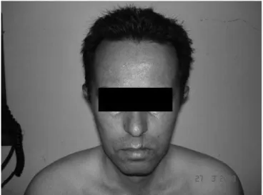
This is a 39 years old male weighing 68 kg, with the diagno-sis of MD1 and cholelithiadiagno-sis, who worked as a salesman, scheduled for videolaparoscopic cholecystectomy, and he was evaluated in the pre-anesthetic outpatient clinic. He had a history of muscular weakness since nine years of age, but only at age 34 when he presented significant dys-phagia, gastroesophageal reflux, and cataract the MD was diagnosed. The patient did not have a history of anesthetic procedures, and he used aloe vera. The family history was significant for his mother who died at age of 50 years due to complications of MD and five siblings with the disease (three sisters and two brothers). He had five children, four healthy boys and a girl with MD. On physical exam, he had typical fascies (Figure 1) with lagophthalmos and frontal baldness, difficulty swallowing that affected speech (dys-phonia), hypotrophy of the distal muscles of the upper and lower limbs (Figure 2 and 3), myotonia of the hands (diffi-culty to relax the hands after a contraction), and Mallampati IV. Laboratorial exams were normal except for CK-NAC of
Figure 1 – Facies of a patient with Steinert Disease
271.0 U.L-1 (normal range for men from 24 to 204 U.L-1). The electrocardiogram showed left branch block, isolated ventricular PVCs, left ventricular overload, and diffuse changes in ventricular repolarization. The chest X-ray and echocardiography were normal. The patient was evaluated by a cardiologist and cleared for the anesthetic-surgical procedure; cardiologic medications were not prescribed. His physical status was classified as ASA 3. The patient received ranitidine (150 mg) and metochlopramide (10 mg) 90 minutes before surgery, but not other pre-anesthetic medication. General anesthesia was induced with midazo-lam (5 mg), remifentanil (100 µg), and rocuronium (50 mg), being intubated with rapid sequence intubation and poste-rior introduction of an orogastric tube for aspiration. Anes-thesia was maintained with O2 and medicinal air at 50%, and continuous infusion of propofol (100 µg.kg-1.min-1) and remifentanil (0.15 µg.kg-1.min-1). Monitoring consisted of pulse oximetry, non-invasive blood pressure, electrocar-diogram, capnography, monitoring of muscular relaxation using the train-of-four in the adductor pollicis muscle, and body temperature. The patient underwent videolaparosco-py with insufflation with carbon dioxide to induce pneumo-peritoneum, but maintaining the intra-abdominal pressure below 15 mmHg. An electrical scalpel was used to facili-tate hemostasia. Body temperature was maintained with an electrical blanket. Thirty minutes before the expected
end of the surgery, subcutaneous tramadol (100 mg) and intravenous dypirone (2 g) and ketoprofen (100 mg) were administered. The surgical procedure was completed in 90 minutes, without intercurrences. At that time the patient did not show any response to the train-of-four. We waited for about 20 minutes until the development of at least one re-sponse and then the curarization was reversed with atro-pine (1.0 mg) and prostigmine (2.0 mg). He then showed four equal responses to the train-of-four and responded to verbal commands to open his eyes and mouth. However, his respiratory pattern was not satisfactory. When the pa-tient was regaining consciousness, he reacted to the pres-ence of the ET tube and he was extubated. Pulse oximetry was 98%, but after five minutes he showed progressive decrease in oxygen saturation, without improvement of the respiratory function. His extremities became cold and cy-anotic, he developed muscular hypertonia, lost conscious-ness, and it was difficult to ventilate the patient with the face mask. Tracheal intubation was impossible due to gen-eralized hypertonia. At that time, blood was drawn for ABGs and electrolytes. The laryngeal mask was used, allowing adequate ventilation and oxygenation. Arterial blood gas-es showed significant cyanosis (pH= 7.17, PaCO2= 77.9 mmHg, PO2= 125.3 mmHg, HCO3 = 28.2 mEq.l-1, and base excess = –2.3), but the level of electrolytes did not change. Myotonia was diagnosed and treated with midazolam (5 mg) and he remained on controlled ventilation with the laryngeal mask. The patient was transferred to the post-anesthetic recovery room because ICU beds were not available. He regained consciousness slowly and mechanical ventilation continued during this time. After five hours, the laryngeal mask was removed without intercurrences. The patient was transferred to a regular room and discharged from the hos-pital after three days.
DISCUSSION AND REVIEW OF THE LITERATURE
Muscular dystrophies belong to a group of hereditary diseases characterized by progressive muscular weakness. There are several classification criteria based on genetic transmission, age of onset of symptoms, muscles involved, and speed of progression.
of cases. Although a neonatal type does exist most patients develop symptoms of MD1 in adult life.
The disease is characterized by muscular involvement and several associated systemic manifestations. Prognosis depends mainly on the cardiac and respiratory repercus-sions.
Muscular involvement: Myotonia can be seen in several neuromuscular disorders. This word describes a persistent muscle contraction observed after termination of voluntary contraction or stimulation. The patient is not capable of relax-ing the muscle after usrelax-ing it. Initially, the muscular involve-ment manifests as myotonia and atrophy, predominantly in distal portions of the limbs, but it can affect the muscles of the face (facial and temporal muscles) and larynx, and the respiratory muscles. It occurs due to an intrinsic change in the muscle, and not in the peripheral nerve or neuromuscu-lar junction. This can be demonstrated since myotonia is not abolished by peripheral nerve blocks or neuromuscular block-ers. Its mechanisms have not been defined. Evidence sug-gests damage of chloride or sodium channels in the muscular membrane. It has been suggested that complete deactivation of sodium channels and an increase in the activity of calcium-dependent potassium channels are contributing factors to in-crease the excitability and susceptibility to myotonia in MD5. Alternatively, a dramatic reduction in transmembrane chloride conductance can also occur. The primary involvement of pe-ripheral nerves is also controversial, and some authors have suggested that weakness and muscular dystrophy are conse-quences of a polyneuropathy6.
Cardiac involvement: It is represented mainly by deterio-ration of the conduction system of the heart, tachycardias, cardiomyopathy, and valve diseases7. First degree AV block with prolonged PR interval is the most common conduction defect in patients with MD1 and present in the ECG of more than 40% of the patients. The most common arrhythmia is atrial in origin, but mono or polymorphic ventricular tachycar-dia can also be seen. Delayed conduction in the His bundle can lead to the development of severe arrhythmias secondary to reentry. Severe rhythm disorders, such as fatal ventricular tachycardia, can develop early in patients with congenital or infantile MD as well as in asymptomatic teenagers with none or little disease manifestation. They can develop sudden car-diac arrest related mainly to physical exertion8. Heart failure is present in a significant proportion of patients, but it is not clinically apparent due to the limited physical activity. For this reason, echocardiography is an important exam to quantify the degree of disruption of the cardiac function. Cardiac le-sions are progressive and they can evolve for more severe types faster than the evolution of the disease itself, and, in those cases, little correlation between the cardiac and mus-cular skeletal disease is observed. Therefore, those patients should be followed continuously, paying special attention for cardiovascular manifestations to make the best treatment de-cisions, and especially for the choice of anesthetic technique and drugs.
Pulmonary involvement: Pulmonary complications are multifactorial and frequent causes of morbimortality in MD. Patients show weakness of the respiratory muscles, and altered central control of the respiration with changes in respiratory mechanisms, leading to global hypoventilation, microatelectasis, and reduction in pulmonary compliance. Ventilatory response to carbon dioxide is reduced, which might have a central origin or be attributed to a reduction in the contractile strength of the diaphragm9. This function is further diminished in upper abdominal surgeries. Several studies have suggested that central control mechanisms of respiration are normal in patients with MD9,10. Chronic hypercapnia is a common finding and can be seen even in the presence of minimal signs of peripheral muscular weak-ness. Along with hypoventilation, patients have day time somnolence, which is also not related to the symptoms of muscular weakness, but it can be due to changes in the respiratory center or sleep disorders characterized mainly by the presence of central or obstructive sleep apnea. Mus-cular weakness can affect the diaphragm and other respi-ratory muscles causing difficulty to cough and reduction in functional residual capacity. Weakness of the abdominal muscles that despite showing increased work during res-piration is not effective in improving respiratory parameters also contributes for the respiratory complications. Increased sensitivity to sedative and anesthetic drugs can be seen.
Gastrointestinal manifestations: Gastrointestinal manifes-tations are present in 80% of the cases, being considered im-portant for the quality of life of MD patients. The most common symptoms include: nausea, vomiting, dysphagia, early satiety (due to a reduction in gastric emptying time), and gastroe-sophageal reflux11. Both myopathic weakness and myotonia of the oropharyngeal muscles are highly important in the dys-function during the oral and pharyngeal phases of swallowing in patients with MD12. Cholelithiasis is a common finding due to the increase in vesicular biliary sphincter. For unknown rea-sons, liver function tests are abnormal13.
Endocrine involvement: It can include hypothyroidism, hy-pogonadism, altered secretion of the growth hormone, and abnormalities in glucose metabolism and insulin (frequently associated with diabetes mellitus)10. Asymptomatic female patients can also develop infertility.
Anesthesia Planning and Perioperative Factors Associ-ated with the Development of Myotonia
Several precautions should be taken not only during anesthe-sia, but also in the postoperative period in patients with Stein-ert disease to prevent known complications. The development of myotonia represents an important problem for anesthesia because, if laryngeal and respiratory muscles are involved, in-tubation can be difficult or even impossible17. Potential trigger factors include the surgery, hypothermia, tremors, electrical or mechanical stimulation during or after the surgery, drugs (clo-fibrate, propranolol, potassium), and anesthetic agents, such as succinylcholine, and anticholinesterase drugs18. Treatment is mainly preventive, avoiding all triggering factors. A protocol of safe anesthetics should be adopted. The use of electrical scalpel should be avoided. Body temperature should be moni-tored closely to minimize the risk of tremors.
The anesthetic technique of choice remains uncertain. When-ever possible, peripheral nerve block or neuroaxis block is the technique of choice19. When general anesthesia is indicated, extreme care should be taken during all phases of anesthesia. During anesthetic induction, thiopental is relatively contraindi-cated due to more prolonged respiratory depression. Propofol has been used both for induction and maintenance of anes-thesia, but it can also be a problem. Changes in sensitivity, which may be increased or decreased, in patients with MD have been reported in the literature, and, consequently, it can be difficult to calculate the proper dose20,21.
Although a direct relationship between MD and malignant hyperthermia has not been reported in the literature, depo-larizing neuromuscular blockers represent a particular prob-lem, since they can have unpredictable effects22. Succinyl-choline seems to have a double effect in myotonic patients. It can cause a “normal” neuromuscular blockade, but it can affect the muscles directly causing muscular contraction and enough hyperkalemia to lead to cardiac arrest. Besides, it can initiate a generalized myotonic response, resulting in difficulty in tracheal intubation and ventilation23. Therefore, the use of succinylcholine in patients with myotonic dystrophy should be avoided, although some cases of administration of this drug in emergency situations in patients with MD without complica-tions have been reported in the literature24. Since myotonia is caused by a primary defect in the musculature, the use of non-depolarizing agents does not abolish generalized con-traction.
Non-depolarizing neuromuscular blockers have an unpredict-ably long action in patients with MD, and the cases reported in the literature suggest a reduction in the dose and use of drugs with intermediate duration, especially atracurium22,25. Nish et al.26 calculated the effective dose for 50% and 90% (ED
50 and ED90 respectively) of the patients to achieve neuromuscular blockade of vecuronium for the orbicularis, adductor pollicis, and flexor hallucis muscles in a patient with MD undergoing general anesthesia with a laryngeal mask and maintenance with propofol, nitrous oxide, and fentanyl. The ED50 for the orbicularis, adductor pollicis, and flexor hallucis muscles was 7.77 (3.10-16.8), 28.3 (20.7-43.3), and 29.5 (11.0-85.6) µg.kg-1, respectively (p < 0.01), and ED
90 of 35.7 (14.8-66.5),
51.8 (29.3-145.0), and 50.6 (5.29-642.0) µg.kg−1, respectively (p < 0.01), indicating a marked increase in susceptibility only for the orbicularis muscle. Other authors27 had already shown an increase in muscular sensitivity to vecuronium, especially of the face muscles. Those drugs do not change the muscu-lar tension triggered by the hyperexcitability of the muscumuscu-lar membrane in myotonia and metabolic exhaustion in malig-nant hyperthermia. It is not known whether the administration of anticholinesterase agents induces myotonia, although the unpredictability of its action has been reported28. Those pa-tients also show increased sensitivity to opioids and their dose should be decreased29.
As for halogenated anesthetics, it has been questioned whether all myopathies are associated with an increased risk of malignant hyperthermia (MH). In 2004, it was recognized that approximately 50% of the patients identified as being sus-ceptible to malignant hyperthermia by the contracture test had mutations of the ryanodine receptor, which is responsible for the development of this complication30. Several studies have shown that 30-50% of the individuals susceptible to MH have myopathological changes31.
Takhar et al.18 reported a case of videolaparoscopic cholecys-tectomy using alfentanil, propofol and sevoflurane, and nitrous oxide without neuromuscular blockers, resulting in elevated abdominal insufflation pressures around 20 mmHg. Complica-tions related with the anesthetic technique were not observed. The use of general anesthesia has been reported with diaz-epam, propofol, rocuronium, isoflurane, and morphine32. Due to the doubts, it is prudent to avoid drugs that could potentially trigger malignant hyperthermia in patients with myotonic dys-trophy33.
Large size surgeries in patients with Steinert disease have also been reported in the literature. Gelsomino et al.34 re-ported a series of six patients with myotonic dystrophy who underwent cardiac surgery with hypothermic extracorporeal circulation. Anesthesia was maintained with midazolam, remifentanil, propofol, and atracurium. Complications were not reported. Propofol, sufentanil, atracurium, and clonidine were used successfully in another patient undergoing car-diac surgery35.
halogenated, hypnotic, and opioid drugs as well as neuro-muscular blockers can induce hypoventilation and respiratory failure. Thus, ventilatory assistance is extremely important in the immediate postoperative period. Mechanical ventilation with tracheal intubation has been used for ventilatory support, although it can increase the risk of pulmonary infection, even for short periods. Besides, sedation is required to prevent the discomfort of the tracheal tube. This can prolong the recov-ery time of the respiratory function. The laryngeal mask mini-mized the need for sedation, since it is less traumatic for the airways and cardiovascular system. It is better tolerated by the patient, allowing calmer arousal, attenuating airways and hemodynamic reflexes. Potential complications of the laryn-geal mask, such as hypopharynlaryn-geal trauma and aspiration, have been reported37,38. We considered the possibility of as-piration of gastric contents during surgery, and maintaining the torso of the patient elevated in the recovery room was responsible for making the procedure safer regarding the risks of aspiration, since complications were not observed in the late postoperative period. Other ventilation modalities, such as positive pressure in the airways (BIPAP) with face mask, have been effective in improving oxygenation and reducing hypercapnia39. Postoperative BIPAP has also been reported in conjunction with the laryngeal mask40.
In view of what happened during anesthesia of this patient with MD, we conclude that deep knowledge of the disease, which is multisystemic and demands careful anesthetic plan-ning by anticipating complications, is recommended.
REFERÊNCIAS – REFERENCES
01. Tramonte JJ, Burns TM – Myotonic dystrophy. Arch Neurol, 2005;62:1316-19.
02. Day JW, Ranum LPW – Genetics and molecular pathogenesis of the myotonic dystrophies. Curr Neurol Neurosci Rep, 2005;5:55-59. 03. Mahadevan M, Tsilfidis C, Sabourin L et al. – Myotonic dystrophy
mu-tation: an unstable CTG repeat in the 3’ untranslated region of the gene. Science, 1992;255:1253-1255.
04. Kornblum C, Lutterbey G, Bogdanow M et al. – Distinct neuromus-cular phenothypes in myotonic dystrophy types 1 e 2: a whole body highfield MRI study. J Neurol, 2006;253:753-761.
05. Luek JD, Mankodi A, Swanson MS et al. – Muscle chloride channel dysfunction in two mouse models of myotonic dystrophy. J Gen Phys-iol, 2007;129:79-94.
06. anaite PA, Gantelet E, Kraftsik R et al. – Myotonic dystrophy transgen-ic mtransgen-ice exhibit pathologtransgen-ic abnormalities in diaphragm neuromuscular junctions and phrenic nerves. J Neuropathol Exp Neurol, 2008;67:763-772.
07. Sovari AA, Bodine CK, Farokhi F – Cardiovascular manifestations of myotonic dystrophy-1. Cardiol Rev, 2007;15:191-194.
08. Bassez G, Lazarus A, Desguerre I et al. – Severe cardiac arrhyth-mias in young patients with myotonic dystrophy type 1. Neurology, 2004;63:1939-1941.
09. Ugalde V, Walsh S, Abresch RT et al. – Respiratory abdominal mus-cle recruitment and chest wall motion in myotonic muscular dystrophy. J Appl Physiol, 2001;91:395-407.
10. Begin P, Mathieu J, Almirrall J et al. – Relationship between chronic hypercapnia and inspiratory –muscle weakness in myotonic dystro-phy. Am J Respir Crit Care Med, 1997;156:133-139.
11. Ronnblom A, Andersson S, Hellstrom PM et al. – Gastric emptying in myotonic dystrophy. Eur J Clin Invest, 2002;32:570-574.
12. Ertekin C, Yüceyar N, Aydogdu Ï et al. – Electrophysiological evalu-ation of oropharyngeal swallowing in myotonic dystrophy. J Neurol Neurosurg Psychiatry, 2001;70:363-371.
13. Heatwole CR, Miller J, Martens B et al. – Laboratory abnormalities in ambulatory patients with myotonic dystrophy type 1. Arch Neurol, 2006;63:1149-1153.
14. Modoni A, Silvestri G, Pomponi MG et al. – Characterization of the pattern of cognitive impairment in myotonic dystrophy type 1. Arch Neurol, 2004;61:1943-1947.
15. Antonini G, Soscia F, Giubilei F et al. – Health-related quality of life in myotonic dystrophy type 1 and its relationship with cognitive and emotional functioning. J Rehabil Med, 2006;38:181-185.
16. Schulz PE, McIntosh AD, Kasten MR et al. – A role for myotonic dystrophy protein kinase in synaptic plasticity. J Neurophysiol, 2003;89:1177-1186.
17. Mahr A, Attof Y, Flamens C. et al. – Prise en charge anesthésique des patients porteurs de myotonie de Steinert: à propos de deux cas cliniques. Ann Fr Anesth Reanim, 2009;28:161-164.
18. Takhar AS, Thaper A, Byrne A et al. – Laparoscopic cholecystectomy in a patient with myotonic dystrophy. J R Soc Med, 2004;97:284-5. 19. Araújo FS, Bessa Jr RC, Castro CH et al. – Anestesia no
pacien-te com doença de Spacien-teinert. Relato de caso. Rev Bras Anespacien-tesiol, 2006;56:649-653.
20. Speedy H – Exaggerated physiological response to propofol in myo-tonic dystrophy. Br J Anaesth, 1990;64:110-112.
21. Morimoto Y, Mii M, Hirata T et al. – Target-controlled infu-sion of propofol for a patient with myotonic dystrophy. J Anesth, 2005;19:336-338.
22. Parness J, Bandschapp O, Girard T – The myotonias and susceptibil-ity to malignant hyperthermia. Anesth Analg, 2009;109:1054-1064. 23. Larach MG, Rosenberg H, Gronert GA et al. – Hyperkalemic cardiac
arrest during anesthesia in infants and children with occult myopa-thies. Clin Pediatr (Phila), 1997;36:9-16.
24. Tomlison S, Macartney I, Lam S – Dystrophica myotonia and suxam-ethonium. Anaesthesia, 1999;54:1234.
25. Nightingale P, Healy TEJ, McGuinness K – Dystrophia myotonica and atracurium. A case report. Br J Anaesth,1985;57:1131-5.
26. Nishi M, Itoh H , Tsubokawa T et al. – Effective doses of vecuronium in a patient with myotonic dystrophy Anaesthesia, 2004;59:1216-1218. 27. Diefenbach C, Lynch J, Abel M et al. – Vecuronium for muscle
relaxa-tion in patients with dystrophia myotonica. Anesth Analg, 1993;76:872-874.
28. Buzello W, Krieg N, Schlickewei A – Hazards of neostgmine in pa-tients with neuromuscular disorders. Report of two cases. Br J Anaes-th, 1982;54:529-534.
29. Grimsehl K, Wilson E – Remifentanil in myotonic dystrophy – avoid-ing the use of muscle relaxants and long actavoid-ing opioids. Internet J Anesthesiol, 2000;4(1). Disponível em: <http://www.ispub.com/jour-nal/the_internet_journal_of_anesthesiology>.
30. Sei Y, Sambuughin N, Muldoon S – Malignant hyperthermia genetic testing in North America, Working Group Meeting. Bethesda, Mary-land, September 4-5, 2002. Anesthesiology, 2004;100:464-465. 31. Payen JF, Bosson JL, Brambilla E et al. – Histological support for
the difference between malignant hyperthermia susceptible (MHS), equivocal (MHE) and negative (MHN) muscle biopsies. Br J Anaesth, 1997;79:327-331.
32. Ioscovich A, Barth D, Briskin A – Biphasic intermittent positive airway pressure (BIPAP) ventilation support in the postoperative period for patients with myotonic dystrophy. Internet J Anesthesiol, 2006;10(2). Disponível em: <http://www.ispub.com/journal/the_internet_journal_ of_anesthesiology>.
33. Wappler F – Malignant hyperthermia. Eur J Anaesthesiol, 2001;18:632-652.
35. Klompe L, Lancé M, van der Woerd D et al. – Anaesthesiological and ventilatory precautions during cardiac surgery in Steinert’s disease. J Card Surg, 2007;22:74-75.
36. Mathieu J, Allard P, Gobeil G et al. – Anesthetic and surgical complica-tions in 219 cases of myotonic dystrophy. Neurology, 1997;49:1649-1650.
37. McCrory CR, McShane AJ – Gastroesophageal reflux during spon-taneous respiration with the laryngeal mask airway. Can J Anaesth, 1999;46:268-270.
38. White RJ, Bass S – Anaesthetic management of a patient with myo-tonic dystrophy. Paediatr Anaesth, 2001;11:494-497.
39. Aguilo R, Togores B, Pons S et al. – Noninvasive ventilatory support after lung resectional surgery. Chest, 1997;112:117-121.
40. Groudine SB, Lumb PD, Sandison MR – Pressure support ven-tilation with the laryngeal mask airway: a method to manage se-vere reactive airway disease postoperatively. Can J Anaesth, 1995;42:341-343.
RESUMEN
Bisinotto FMB, Fabri DC, Calçado MS, Perfeito PB, Tostes LV, Sousa GD – Anestesia para Colecistectomía Videolaparoscópica en Pacien-te Portador de Enfermedad de SPacien-teinert. Relato de Caso y Revisión de la Literatura.
JUSTIFICATIVA Y OBJETIVOS: Las distrofias miotónicas son
en-fermedades neuromusculares de transmisión autosómica dominante. Entre ellas está la distrofia miotónica tipo 1 (DM1), o enfermedad de
Steinert, que es la más común en el adulto y además de la involucra-ción muscular, presenta manifestaciones sistémicas importantes. La DM1 representa un reto para el anestesiólogo. Los pacientes presen-tan una mayor sensibilidad a los fármacos anestésicos y complicacio-nes, principalmente cardíacas y pulmonares. Además de eso, existe la posibilidad de presentar hipertermia maligna y crisis miotónica. Se ha descrito el caso de un paciente que tuvo una complicación pul-monar importante después de haber sido sometido a la anestesia general.
RELATO DEL CASO: Paciente de 39 años, portador de DM1,
so-metido a la anestesia general para colecistectomía videolaparos-cópica. La anestesia fue venosa total con propofol y remifentanil y rocuronio. El procedimiento quirúrgico de 90 minutos no presentó intercurrencias, pero después de la extubación, el paciente presen-tó insuficiencia respiratoria y crisis miopresen-tónica, que hizo la intubación traqueal imposible. Se utilizó la máscara laríngea, que posibilitó la oxigenación adecuada, y la ventilación mecánica se mantuvo hasta la recuperación total de la actividad respiratoria. Evolucionó sin otras complicaciones.
CONCLUSIONES: La DM1 es una enfermedad que presenta varias