Rev Bras Anestesiol ARTIGO CIENTÍFICO
2010; 60: 2: 144-153 SCIENTIFIC ARTICLE
* Recebido (Received from) do Serviço de Anestesiologia e Neurologia do Hospital Universitário Antonio Pedro (HUAP) da Universidade Federal Flumin-ense (UFF)
1.Anestesiologista; Corresponsável CET/SBA HUAP/UFF 2. Professor Titular de Neurologia da UFF
3. Anestesiologista do HUAP-UFF; Doutora em Patologia Experimental – UFF 4. Médica do Ambulatório de Neuropatia Periféricas da UFF; Mestre em Neu-rologia – UFF
5. ME2 em Anestesiologia
6. Médico; ME1 da CNRM do HUAP-UFF
Apresentado (Submitted) em 31 de maio de 2009
Aceito (Accepted) para publicação em 24 de dezembro de 2009 Endereço para correspondência (Correspondence to): Dr. Osvaldo J. M. Nascimento
Rua Siqueira Campos, 53/1204 Copacabana
22031-071 Rio de Janeiro, RJ
Perfil da Dor Neuropática: a Propósito do Exame Neurológico
Mínimo de 33 Pacientes *
Neuropathic Pain Profile: the Basic Neurological Exam of 33 Patients
Marco Antonio Cardoso de Resende, TSA 1, Osvaldo José Moreira Nascimento 2, Anna Amélia Silva Rios 3, Giseli Quintanilha 4, Luís Eduardo Sacristan Ceballos 5, Fernando Paiva Araújo 6
SUMMARY
Resende MAC, Nascimento OJM, Rios AAS, Quintanilha G, Sacris-tan Ceballos LE, Araújo FP – Neuropathic Pain Profile: the Basic Neu-rological Exam of 33 Patients.
BACKGROUND AND OBJECTIVES: Very few texts in the literature approach the neurologic exam of patients with neuropathic pain (NP). The objective of this study was to evaluate the profile of patients with NP through the neurological exam.
METHODS: This is an observational study that followed-up patients with NP for one year. The neurologic exam was evaluated at the ou-tpatient clinic and through prospective analysis. Patients whose pain severity was equal or greater than six on the Visual Analogue Scale were included in this study.
RESULTS: Burning pain predominated, affecting 54.5% of the pa-tients. Unlike multifocal neuropathy (15.15%), distal and symmetri-cal polyneuropathy was the predominant clinisymmetri-cal-topographic pattern (48%). The thermoalgic and tactile modalities of the sensorial exam were affected the most, followed by changes in motor function and deep tendon reflexes, and proprioception. Although NP does not have specific signs and symptoms, burning pain is attributed to the invol-vement of thin nerve fibers and thermoalgic pain is typical of those changes.
CONCLUSIONS: History and physical exam findings are key fac-tors in the diagnosis of NP. The log of changes in the physical exam should emphasize the involvement observed, guiding the diagnostic and therapeutic approach, curative or palliative.
Keywords: DIAGNOSTIC EXAMS; PAIN: neuropathic
RESUMO
Resende MAC, Nascimento OJM, Rios AAS, Quintanilha G, Sacris-tan Ceballos LE, Araújo FP – Perfil da Dor Neuropática: a Propósito do Exame Neurológico Mínimo de 33 Pacientes.
JUSTIFICATIVA E OBJETIVOS: Há poucos textos na literatura a li-dar com o exame neurológico do paciente com dor neuropática (DN). O objetivo deste estudo foi avaliar o perfil de pacientes com DN atra-vés de exame clínico neurológico.
MÉTODO: Em estudo observacional, uma série de casos de pacientes com DN foi acompanhada no período de um ano. A avaliação do exame neurológico foi efetuada durante visita ao ambulatório e através de aná-lise prospectiva. Foram incluídos pacientes cuja intensidade da dor era igual ou maior que seis, segundo a Escala Analógica Visual.
RESULTADOS: A dor em queimação predominou como descritor em 54,5% dos pacientes. A polineuropatia foi o padrão clínico-topográfico predominante (48%) com padrão distal e simétrico, em oposição a qua-dros de neuropatia multifocal (15,15%). As modalidades termoalgésica e tátil do exame de sensibilidade foram as mais comprometidas, logo acompanhadas por alterações motoras e reflexos profundos, enquan-to modalidades de sensibilidade proprioceptiva vieram a seguir. Apesar de nenhum sinal ou sintoma ser específico de DN, a queimação como sintoma costuma ser atribuída ao acometimento de fibras finas, assim como o padrão típico destas é a alteração térmico-dolorosa.
CONCLUSÕES: A história e os achados do exame físico são a chave para o diagnóstico de DN. O registro das alterações encontradas ao exame deve ressaltar o comprometimento observado e assim nortear a abordagem diagnóstica e terapêutica, se curativa ou paliativa.
Neuropathic Pain Profile: the Basic
Neurological Exam of 33 Patients
Marco Antonio Cardoso de Resende, TSA, M.D.; Osvaldo José Moreira Nascimento, M.D.; Anna Amélia Silva Rios, M.D.; Giseli Quintanilha, M.D.; Luís Eduardo Sacristan Ceballos, M.D.; Fernando Paiva Araújo, M.D.
INTRODUCTION
Neuropathic pain (NP) is one of the most prevalent s chro-nic pain syndromes. Patients with predominantly neuropathic pain are older, and pain is more severe and frequent than other types of chronic pain. Besides, it is associated with worse indices of quality of life and general health status of patients1,2. the etiology of NP is diverse, and it is classified,
according to the location of the lesion or inflammation in the nervous system, in peripheral or central3. Non-resolved
tis-sue damage leads to persistent pain and it is estimated that central sensitization is responsible for secondary hyperalgia and tactile allodynia, which are common in inflammatory and neuropathic processes4.
Woolf and Mannion have suggested that the progress in the treatment of peripheral neuropathic pain relies more on the identification of the mechanisms and not on etiological factors and nature of the symptoms5. However, the search of several
the-NEUROPATHIC PAIN PROFILE: THE BASIC NEUROLOGICAL EXAM OF 33 PATIENTS
rapeutic decision, helping anesthesiologists daily. Understan-ding the reason of the pain should come before the choosing among infiltrations, anesthetic blocks, oral drugs, and suppor-tive treatment.
In some patients, it is difficult to determine the nature of the pain, which can be central, peripheral neuropathy, nocicepti-ve, or psychogenic. The coexistence of more than one type of pain, such as that seen in ataxic and paretic patients, who depend enormously on accessory muscles, which trigger no-ciceptive pain, can represent a challenge for the differential diagnosis.
Very few texts in the literature focus specifically on the neuro-logical exam of NP6-7. The neurological exam is not an exclu-sive responsibility of the neurologist, but, as suggested by De Jong, it is an integral component of the medical diagnosis8. Tracking methods for identification of NP, such as the Leeds Assessment of Neuropathic Symptoms and Signs (LANSS) and Douler Neuropatique en 4 questions (DN4), use scores based on symptoms and data of the clinical exam9. In 2008, the lack of a tool for the specific diagnosis generated a propo-sal by Treede et al. for the classification of NP as “possible”, in the hypothetical sense, “probable”, and “definitive”, in which the last two require confirmation by the neurological exam10. The objective of the present study was to evaluate patients with NP, relying more on the conventional clinical neurological exam and not only on symptoms. Analyzing the profile ob-served and applying flow charts, we can contribute with the anesthesiologist, adding data on how to recognize NP and evaluate its clinical presentation.
METHODS
This is an observational, descriptive study of patients with NP followed-up at the Peripheral Neuropathies and Neuropathic Pain Outpatient Clinic or admitted to Hospital Antonio Pedro of Universidade Federal Fluminense, and approved by the Research Ethics Committee. Data was collected from March 2006 to March 2007, and it was based on the assessment of the classical neurologic exam proposed by De Jong8, which was applied through flow charts (1 and 2). The reproducibility of the initial exam was analyzed every three months by the same physicians, two neurologists (OJMN and GQ) and one anesthesiologist (MACR). We used the Visual Analogue Sca-le (VAS) to assess the severity of NP; to be included in the study, pain scores had to be equal to or higher than six, which was considered severe pain by the methodology used11, and patients with three or more clinical disorders and incomplete medical charts were excluded.
Some material was available to evaluate superficial sensitivi-ty, such as cotton and brushes, blunted stylet, and test tubes with hot and cold water. Tactile, thermal, and painful sensiti-vity were analyzed. Besides location, the description of the exam should indicate and grade any changes observed. The following were evaluated during the deep sensorial exam: bratory (pallesthesia), evaluated by a tuning fork of 128 vi-brations/sec; sensitivity to pressure (baresthesia), by digital or
manual compression of the muscles or any area of the body; and kinetic-postural sensitivity, by gentle dislocation of body segments (foot, thumb) and recognition of positioning. Bilate-ral deep tendon reflexes (proprioceptive or myotatic) were in-vestigated by striking the tendon of the flexor of the digits and triceps muscles, as well as the Achilles and patellar tendons with a percussion hammer. To evaluate muscle strength, a scale for manual muscular test, according to the Medical Re-search Council (MRC), with scores of 0 to 5 for each muscle group tested (0, absence of movement; 1, tonus present; 2,
Main complaint
Pain distribution with plausible neuroanatomy or History suggestive of relevant injury or disease
Features
A – Presence of damaged nervous structures: mononeuropathy, multiple mononeuropathy,
polyneuropathy, radiculopathy, plexopathy, polyradiculoneuropathy, others.
Neuropathic pain
CONFIRMED
B – Neurologic exam showing: sensorial signs (for Example: allodynia); and/or thermoalgesic hypo/anesthesia; and/or autonomic dysfunction
History Location
Neurologic Exam
Neuropathic pain PROBABLE
PAIN
Basic neurologic exam neuropathic pain
Kinetic-postural
History Physical exam
Sensorial Mobility Reflexes
Exteroceptive Proprioceptive Strength
Thermal
Tactile Pressure Ambulation
Pain Vibratory
movement on the same plane; 3, overcomes gravity without resistance; 4, overcomes gravity against resistance; and 5, normal muscle strength) was used. Muscle tone and coordi-nation were also assessed.
The following parameters were analyzed: gender, age, du-ration of the symptoms, distribution and number of patients for each group of clinical disorders (etiology), the presen-ce of free descriptors for pain in those disorders, and the clinical-topographic neurologic pattern observed in each patient. In this study, patients referred more than one des-criptor, and they were asked to name the one that predo-minated.
RESULTS
Thirty-three patients with neuropathic pain were identified. Pain was isolated or associated with sensorial symptoms pre-sent in other disorders, observed on the neurologic exam over one year (Table 1).
Patients had a mean age of 51 years, with mean symptom dura-tion of 57 months at the onset of the study, but one patient pre-sented pain for 324 months due to an injury of the sciatic nerve. When patients were divided in groups that identified the cau-ses of NP, a higher frequency was observed in patients with metabolic disorders (Table 2). Among those metabolic condi-tions, diabetes mellitus was present in nine out of 11 cases. Other metabolic causes included hypothyroidism and chronic renal failure.
Infections were the second group in frequency. They were seen in seven cases, two with hepatitis (C virus and the other by the B virus); two cases of leprosy; one case of herpes zoster; one case of HTLV-1; and one case of herpes simplex infection.
Among idiopathic neuropathies, we had one case of plexo-pathy with throbbing pain and proximal atrophy of the left upper limb; one case of complex regional pain syndrome type I (CRPS-I); and one case of trigeminal neuralgia.
Two cases were considered secondary to direct trauma of a peripheral nerve. One was due to a bullet in the topography of the right sciatic nerve, and the other due to surgical injury of the saphenous nerve during resection of a hamartoma. Demyelinating diseases were represented by one case of Guillain-Barré syndrome and one case of chronic inflammatory demyelinating polyradiculopathy associated with neurofibromas. The cases of NP secondary to degenerative process of the spine included one case of a herniated disk in L5-S1 that was not treated surgically, and one with pain after laminectomy of two segments, L4-L5 and L5-S1.
Two patients had toxic neuropathy. One was due to long-stan-ding alcohol abuse with nutritional deficiency, and the other case was attributed to chronic heavy metal toxicity (lead) in a patient with an intra-articular bullet for 13 years.
Several conditions were listed under the designation of “others”, such as amyloidosis (hereditary); oncologic pain, with characteristics of mixed pain (neuropathic and nocicepti-ve), secondary to abdominal lymphoma; central pain in a pa-tient with sequela of a stroke and thalamic pain; and one case of systemic lupus erythematosus (SLE), in which sural nerve biopsy showed vasculitis and focal necrosis.
Burning pain affected 18/33 (54.4%) patients, followed by tin-gling/burning pain in 8/33 (24.2%) (Table 3). Long-standing pain was associated with burning pain, and metabolic neuro-pathy affected 7/11 (63.6%).
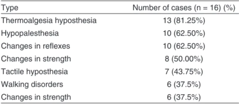
Cases of polyneuropathy predominated and they were asso-ciated with severe pain and hypoesthesia in glove and sock distribution, with predominance of sensorial complaints in the lower limbs and symmetrical weakness and decreased deep tendon reflexes (Tables 4 and 5). Those cases were associated with lower VAS (7) when compared to cases of mononeuropathy and radiculopathy, but higher than the score of patients with multiple neuropathy. Patients in this group had pain for longer time than that associated with other patterns.
All five patients with multiple mononeuropathy were females with the following diagnosis: CRPS, SLE, leprosy, diabetes mellitus, and hepatitis C. Besides the multifocal character of the nerve lesion, the onset of symptoms was not always well defined and simultaneous, but progressive and asymmetrical. Tactile-painful hypoesthesia was the most common characte-ristic of this group.
Two out of five cases of mononeuropathy were trauma-rela-ted. The others included herpes simplex, even without detec-table lesions, trigeminal neuralgia, and leprosy. Dysesthesia, abnormal or uncomfortable sensation, with or without stimuli, was the most common characteristic.
Cases of radiculopathy related with herniated disks involved L5-S1 lesion with compromised Achilles reflex and decreased dorsiflexion of toes and foot eversion. In those cases, changes in tendon reflexes and strength predominated. However, other causes of radiculopathy, such as post-herpetic neuralgia and Table 1 – Clinical and Demographic Characteristics of the
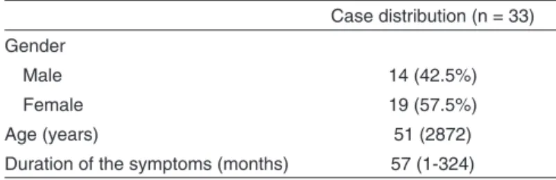
Study Patients
Case distribution (n = 33) Gender
Male 14 (42.5%)
Female 19 (57.5%)
Age (years) 51 (2872) Duration of the symptoms (months) 57 (1-324)
Table 2 – Patient Distribution According to Etiology
Number of cases Neuropathies secondary to metabolic disorders 11 (34%) Infectious neuropathies 7 (21%) Idiopathic neuropathies 3 (9%) Toxic neuropathies 2 (6%) Trauma-related neuropathies 2 (6%) Neuropathies due to degeneration of the spine 2 (6%) Demyelinating neuropathies 2 (6%)
NEUROPATHIC PAIN PROFILE: THE BASIC NEUROLOGICAL EXAM OF 33 PATIENTS
diabetes mellitus with involvement of thoracic segments, were associated with significant allodynia.
Diabetes was the most common disorder among the poly-neuropathies, but it was also associated with multiple mo-noneuropathy and radiculopathy. Central NP was present in only two patients – thalamic pain and HTLV-1 myelopa-thy (6%).
DISCUSSION
Pain, or the fifth vital sign, should be recognized and treated by the physician, regardless of the subspecialty. However, some professionals, such as anesthetists, are trained, throu-gh procedures, to prevent their patients from feeling pain. The fundamental question is to incorporate elements that allow those specialist to understand the complex nature, but com-pletely distinguishable, of the presentation of NP through a basic neurological exam. Despite the diversity of the diseases involved, there are defined patterns of sensorial-motor
topo-graphy whose identification establishes limits for possible di-sorders12.
Although only 33 cases of NP were included in this study, they are representative in this context, although they lack statisti-cal significance. A VAS equal or greater than six centimeters, chosen to select patients for this study, is a limiting factor that should be considered regarding the size of the cohort. Pain greater than six is considered very severe. We consider the VAS a method that is both easy to use and that has a high de-gree of reproducibility in our study population. Questionnaires and more complex assessments, which evaluate the quality of life and mood status, did not fit the proposed study due to the variability of the related comorbidities.
The study population had a predominance of females and, although this data is not consistent as an epidemiological profile due to the absence of better limits, confirmed the existing evidence of the greater tendency of chronic pain to affect women13. The age distribution, with a mean of 51 years, confirms the increased prevalence of NP with higher longevity of patients, especially regarding the possibility of comorbidities like diabetes mellitus. In the literature, both the female gender and older age are listed as risk factors for chronic pain14,15.
Studying pain in the elderly, Helme considered pain for longer than three months as chronic pain and acute pain as that las-ting for less than three months16. The time between the onset of the symptoms and the first time patients were seen at the clinic ranged from one to five years in 58% of the patients. Adding the percentage of patients with pain for five to ten ye-ars and for more than ten yeye-ars, this proportion rose to 82% of the cases.
Although NP does not have specific signs or symptoms, identification of pain descriptors was useful since it revealed characteristics of the study population. Some studies have re-commended the use of key words and verbal descriptors to qualify the symptoms as tracking tools for NP9,17,18. Burning is usually attributed to the involvement of thin fibers, tingling to large fibers, and stabbing pain to a combination of neuropathy of thin and large fibers12.
Change in thermoalgic sensitivity is the typical pattern of thin fibers neuropathy19. It usually affects patients older than 50 years with decreased pin prick sensitivity on the feet with centripetal extension up to the knees, but very seldom above this level. When thin and large fibers are involved, decreased proprioception and muscular stretching reflexes, along with muscle weakness, are observed in latter stages of painful pe-ripheral neuropathies20,21.
The lesion in the peripheral nerve system can be predominan-tly axonal or demyelinating, affecting sensorial or motor ner-ves, thin and large fibers. Injuries and degenerative processes of the Schwann cells can be secondary to moderate degrees of ischemia, but severe ischemia causes axonal injury with Wallerian degeneration and pain, while demyelinating neuro-pathies usually are associated with less pain when compared to those secondary to axonal damage8.
Table 3 – Free Pain Descriptors
Type Number of cases (%)
Burning pain 18 (54.5%)
Tingling 8 (24.3%)
Stabbing pain 4 (12.1%) Throbbing pain 2 (6.1%)
Shock-like 1 (3.0%)
Table 4 – Clinical-Topographic Neurologic Pattern
Type Number of cases (%)
Polyneuropathy 16 (48.48%) Multiple neuropathy 5 (15.15%) Mononeuropathy 5 (15.15%) Radiculopathy 4 (12.12%)
Plexopathy 1 (3%)
Myelopathy 1 (3%)
Thalamic pain 1 (3%)
Table 5 – Characteristics of Patients with Polyneuropathy on Physical Exam
Bulb-shaped neuromas are due to disorganized axonal re-generation secondary to partial or complete nerve lesion22. When stimulated by pressure, tension, and/or hypoxia, they become painful. Surgical and non-surgical injury of peripheral nerves with neuroma formation is one of the most frequent causes of NP.
This study showed a heterogenous group of causes of NP with homogenous clinical-topographic pattern. On neurolo-gical exam, polyneuropathies were more prevalent (48.4%), while multifocal and focal mononeuropathies were responsi-ble for 30.3% of the cases, and radiculopathies for 12%. We did not observe isolate NP, without other findings on physical exam. All patients had associated manifestations; pain distri-bution allowed the topographical characterization, showing a predominance of thermoalgesia, indicating involvement of thin nerve fibers.
Autononomic dysfunction was observed in cases of diabetic polyneuropathy, but it was also associated with amyloido-sis and chronic alcohol abuse. Guillain-Barré syndrome was another disease with this same component, which has also been observed by other authors23. Complex regional pain syn-drome type I, interpreted as multiple mononeuropathy of the upper limb, is another example of significant dysautonomia with changes in sudoresis and vasomotor, as well as edema and trophic changes.
Sensorial, motor, and deep tendon reflexes evaluation with the basic neurologic exam identified clinical-topographic patterns of NP. Thus, we should try to decrease the ten-dency, in medical practice, to define any clinical presenta-tion of pain as NP24. Questionnaires and methods, such as LANSS and DN4, consider both symptoms and the physical exam, like the presence of allodynia and touch threshold. However, pain as a consequence of injury or disease of the somatosensory system can only be confirmed through the neurologic exam25.
Changes in NP, such as variation in anatomical distribution, would be seen with a larger number of patients and differen-tiated clinical presentation for analysis. Evaluation at the neu-rology clinic might have shown differences in predominating causes, mainly those related with radiculopathies, due to the higher number of clinical-surgical patients with involvement of the cervical or lumbar spine, and would most certainly indica-te a greaindica-ter percentage of compressive or non-compressive myelopathies. Polyneuropathy was present in the majority of the cases; this result is in agreement not only with metabolic disorders, but with other clinical conditions – infectious, toxic, demyelinating – seen among us.
The predominance of patients with metabolic disorders in the neurology outpatient clinic could be interpreted as a bias, but it can also indicate the difficulty to maintain adequate control of blood glucose levels in a large proportion of diabetics and even in patients with glucose intolerance who develop neuropathy. Strict preoperative evaluation should be an integral part of anesthesiology. Understanding neurological changes helps the diagnosis and treatment of patients with pain. History and
the physical exam represent key factors in the diagnosis of NP. When recording the changes observed in the physical exam, the degree of involvement should be included to help guide the diagnostic and therapeutic approach, whether cura-tive or palliacura-tive.
REFERÊNCIAS – REFERENCES
01. Smith BH, Torrance N, Bennett MI et al. – Health and quality of life as-sociated with chronic pain of predominantly neuropathic origin in the community. Clin J Pain, 2007;23:143-149.
02. Eriksen J, Jensen MK, Sjogren P et al. – Epidemiology of chronic non-malignant pain in Denmark. Pain, 2003;106:221-228.
03. Kraychete DC, Gozzani JL, Kraychete AC – Dor Neuropática – Aspec-tos Neuroquímicos. Rev Bras Anestesiol, 2008;58:492-505. 04. Woolf CJ – Central sensitization: uncovering the relation between pain
and plasticity. Anesthesiology, 2007;106:864-867.
05. Woolf CJ, Mannion RJ – Neuropathic pain: aetiology, symptoms, mechanisms, and management. Lancet, 1999;353:1959-1964. 06. Marquez JO – Exame Clínico, em: Drummond JP, Marquez JO – Dor
Neuropática. Curitiba, Evidence, 2005;97-103.
07. Amâncio EJ, Teixeira MJ – Dor Central, em: Drummond, JP, Marquez JO – Dor Neuropática. Curitiba, Evidence, 2005;199-230.
08. Campbell WW – De Jong, o Exame Neurológico. 6ª Ed, Rio de Janei-ro, Guanabara Koogan, 2007.
09. Bennett MI, Attal N, Backonja MM et al. – Using screening tools to identify neuropathic pain. Pain, 2007;127:199-203.
10. Treede RD, Jensen TS, Campbell JN et al. – Neuropathic pain: re-definition and a grading system for clinical and research purposes. Neurology, 2008;70:1630-1635.
11. Scott J, Huskisson EC – Graphic representation of pain. Pain,1976; 2:175-184.
12. Ropper AH, Brown RH – Adams and Victors Principles of Neurology, 8th Ed, New York, McGraw-Hill, 2005;1110-1177.
13. Fillingim RB – Sex, Gender and Pain. Progress in Pain Research and Management, Seattle, IASP Press, 2000;17.
14. Hans G, Masquelier E, De Cock P – The diagnosis and manage-ment of neuropathic pain in daily practice in Belgium: an obser-vational study. BMC Public Health, 2007; 7:170. Disponível em http://www. biomedcentral.com/1471-2458/7/170. Acessado em 10/03/2008.
15. Lachaine J, Gordon A, Choiniere M et al. – Painful neuropathic di-sorders: an analysis of the Regie de l’Assurance Maladie du Quebec database. Pain Res Manag, 2007;12:31-37.
16. Helme RD, Gibson SJ – Pain in the elderly, em: Jensen TS, Turnei JA, Wiesenfeld-Hallin Z – Proceedings of the 8th World Congress in Pain. Seattle, IASP Press,1997;919-44.
17. Boureau F, Doubrere JF, Luu M – Study of verbal description in neu-ropathic pain. Pain, 1990;42:145-152.
18. Bouhassira D, Attal N, Alchaar H et al. – Comparison of pain syn-dromes associated with nervous or somatic lesions and development of a new neuropathic pain diagnostic questionnaire (DN4). Pain, 2005;114:29-36.
19. Gooch CL – Neuropathic Pain, em: Rowland LP – Merritt‘s Neurology, 11 Ed, Lippincott Williams & Wilkins, 2005;545-551.
20. Mendell JR, Sahenk Z – Clinical practice. Painful sensory neuropathy. N Eng J Med, 2003;348:1243-1255.
21. Nascimento OJM – Polineuropatias Dolorosas, em: Drummond JP, Marquez JO – Dor Neuropática. Curitiba, Evidence, 2005;159-170.
NEUROPATHIC PAIN PROFILE: THE BASIC NEUROLOGICAL EXAM OF 33 PATIENTS
23. Mendell JR, Kissel JT, Cornblath DR – Evaluation of the Patient with Peripheral Neuropathy: the Challenges, em: Mendell JR, Kissel JT, Cornblath DR – Diagnosis and Management of Peripheral Nerve Dis-orders. Oxford, Oxford University, 2001;3-9.
24. Backonja MM – Defining neuropathic pain. Anesth Analg, 2003;97: 785-90.
25. Schestatsky P, Nascimento OJM – O que os neurologistas ge-rais devem saber sobre dor neuropática? Arq neuropsiquiatr, 2009;67(3a):741-749.
RESUMEN
Resende MAC, Nascimento OJM, Rios AAS, Quintanilha G, Sacris-tan Ceballos LE, Araújo FP
JUSTIFICATIVA Y OBJETIVOS: Existen pocos textos en la literatura
que aborden el examen neurológico del paciente con dolor neuropáti-co (DN). El objetivo de este estudio fue evaluar el perfil de pacientes con DN a través de examen clínico neurológico.
MÉTODO: En un estudio observacional, una serie de casos de
pa-cientes con DN tuvo un seguimiento en el período de un año. La evaluación del examen neurológico fue efectuada durante una visita
al ambulatorio y a través de un análisis prospectivo. Se incluyeron pacientes cuya intensidad de dolor era igual o mayor que seis, según la Escala Analógica Visual.
RESULTADOS: El dolor en quemados predominó como
descrip-tor en un 54,5% de los pacientes. La polineuropatía fue el están-dar clínico-topográfico predominante (48%) con estánestán-dar distal y simétrico, en oposición a los cuadros de neuropatía multifocal (15,15%). Las modalidades termoalgésica y táctil del examen de sensibilidad fueron las más comprometidas, y venían acompaña-das de alteraciones motoras y reflejos profundos, mientras que las modalidades de sensibilidad proprioceptiva venían después de aquellas. A pesar de que no había ninguna señal o síntoma específico de DN, la quemadura como síntoma acostumbra a ser atribuida al acometimiento de las fibras finas, como también su estándar típico es la alteración térmico-dolorosa.
CONCLUSIONES: La historia y los descubrimientos del examen