Clinical and microbiological study of otitis externa in sheep
M. I. Al-Farwachi and M. M. Al-Hassan
Department of Internal and Preventive Medicine, College of Veterinary Medicine, University of Mosul, Mosul, Iraq
(Received June 10, 2007; Accepted June 26, 2008)
Abstract
In this study one hundred Awassi sheep were examined clinically and bacteriologically for isolation and identification of the bacterial agents of otitis externa in sheep. The main clinical signs appeared included weakness, pale mucus membrane, auricular discharge, cough, anorexia, emaciation, and nasal discharge. Results revealed isolation of bacteria from (45%) examined swabs. The most being from right ear. Younger animals were more frequently infected than older animals. Pseudomonas aeruginosa, Staphylococcus aureus, Mannheimia haemolytica, Staph. epidermidis, Pasteurella multocida, Streptococcus spp., Acintobacter spp., Escherichia coli and Klebsiella pneumonia were isolated. The results revealed that the most bacterial isolates were resistance to the bactericidal effect of the normal serum included Streptococcus pneumonia, Staphylococcus aureus, Mannheimia haemolytica. While the most bacterial isolates were produced hydroxymate siderophore included Staphylococcus aureus, Mannheimia haemolytica, Streptococcus pneumonia. The obtained results indicated to the importance of determination of serum resistance as a bacterial virulence factor in otitis externa in sheep.
Keywords: Otitis externa; Bacterial infection; sheep
!
"#
!"
# $%&
'(
)
* %
+
,-( . / +
0
! 12(
3
/
1
%.
4
5
6 ( 0 -7
)
/ 1
/ 28.
"2
)
0 , 9
'&
& %.
:
#
/
0
" ; < /7"9
/
)
3-
/ -2
#=
&9(
)
> . :
4
6 ( 0 2
' +
3< ;
/
?@
A
/
">
0
)
;
%.
0
0.3
'7
)
,
$ B
0
"
/
. /
$
0
"
+
4
/ " = 5 3 ' +
0 3<
)
/ &% / # =
0
)
'# / "
9
(
)
0
2
)
#
(
9
(
)
"
0
)
" = ' (
( .
)
/
=
2 .
)
/
9
4
0 -7
"
0
' +
0
( =
+C( / =
/ +
0.3
+
6 (
)
,
0
/ &% / # =
)
'# / "
9
(
4
' + ,
0
#
0
# - / (
/ +
0.3
+
"
/ &% / # =
0
)
'# / "
9
(
)
"
0
4
,
/, = #, #"( /, & 6 ( 0 +
)
D ,(
#
-/
1
%.
-( . /
/ 3
' +
/< # #"(
# 0
Introduction
Otitis externa is an inflammation of the external auditory canal (1). Otitis is common in calves and pigs but young and adult of all species are susceptible (2), especially in lambs (1,3). Although affected animals remain alert, strong, while in cases of bilateral ear infection a tremor observed, resembling that seen with cerebellar diseases (4). Clinical signs such as an exudates in conjunction with the isolation of a particular bacterial species in large number, are of significance in most cases and may indicate the presence of pathogen (4, 5). In advanced clinical cases there can be irreversible and fatal neural lesion (2). Ear infection in calves and lambs has been associated with concurrent respiratory diseases and mixed infection (1,5). Otitis externa has a multifactorial etiology and bacteria play an important role in otic diseases (1,3,6). Most of the bacteria incriminated in ear infections including Staphylococcus spp, Pseudo-monas spp, Escherichia spp, Mannheimia haemolytica, Pasteurella multocida and Proteus spp can be recovered occasionally, usually in small numbers from healthy ears (1,4,6-8). Iron is essential for almost all living organisms, including bacteria. The ability of pathogenic bacteria to acquire iron in hosts is absolutely essential for bacterial growth and infection (9,10). In animal hosts, iron is usually bound to proteins such as transferrin and lactoferrin in extracellular fluid and to ferritin, hemoglobin, and heme-containing enzymes in cells (11,12). To utilize such complexes as iron sources, bacteria generally possess some sophisticated mecha-nisms, which include an iron uptake system mediated by high-affinity iron chelators called siderophores and a system for heme uptake via specific receptors (11,13). The siderophore defined as low iron induced virtually ferric specific ligands, is widely distributed in microbial species (14), where they function in the sequestration and transport of iron. Siderophore is a component of the virulence of clinical isolates E. coli and certain other microorganisms pathogenic to man and animals. The serum resistance beside the hydrooxymate siderophore and other factors which are counted as strong indicators for bacterial virulence (14-16). In Iraq there is very little records on the prevalence of otitis externa in sheep. The present study was, therefore designed to reveal the importance of some bacteria in sheep otitis externa by the determination of their virulence factors with refernce to serum resistance as well as production of siderophore.
Materials and Methods
Animals
A total of 100 Awassi sheep of both sexes (40 male, and 60 female), 1 month -1 year old were used in this study. The number of the animals examined included 53 sheep were presented for treatment from different diseases and 47
animals referred to the Veterinary Clinic from farms around Mosul city. The routine clinical examinations were carried out for each animals (2).
Samples
Both external ears canals were swabbed with sterile cotton–tipped applicators. Swabs were inoculated on peptone water and incubated at 37Cº for 24 hours.
Routine bacterial culture was performed in all samples by streaked on nutrient agar, blood agar, chocolate agar and MacConkeys agar plates and incubated at 37Cº for 24-48 hours. The isolated colonies were identified
mor-phologically, culturally and biochemically (17, 18, 19).
Determination of serum resistance
For determination of serum resistance of the isolates a pooled serum of ten healthy sheep were used. The sera were sterilized by 0.22 mm Millipore membrane filter and kept at 30º C. Serum resistance was conducted according (15). In brief, 100 µl of 24 hours old culture of each of the 120 isolates inoculated in 5 ml tryptic soy broth .After incubation at 37º C, their optical densities were adjusted to 0.3 at 540 nm wave length using spectrophotometer (CECIL CE 1010, England).
Twenty five microliters were mixed with 2.5 ml of sterile sheep serum. For determination of their count, aliquots were serially diluted before and after 1 hour incubation at 37ºC in an orbital shaking water bath. The viable bacterial count of each isolate was determined by computing the average of a replicate of 2 successive dilutions. The stain was determined as serum resistant when its count was increased or as serum sensitive when the count was decreased.
Determination of hydroxymate siderophore production
Determination of hydroxymate siderophore production was based on the technique described by (20). In brief, to 1 ml of the supernatant bacterial culture in its stationary phase, 1 ml H2So4 (3 M) was added. After 4 hours hydrolysis time 120º C, 1.55 ml of 35% CH3CooNa, 1 ml of each of 1% sulfanilic acid, 1.3% iodine (for oxidation), 2% sodium arsenite and 0.3% -naphtylamin were added. Development of red coloration (in darkness) after 30 minutes would indicate the hydro-xylmate siderophore production. The data were analyzed statistically using analysis of variance, the level of significance was at P< 0.05.
Results
The results also shows that the percentage of the infected swabs were (45%), (12 bacterial isolates from healthy animals, sub clinical cases), 60 (30%) from right ear canal, 30 (15%) from left ear canal. (Table 2) .Younger animals were more frequently infected (37.5%) than older animals (7.5%) (Table 3).
Table 1: Clinical sings in sheep suffered from otitis externa.
% Number of
affected animals Clinical signs
80 60
Weakness
66.7 50
Pale mucus membrane
66.7 50
Auricular discharge
53.3 40
Cough
53.3 40
Anorexia and emaciation
53.3 40
Bilateral nasal discharge
Table 2: The percentages of infected swabs from ear canal in sheep.
Number of infected swabs Number of
examined swabs Site of ear
swabs
30* 60
100 Right ear
15 30
100 Left ear
45 90
200 Total
* Significantly P< 0.05.
Table 3: Age and percentage of the infected sheep with otitis externa.
Number of infected swabs Number of
examined swabs Age
37.5* 75
100 1 – 6
month
7.5 15
100 6
month-1 year
45.0 90
200 Total
* Significantly P< 0.05.
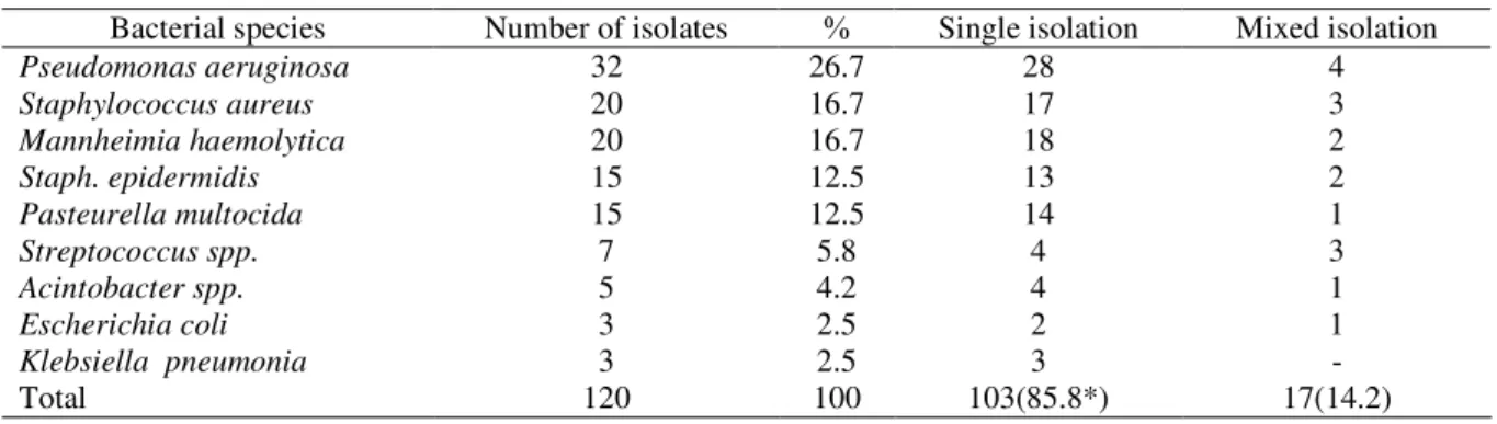
The bacterial species isolated from external ear canal included Pseudomonas aeruginosa were most prevalent (26.7%), followed by Staphylococcus aureus, and Mannheimia haemolytica (16.7%), but Staph. epi-dermidis and Pasteurella multocida were isolated at (12.5%), while Streptococcus spp, Acintobacter spp., Escherichia coli and Klebsiella pneumonia were isolated at (5.8, 4.2, 2.5%) respectively. From total of 200 ear swabs, 120 isolates showed that the single isolation was more frequently than mixed isolation. (Table 4 and 5).
The results of resistance of the bacterial isolates from external ear canal of sheep to normal bactericidal effect of sheep normal serum revealed that 73 bacterial isolates from the total numbers (120 bacterial isolates) were resistance to normal sheep serum. Streptococcus spp, Staphylococcus aureus, Mannheimia haemolytica, Pseudomonas aeruginosa, Pasteurella multocida were resistance to normal sheep serum at 85.7%, 80.0%, 70.0%, 62.5%, 60.0% respectively. While only 30 % of E. coli and Klebsiella pneumonia and 13.0% of Staph. epidermidis isolates were resistance to normal serum (Table 6).
Table 4: Bacterial species isolates from ear canal in sheep.
Mixed isolation Single isolation
Number of isolates Bacterial species
4 28
26.7 32
Pseudomonas aeruginosa
3 17
16.7 20
Staphylococcus aureus
2 18
16.7 20
Mannheimia haemolytica
2 13
12.5 15
Staph. epidermidis
1 14
12.5 15
Pasteurella multocida
3 4
5.8 7
Streptococcus spp.
1 4
4.2 5
Acintobacter spp.
1 2
2.5 3
Escherichia coli
- 3
2.5 3
Klebsiella pneumonia
17(14.2) 103(85.8*)
100 120
Total
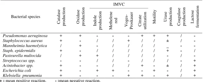
Table 5: Biochemical reactions characteristic of bacterial species isolates from ear canal in sheep. IMVC L ac to se fe rm en ta ti o n C o a g u la se p ro d u ct io n U ra se p ro d u ct io n M o ti li ty C it ra te u ti li za ti o n V o g es - P ro sk a u er M et h el e n e re d In d o le p ro d u ct io n O x id as e p ro d u ct io n C at al as e p ro d u ct io n Bacterial species Pseudomonas aeruginosa Staphylococcus aureus Mannheimia haemolytica Staph. epidermidis Pasteurella multocida Streptococcus spp. Acintobacter spp. Escherichia coli Klebsiella pneumonia
+ : mean positive reaction. - :mean negative reaction.
Table 6: Resistance of bacterial isolates from external ear canal to the normal bactericidal effect of sheep serum.
% Number of sensitive
isolates Number of resistance
isolates Bacterial species 14.3 1 85.7 6 Streptococcus spp. 20.0 4 80.0 16 Staphylococcus aureus 30.0 6 70 14 Mannheimia haemolytica 37.5 12 62.5 20 Pseudomonas aeruginosa 40.0 5 60.0 10 Pasteurella multocida 40.0 2 60.0 3 Acintobacter spp. 70.0 2 30.0 1 Escherichia coli 70.0 2 30.0 1
Klebsiella pneumonia
87.0 13 13.0 2 Staph. Epidermidis 39.2 47 60.8 73 Total
The most isolates of Staphylococcus aureus 95.0%, and 80.0%, 71.4% of the Mannheimia haemolytica and Streptococcus spp. isolates respectively were produced hydroxymate siderophore. A 66.7% of Pasteurella multocida, 65.6% of Pseudomonas aeruginosa, 60% of the Acintobacter spp. isolates were produced sidero-phore, while only 33.3% of the E. coli and Klebsiella pneumonia isolates produced siderophore (Table 7).
Table 7: Types and numbers of bacterial isolates produced hydroxymate siderophore. Number of isolates produced Bacterial species 95.0 19 Staphylococcus aureus 80.0 16 Mannheimia haemolytica 71.4 5 Streptococcus spp. 66.7 10 Pasteurella multocida 65.6 21 Pseudomonas aeruginosa 60.0 3 Acintobacter spp. 33.3 1 Escherichia coli 33.3 1
Klebsiella pneumonia Staph. epidermidis
60.3 76
Discussion
The present study aimed to determine the clinical signs, isolation and identification of the bacterial species of otitis externa in sheep , in addition to evaluate their serum resistance of normal sheep serum and production of hydroxymate siderophore from bacterial isolates (for determination of bacterial virulence of the isolated bacteria). The main clinical signs appeared included general weakness, pale mucus membrane, auricular discharge, cough, anorexia, and emaciation, nasal discharge. This sings were attributed to the chronic otitis and/or upper respiratory infection (2). Ear infection in calves and lambs has been associated with concurrent respiratory diseases and mixed infection (1,4,21).
Pugh (1) reported that little is known about otitis in goat and sheep compared with the information available on cattle and horses, and many factors can predispose sheep to otitis externa including the anatomic orientation of the ear canal itself. The vertical canal slopes medially into horizontal orientation on the outside of the tympanic membrane, this prevent drainage of debris leads to accumulate it. The skin lining of the external ear canal has a large numbers of glands, these include modified a porcine glands which produce large amounts of sec-retions which provide good suitable media for irritation and infection (1, 2). The results revealed isolation of a percentage of 45% from examined samples, most of isolates were from right ear. Many bacterial species commonly inhabit the ear canal, and can become secondary opportunistic invaders when conditions are favorable (1,4,6), and it probably ascended from the pharynx through the auditive tubes into, the tympanic cavities (21). In this study Pseudomonas aeruginosa was prevalent bacteria, followed by Staphylococcus aureus, Mannheimia haemolytica, while Escherichia coli and Klebsiella pneumonia were isolated in lower percentage (1,3,4,6) recorded many pyogenic bacterial species causes otitis externa in animals. The results revealed that the most bacterial isolates were resistance to the bactericidal effect of the normal serum included Streptococcus pneumonia, Staphylococcus aureus, Mannheimia haemolytica. While the most bacterial isolates were produced hydroxymate siderophore included Staphylococcus aureus, Mannheimia haemolytica, Streptococcus pneumonia. The virulence of bacterial isolates from external ear swabs was correlated to some other different factors like the hemolytic activity and aerobatic production (22,23). The serum resistance beside the hydrooxymate sidero-phore and other factors are counted as strong indicators for bacterial virulence (14,15,16). The encapsulated avian strains of Pasteurella multocidawere recorded to resist the bactericidal action of serum, whereas the uncapsulated were not (15). The Coagulase-negative Staphylococcus could not produce
siderophore, and therefore the ability of siderophore mediated iron uptake would contribute to the increased pathogenesity of Staphylococcus aureus (24). However, siderophore production may be increased in the presence of some antibiotic even in their minimum inhibitory concentration (25).
In conclusion, the present study indicated, the importance of some bacteria in sheep otitis and the determination of their virulence factors with reference to serum resistance as well as the production of siderophore is important for better understanding of the bacterial infection in sheep..
Acknowledgment
This study was supported by the College of Veterinary Medicine, University of Mosul, Mosul, Iraq.
References
1. Pugh DG. Sheep and Goat Medicine. 4th ed. Philadelphia: WB
Saunders Com. 2000.
2. Radostits OM, Gay CC, Blood DC and Hinchcliff KW. Veterinary
medicine: A textbook of the diseases of Cattle, Sheep, Pigs, Goats and
Horses. 9th ed. Philadelphia: WB Saunders Com. 2000.
3. Ismail SF. Studies on some surgical affections of the external ear in farm animals. Assuit Vet Med J 1994; 32: 177-187.
4. Duarte ER and Hamdan JG. Otitis in cattle, An aetiology review. J Vet
Med 2004; 5: 1 – 7 .
5. Yeruham D, Elad ML. Clinical and Microbiological study of an otitis
media outbreak in calves in a dairy herd. J Vet Med 1999; 46: 145-150.
6. Harry H, Mada C, Rober P. Update on antimicrobioal susceptibilities bacterial isolates from canine and feline otitis externa. Can Vet J 2006; 47: 253-255.
7. Morgan K. Otitis externa associated with Pseudomonas aeruginosa:
Clinical and epidemiology study. Vet Rec 1992; 130: 530-532.
8. Watson PJ, Watabe M, Moore JE. Purulent rhinitis and otitis caused by
Pseudomonas aeruginosa in sheep showed with contaminated shower wash. Vet Rec 2003; 153: 704-707.
9. Bullen JJ. The significance of iron in infection. Rev Infect Dis 1981; 3: 1127-1138.
10.Marinez JL, Delgado-Iribarren A, Baquero F. Mechanisms of iron
acquisition and bacterial virulence. FEMS Microbiol Rev 1990; 75: 45-56.
11.Neilands JB. Iron absorption and transport in microorganisms. Annu Rev Nutr 1981; 1: 27-46.
12.Wooldridge KG and Williams PH. Iron uptake mechanisms of
pathogenic bacteria. FEMS Microbiol Rev 1993; 12: 325–348. 13.Govan JRW, Deretic V. Microbial pathogenesis in cystic fibrosis:
mucoid Pseudomonas aeruginosa and Burkholderia cepacia.
Microbiol Rev 1996; 60: 539-574.
14.Neilands JB. Siderophores. Arch Biochem. Biophys 1993; 302: 1-3.
15.Hansen LM, Hirsh DC. Serum resistance correlated with encapsulated
of avian strain of Pasteurella multocida. Vet Microbiol 1989; 21:
177-184.
16.Opal S, Cross A and Gemski PK. Antigen and serum sensitivity of
rough Escherichia coli. Infect Immun 1982; 37: 956-960.
17.Cater GR, Chengappa MM. Microbial Diseases. Aveterinarians Guide
18.Quinn PJ, Carter ME, Markey B, Carter GR. Clinical Veterinary Microbiology. London: Wolfe, Mosby 1994: 95-101.
19.Koneman EW, Stephen DA, Dowell JR, Sommers SM. Color Atlas
and Textbook of Diagnostic Micobiology. Philadelphia: Lippenott 1983.
20.Ou Said AM, Contrepois MG, Vartaniam MD, Giradeau JP.
Virulence factors and markers in Escherichia coli from calves with bacteraemia. Am Vet Res 1988; 49: 1657-1660.
21.Jensen R, Pierson RE, Weibel JL, Tucker JO, Swift BL. Middle ear infection in feedlot lambs. J Am Vet Med Assoc 1982; 181: 805-807. 22.Hirsh D, Kikham C, Wilson WD. Characteristic of Escherichia coli
isolated from septic foals. Vet Microbiol 1993; 34: 123-130.
23.Johnson JR. Virulence factors in Escherichia coli urinary tract infections. Clin Microbiol Rev 1991; 4: 80-128.
24.Lindsay JA, Riley TV. Staphylococcal iron requirements, siderophore
production, and iron regulated protein expression. Infect Immun 1994; 62: 2309-2314.
25.Courcol RJ, Lambert PA, Fournier P, Martin GR. Effect of iron depletion and sub-inhibitory concentration of antibodies on
siderophore production by Staphylococcus aureus. J Antimicrob