Diversity, toxicity and
biotechnological
potential of subaerial
cyanobacteria
João Paulo Moreira da Silva
Mestrado em Biologia e Gestão da Qualidade da Água
Departamento de Biologia2017/2018
Orientador
Vitor Manuel Capela Ramos Doutorado
Centro Interdisciplinar de Investigação Marinha e Ambiental (CIIMAR)
Coorientador
Vitor Manuel de Oliveira e Vasconcelos, Professor Catedrático
Faculdade de Ciências da Universidade do Porto (FCUP)
Todas as correções determinadas pelo júri, e só essas, foram efetuadas. O Presidente do Júri,
Agradecimentos
Gostaria de expressar o meu agradecimento a todas as pessoas que contribuiram e me apoiaram na realização desta dissertaçâo.
Agradeço ao Professor Vitor Vasconcelos por me ter dado a oportunidade de realizar a minha dissertação no laboratório Blue Biotechnology and Ecotoxicology (BBE) onde pude expandir os meus conhecimentos cientificos.
Agradeço ao Doutor Vitor Ramos pela orientação, pelo apoio e sugestões para o planeamento e realização dos trabalhos.
A todos os colegas de laboratório do BBE que sempre se disponibilizavam para tirar dúvidas sempre que estas surgiam. Em especial á colega Raquel Castelo Branco, que inúmeras vezes se disponibilizou para ajudar na parte molecular do trabalhos, tirando dúvidas e dando dicas. À colega de mestrado Rita Figueiredo pela amizade e pelos momentos de entre ajuda quando as dificuldades surgiam.
À Ana Matos e Cidália Gomes do laboratório Evolutionary Genomics and Bioinformatics (EGB) pelas dicas que deram acerca da realização de electroforeses.
Ao pessoal do laboratório Bioremediation and Ecosystems Functioning (ECOBIOTEC), pelo empréstimo do vortex e do adaptador horizontal para a extração de DNA das amostras ambientais.
À minha namorada, Mélanie Barbosa, que me aconselhou e me apoiou durante este percurso. E que cujo carinho me animou nos piores momentos.
E agradeço aos meus pais, pela paciência, pelo apoio que me dão, e por incessantemente torcerem por mim.
Este tese foi financiada pelos programas: Projeto FCT UID/Multi/04423/2013 e o Projeto Atlântico Interreg – EnhanceMicroAlgae - High added-value industrial opportunities for microalgae in the Atlantic Area (EAPA_338/2016).
Resumo
Cianobactérias subaéreas são microorganismos que habitam ambientes que podem ser considerados extremos, estão geralmente em contacto direto com o ar e o seu acesso á água está altamente dependente do clima de uma certo local, devido a isso elas estão mais suscetíveis a dessecação, elas podem estar sob luz solar direta experienciando altos níveis de radiação UV e porque elas não estão submergidas, a turvidez da água, que bloqueia parte da radiação é uma camada de proteção que elas não têm ao contrário das cianobactérias aquáticas. Em vez disso, elas têm outros mecanismos que as ajudam a sobreviver nesses tipos de ambientes, elas conseguem produzir vários metabolitos secundários para combater tipos específicos de perigos característicos de ambientes terrestres. Uma vez que esses metabolitos secundários são moléculas bioactivas que podem ter potenciais usos biotecnológicos, e considerando a diferença de condições ambientais entre ambientes aquáticos e terrestres, as cianobactérias subaéreas podem ser consideradas um bio-recurso inexplorado que pode ser fonte de um diferente conjunto de compostos bioativos, alguns dos quais podem ser tóxicos para os humanos. Nessa perspectiva, tapetes de cianobactérias terrestres foram colhidos ao longo da região hidrográfica 2 de Portugal, no norte do pais, e diversas estirpes foram isoladas usando os meios Z8 e BG110. A diversidade das estirpes isoladas foi
determinada através da sua identificação usando uma abordagem polifásica, que consistiu na observação morfológica das estirpes e da amplificação do gene do 16S rRNA para cada uma, e usando as sequências consensus do gene do 16S rRNA uma árvore filógenética de maximum likelihood foi construida para avaliar as suas semelhanças relativamente a outras estirpes e entre elas. O seu potencial biotecnológico foi determinado através da examinação da presença dos genes PKS e NRPS através de amplicação por PCR, e revelou que o gene NRPS tinha uma alta prevalência nas amostras ambientais colhidas e nos isolados obtidos e que o gene PKS tinha muito pouca presença nas amostras ambientais. Para avaliar a sua capacidade de produzir cianotoxinas, uma amplificação por PCR foi feita usando vários primers especifícos que amplificavam genes envolvidos na sintese de cianotoxinas, e apesar de não ter sido detectada a presença de nenhum gene de cyanotoxinas em nenhum dos isolados rastreados, foi descoberto que a presença do gene sxtI, envolvido na sintese da saxitoxina, era muito alta nas amostras ambientais terrestres, e que genes envolvidos na sintese de microcistinas, nodularinas e cilindrospermopsinas foram também detetados, revelando que os tapetes terrestres podem possuir cianobactérias produtoras de cianotoxinas.
Palavras-chave: Cianobactérias, ambientes terrestres, biorecursos, cianotoxinas, compostos bioativos, potencial, biotecnologia.
Abstract
Subaerial cyanobacteria are microorganisms inhabiting environments that can be considered extreme, they are generally in direct contact with air and their access to water is highly dependent on the weather of a certain location, because of that they are more prone to desiccation, they can be under direct sunlight experiencing high levels of UV radiation and because they are not submerged, water turbidity, that blocks part of the radiation is a layer of protection that they don’t have unlike aquatic cyanobacteria. Instead, they have other mechanisms that help them survive in these types of environments, they can produce several secondary metabolites to combat specific types of hazards characteristic of terrestrial environments. Since those secondary metabolites are bioactive molecules that can have potential biotechnological usage, and considering the difference of environmental conditions between aquatic and terrestrial environments, subaerial cyanobacteria can be considered an untapped bioresource that can be the source of a different array of bioactive compounds, some of which may also be toxic to humans. In that perspective, cyanobacterial terrestrial mats were collected along Portugal’s hydrographic region 2, in the north of the country, and several strains were isolated using Z8 and BG110 media. The isolated strains diversity was determined
through their identification them using a polyphasic approach, which consisted of morphological observation of the strains and the amplification of the 16S rRNA gene for each one, and using consensus sequences of the 16S rRNA gene a maximum likelihood phylogenetic tree was built to assess their similarities relatively to other strains and to each other. Their biotechnological potential was determined by examining the presence of the PKS and NRPS genes through PCR amplification, and it revealed that the NRPS gene had a high prevalence both in the environmental samples collected and the isolates obtained and that the PKS gene had very little presence in the environmental samples. To access their capacity to produce cyanotoxins, a PCR amplification was made using several specific primers that targeted genes involved in the synthesis of cyanotoxins, and although it was not detected the presence of any cyanotoxin genes in any of the isolates screened for, it was discovered that the presence of sxtI gene, involved in saxitoxin synthesis, was very high in terrestrial environmental samples, and that genes involved in the synthesis of microcystin, nodularin, and cylindrospermopsin were also detected, revealing that terrestrial mats can host cyanotoxin-producing cyanobacteria. Keywords: Cyanobacteria, terrestrial environments, bioresources, cyanotoxins, bioactive compounds, potential, biotechnology.
Summary:
1. Introduction: ... 1
1.1. Framework: ... 1
1.2. Cyanobacterial secondary metabolite machinery: NRPS and PKS ... 4
1.3. Polyphasic approach ... 5
1.4. Cyanotoxins ... 6
1.5. Aim of the study ... 6
2. Methodology: ... 7
2.1. Sampling ... 7
2.1.1. Sampling locations ... 7
2.1.2. Sample retrieval ... 8
2.1.3. Sample collection process ... 10
2.2. Sample processing: culturing and isolation ... 10
2.3. Morphological observation ... 11 2.4. Molecular methods ... 12 2.4.1. DNA extraction ... 12 2.4.2. PCR screening ... 13 2.4.3. Sequencing ... 16 2.5. Sequence analysis ... 16 2.6. Phylogenetic analyses ... 16
3. Results and Discussion ... 17
3.1. Morphological characterization and strain identification ... 18
3.2. Isolation and diversity analysis ... 21
3.3. Phylogenetic results ... 24
3.4. Toxicity screening ... 28
3.5. Biotechnological potential analysis ... 32
4. Conclusion ... 33
5. References ... 34
List of tables:
Table 1. Summary of cyanotoxins screened in this study. ... 6 Table 2. Description and coordinates of sampling locations. ... 9 Table 3. Target genes and their respective primers, target groups, primer sequences, amplified fragment size and positive controls. ... 14 Table 4. Mastermix preparation volumes for each component of the PCR reaction. ... 14 Table 5. PCR conditions for each primer set. ... 15 Table 6. Isolated strains, their respective identification (Komarek et al., 2014) with each correspondent morphotypes ... 20 Table 7. Environmental samples screening for the presence of genes involved in cyanotoxin production. ... 28 Table 8. Toxic Environmental samples sent for sequencing ... 31 Table 9. PKS and NRPS results for the environmental samples and isolates ... 32
List of figures:
Figure 1. Parque da Cidade satellite aerial view marked with the sampling points. ... 7
Figure 2. Hydrographic region 2 sampling area. ... 8
Figure 3. Diversity of different morphotypes among strains ... 19
Figure 4. Number of strains isolated per order. ... 21
Figure 5. A- ENV003 environmental sample showing a Microcoleus sp. B- Microscopic preparation of biomass collected from the ENV003 agar plate, during the later stages of isolation, showing the Microcoleus vaginatus strain. ... 23
Figure 6. Maximum likelihood (ML) phylogenetic tree based on partial 16S rRNA gene sequence. ... 27
Figure 7. A few sampling sites whose samples showed positive results for the presence of cyanotoxins. ... 30
List of abbreviations
BLASTn Basic Local Alignment Search Tool for nucleotides
CIIMAR Interdisciplinary Centre of Marine and Environmental Research ddH20 Double Distilled Water
dNTPs Deoxynucleotides
eDNA Environmental ribonucleic acid EDTA Ethylenediamine tetraacetic acid EPSs Extracellular polymeric substances gDNA Genomic deoxyribonucleic acid GPS Global Positioning System
LC-MS Liquid chromatography-mass spectrometry
LEGEcc Blue Biotechnology and Ecotoxicology Culture Collection MAAs Mycosporine-like amino acids
mBRCs Microbial biological resource centers
MEGA7 Molecular Evolutionary Genetics Analysis Version 7.0 MgCl2 Magnesium dichloride
ML Maximum likelihood
mRNA Messenger ribonucleic acid
NCBI National Center for Biotechnology Information NRPSs Non-ribosomal polypeptide synthetases PCR Polymerase Chain Reaction
pH Potential of Hydrogen
1. Introduction:
1.1. Framework:
Cyanobacteria, previously known as blue-green algae due to the fact that most cyanobacteria produce phycocyanin pigments that give them a blueish color, belong to a group of gram-negative photosynthetic bacteria whose fossil records, the stromatolites, indicate that they date back to approximately 3.5 billion years ago (Awramik et al., 1983), at a stage in which oxygen was first beginning to develop on planet earth. They are believed to be some of the first organisms to appear on our planet and have accompanied its development throughout many of its stages, including the continental drift and the formation of the continents and oceans as we know them today(Santucci, 2005; Paerl et al., 2000). In that sense, given how much the planet has changed since those 3.5 billion years ago, a number factors, namely UV radiation, and other abiotic factors during that period have in a way contributed to their evolution and to the development of defense mechanisms against the environmental stresses they faced in a wide variety of environments (Garcia-Pichel, 1998).
Their resilience has contributed to their survival and worldwide presence even in the most extreme habitats (Paerl et al., 2000). As a result, they are virtually present anywhere on the planet. They can occur in freshwater environments, like rivers and lakes, and higher salinity environments, like brackish waters, salt waters at sea, and can even endure extreme salinities in salterns (Tkavc et al., 2010). Cyanobacteria can also be found in many terrestrial environments present in biocrust communities on top of other organisms (Singh et al., 2017), on rocks, soil and man-made infrastructures (Vázquez-Nion et al., 2016) and they can also survive in other extreme environments like hot springs (Subudhi et al., 2018), in hot and arid deserts (Alwathnani & Johansen, 2011), cold deserts (Vincent, 2007) or survive in low to no sunlight irradiation conditions like in caves (Vasiliki, 2015).
Their ability to resist a broad range of environmental conditions can be attributed to their metabolic plasticity and to their ability to produce secondary metabolites which can aid them in their survival in a particular environment (Paul et al, 1999; Paerl et al., 2000). They have a remarkable ability to survive in extreme conditions where many other microorganisms would perish, for example, some strains have a huge tolerance to low or to high temperature (Schmidt et al., 2011; Alwathnani & Johansen, 2011), to high salinity (Tkavc et al., 2010), to low and high pH values (Lopez-Archilla et al., 2004; González-Toril et al., 2003), to desiccation (Potts, 1999) and to exposure to high UV radiation or even in lack of sunlight irradiation (Singh et al., 2017; Lamprinou et al., 2015). So, considering the variety of extreme environments throughout our planet and each of
their specific set of harmful/stressful conditions, the microorganisms that are able to inhabit them are more likely to have unusual ways of surviving in them, and are thus considered a good source for finding new bioactive compounds/substances that might have potential for biotechnological uses (Harvey, 2000).
In this work the object of study were subaerial cyanobacteria, which according to Schlichting (1975) can be defined as being subaerial organisms inhabiting any object above the soil, litter or water surface, meaning those directly in contact with air, although some authors include endolithic forms, which refers to those inhabiting partly on or inside the surface, as also being subaerial cyanobacterial (Pentecost & Whitton, 2012).
Subaerial environments can be considered extreme environments to cyanobacterial mainly due to stress factors like temperature, lack or excess of UV radiation and desiccation (Pentecost & Whitton, 2012). Because subaerial cyanobacteria are not submerged in water, and are generally in direct contact with air and exposed to sun radiation their temperature can rise quickly and they are susceptive to lose water quickly and to have to endure long periods of dryness depending on the weather (Pentecost & Whitton, 2012). One particular characteristic that allows subaerial cyanobacteria to survive in an environment with such a low contact with water is their ability to produce extracellular polymeric substances (EPS) which keeps the mats together by working like a glue and prevents loss of water, EPS are produced by most subaerial filamentous cyanobacteria, because of that they are the first colonizers of the terrestrial mats especially in dry locations (Garcia-Pichel & Wojciechowski, 2009). Their relative wetness varies along the year due to seasonal variations in precipitation and temperature, and it can be classified as mesic, for surfaces that remain wet for long periods, and xeric, for surfaces which are rarely wet (Fletcher, 1973). Temperature is another stress that subaerial cyanobacteria have to endure, they experience much wider temperature changes than cyanobacteria in aquatic habitats. (Pentecost & Whitton, 2012). Relative humidity, temperature and intensity of UV irradiation are all factors that are determined by the weather of a geographical location. Each region of the planet has its own climacteric conditions, and the north of Portugal can be classified as a region with a temperate climate (Kottek, 2006). Nutrient availability can limit the growth of cyanobacteria in terrestrial environments where major nutrients like nitrogen (N) can be in deficiency, but heterocystous cyanobacteria have the ability to fix N2 (Wolk et al.,
1994), acting as N2 providers to other microorganisms in terrestrial mats (Belnap, 2002).
Although subaerial cyanobacteria can - much in the same way as aquatic cyanobacteria - produce bioactive secondary metabolites with potential biotechnological usefulness (Lamprinou et al., 2015; Martins et al., 2008), they are still understudied when
in comparison with their aquatic counterparts and are pretty much an unexplored resource with the potential to yield new bioactive compounds, especially in Portugal. The secondary metabolites that cyanobacteria produce are products that rarely have a role in their primary metabolism, growth or reproduction but cyanobacteria have evolved to somehow benefit from their production, depending on the type of habitat (Paul et al, 1999). Cyanobacteria strains may produce specific secondary metabolites that help them survive and resist biotic or abiotic stresses present in a particular environment. For example, it has been shown that subaerial cyanobacteria from caves, which are dark and nutrient-limited environments, have antibacterial activity against human pathogenic bacteria (Vasiliki, 2015). This happens probably as a response to hinder the growth of other microorganisms and give themselves a competitive advantage over the limited resources in caves. It has also been shown that subaerial cyanobacteria present in building rooftops and trees are able to produce mycosporine-like amino acids (MAAs), that are a UV absorbing compounds that act as a defense mechanism against high UV radiation (Singh et al., 2017). Also, most subaerial cyanobacteria have a thick sheath or extracellular matrix that can protect them from desiccation, which can be particularly helpful in terrestrial environments (Potts, 1999). The secondary metabolites besides being beneficial to their survival, have shown potential for biotechnological applications (De la Coba et al., 2009).
During the last decades, cyanobacteria have gained a lot of attention as great sources of bioactive compounds with potential for pharmacological and biotechnological applications, such as antiviral activity (Lopes et al., 2011), anti-carcinogenic (Leão et al., 2013), anti-microbial (Martins et al., 2008), anti-obesity (Castro et al., 2016) and many others. Because they are considered a reliable object of study for the discovery of new drugs and other potentially useful compounds many institutions recognize their importance and make an effort to catalog and organize their cultures in microbial biological resource centers (mBRCs) (Janssens et al., 2010). These are culture collections that are managed to ensure their preservation and to provide publically access to the strains and to their related information. LEGE’s culture collection (LEGE CC) hosted at CIIMAR is a good example of a cyanobacterial biological resource center and it hosts over 380 strains comprising of 46 genera and several of those strains have already shown the capability or potential to produce several bioactive compounds, some of those are toxins (Ramos et al., 2018). LEGE CC strains are derived from many environments, 93% of those are from aquatic environments (2% hypersaline, 46% marine, 11% brackish and 34% freshwater), 3% from terrestrial environments and 4%
with unknown origin. They are also producers of secondary metabolites, such as microcystin, that have toxic effects on human beings (Saker et al., 2005).
1.2. Cyanobacterial secondary metabolite machinery: NRPS and PKS
The synthesis of secondary metabolites in cyanobacteria can occur by deciphering the genetic code on the ribosome or it can happen non-ribosomally on a protein template, via polyketide synthases (PKSs) or non-ribosomal polypeptide synthetase (NRPS) (Shih et al., 2013). They can also be synthetized via a pathway that combines both types. In this case they are called hybrid PKS-NRPS (Fisch, 2013). An example of this is the case of the compound trichloroleucine that is a direct precursor of barbamide, which is the final product in the assembly chain, that displays molluscicidal activity and is produced by a marine strain of cyanobacteria (Chang et al., 2002).
PKSs and NRPSs are big multifunctional protein complexes that have a modular organization, where each module carries all the essential information for recognition, activation and modification of one substrate into the growing peptide chain (Fisch, 2013). Each module can be divided into different domains, each responsible for a specific biochemical reaction. The structure of the final product being assembled depends on the number of those modules and on their organization within each enzyme, so each enzyme is responsible for the production of only one specific type of peptide (Schwarzer & Marahiel, 2001).
Benthic filamentous cyanobacteria are generally a greater source of secondary metabolites than unicellular bacteria (Tidgewell et al, 2010). This is partly due to filamentous and colonial cyanobacteria apparently having larger genomes making them more likely to accommodate PKS and NRPS pathways (Shih et al., 2013). Despite that, smaller sized unicellular cyanobacteria also have the potential to equally produce potentially useful natural compounds, for example, Cyanobium sp. – a small unicellular picocyanobacterium has the potential to produce hierridin B, which is a compound that shows antitumoral activity (Leão et al., 2013). By using molecular methods, in combination with chemical methods like the LC-MS analysis, we can use the NRPS and PKS genes as a method to assess the potential of newly isolated strains to produce bioactive secondary metabolites (Brito et at., 2015)
Most cyanotoxins are synthetized by both of NRPS and PKS complexes, such is the case of, for example, the toxin cylindrospermopsin (Kellman et al., 2006).
NRPS are mega enzymes that function as protein templates that direct the formation of compounds from monomers to molecules, responsible for assembling the non-ribosomal peptides (NRP), derived from the secondary metabolism of mostly microorganisms, in a process that has no need for ribosomes and messenger RNA
(mRNA) (Kastin, 2013). Their production can be attributed to determined gene clusters that encode a particular NRPS that is responsible for assembling only one type of peptide.
It is via this assemblage mechanism that many useful bioactive compounds and toxins are produced in cyanobacteria. For example, it is via NRPS and PKS that both the toxins microcystin and nodularin, which are very similar in structure, are produced (Jungblut & Neilan, 2006). Other secondary metabolites produced non-ribosomally are for example the immunosuppressant cyclosporine and antibiotics such as gramicidin S, tyrocidin A, and surfactins (Kleinkauf & Von Döhren, 1996).
1.3. Polyphasic approach
The classical approach for the identification of cyanobacteria is based solely on their phenotypical characters, mainly based on their morphological characters and it was widely used before several technological advances took place, namely the introduction of electron microscopy and very particularly molecular methods, and since those advances its taxonomical system has been revised several times (Komarek, 2014). But the identification of cyanobacteria based only in morphological characters will not lead to a proper classification, because their shape may vary a lot, they can be simple unicellular organisms or multicellular types that form different types of thallus. Traditional classification would group them together in accordance with their phenotypical similarities but molecular analyses indicate that morphologically similar strains can be phylogenetically distant, which is the case of the cryptic groups described by Komarek (2014).
In fact, the study of phenotypical and molecular characteristics has revealed a few points (Komarek, 2016) that are: that the location of the thylakoids in the cell is somewhat in agreement with clusters derived from molecular sequencing, that the coccoids morphotypes are heterogeneous, and that different groups of unicellular and colonial strains are more related to some clusters of filamentous bacteria than to each other.
According to Komarek (2014) a polyphasic approach should be used to identify cyanobacteria in which molecular sequencing should be the primary method for cyanobacterial identification while being combined with other criteria, like morphological or ecological observations, chosen depending on the nature of the samples and taking into consideration which criteria would prove more helpful in the identification. This molecular approach is based on the sequencing of the 16S rRNA gene, that despite being a much conserved gene, allowing to compare groups of very different organisms,
it also has 9 variable zones that allow distinction inside the same group of organisms and is thus used to distinguish cyanobacteria into the proper taxonomic groups, at least at the genus level (Komarek, 2016).
In subaerial cyanobacteria, taxonomical identification is normally difficult because they generally have a simple morphology, so there is a special emphasis in using a molecular approach which should be employed for the sake of determining whether there is more diversity than indicated by classical taxonomy (Komarek, 2016).
1.4. Cyanotoxins
Many genera of cyanobacteria are known to produce a wide variety of toxic secondary metabolites known as Cyanotoxins, being a major concern for public health (Van Apeldoorn et al., 2017). They are usually associated with harmful algal blooms in aquatic environments where cyanobacteria grow very rapidly due to eutrophication while at the same time producing cyanotoxins. This phenomenon usually is attributed to anthropogenic causes, such as nutrient pollution (Heisler et al., 2008) and is also associated with the global temperatures increase that seem to favor their growth (Paul
et al., 2008). Molecular methods based on the detection of the gene involved in their
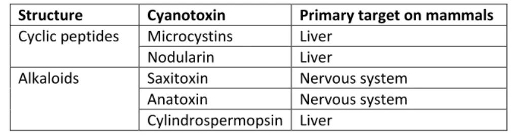
production can be employed for monitoring their presence in the environment as an early warning signal (Moreira et al., 2014), but that does not necessarily mean the cyanotoxin is being produced. The presence of the cyanotoxins in the environment should be complementarily quantified through chemical analytical or immunological methods. The table 1 summarizes the cyanotoxins screened for in this study:
Table 1. Summary of cyanotoxins screened in this study.
Structure Cyanotoxin Primary target on mammals Cyclic peptides Microcystins Liver
Nodularin Liver
Alkaloids Saxitoxin Nervous system Anatoxin Nervous system Cylindrospermopsin Liver
They also have shown to have toxic effects on other animals (e.g Puerto et al., 2011) and plants (Freitas et al., 2015).
1.5. Aim of the study
Subaerial cyanobacteria are an understudied group when compared to aquatic cyanobacteria, which inhabit in ecological conditions that can be considered extreme. This exploratory work was conducted with the aim to isolate, identify and asses their diversity of in the north of Portugal, by mean of a culture-dependent, polyphasic approach. At the same time, I aimed to evaluate the biotechnological potential of subaerial cyanobacteria by screening for PKS and NRPS, and to check their ability for
producing cyanotoxins. In both cases, a PCR-based approach was followed by using the DNA from the environmental samples (eDNA) and the isolates (gDNA).
2. Methodology:
2.1. Sampling
2.1.1. Sampling locations
Two areas in the north of Portugal were selected to collect the samples, both in close proximity to freshwaters bodies, less than 300 meters from a body of water. One was an urban park called “Parque da Cidade” located in Porto and the other was Portugal’s Hydrographic Region 2 (RH2- Região Hidrográfica 2 in Portuguese), both locations situated in the north of Portugal. Portugal’s climate in the north is classified as Csb according to Kottek et al. (2006), meaning it has a warm temperate climate with a dry and warm summer.
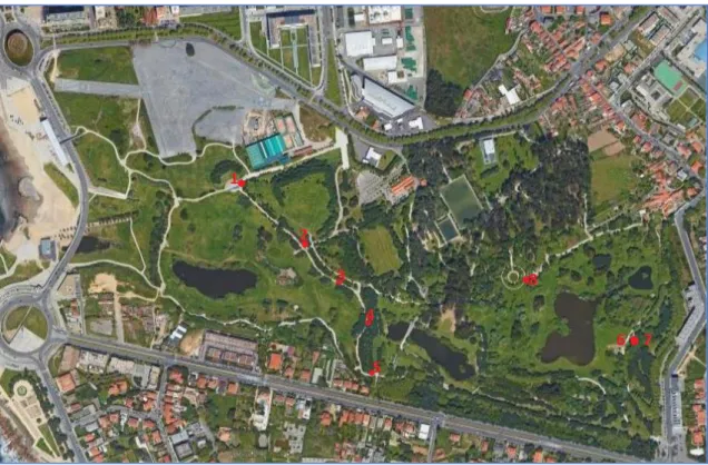
“Parque da Cidade” has an area of 83 hectares and is located near the coast in the north of Portugal, it has 3 lakes connected via underground pipes and their water is used to feed the irrigation system of the park. The samples were collected from several surfaces along the pathways throughout the park and in proximity to the 3 lakes. “Parque da Cidade” is located just a few kilometers south of the Leça river, whose hydrographic basin incorporates the RH2 region.
The figure 1 shows the satellite aerial view of the park with the sampling points marked on it.
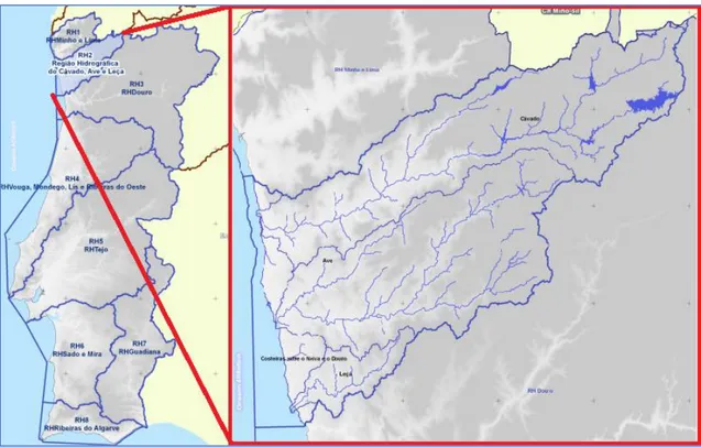
The RH2 region has an area of 3 400 km2 and comprises mainly of 3 hydrographic
sub-basins of 3 rivers and their affluents, they are the Cávado, Ave and Leça sub-basins, the region also comprises of the basins of smaller coastal streams along the coast of the region. The samples were collected on several surfaces but always in close proximity to rivers. The figure 2 shows the area and delimitation of the RH2 region.
Figure 2. Hydrographic region 2 sampling area.
2.1.2. Sample retrieval
The samples were collected on the 4th of January 2018, during winter, in “Parque
da Cidade” where 8 samples were retrieved, and in the RH2 region on the days 21st
March and 4th of April 2018, when 11 and 9 samples were retrieved, respectively. The
samplings were performed during an atypical winter/early spring season, when precipitation in the north of Portugal has hit historical records. Overall, 28 samples were collected in both sampling locations. The samples were collected during a seasonal period of the year which has the highest annual precipitation and the lowest annual temperatures of the year. The sampling days, especially on the 4th of January, were rainy
with only mild raining on the days 21 of March and 4 of April, as such the majority of the surfaces chosen to collect the samples were well wet which facilitated the sampling process.
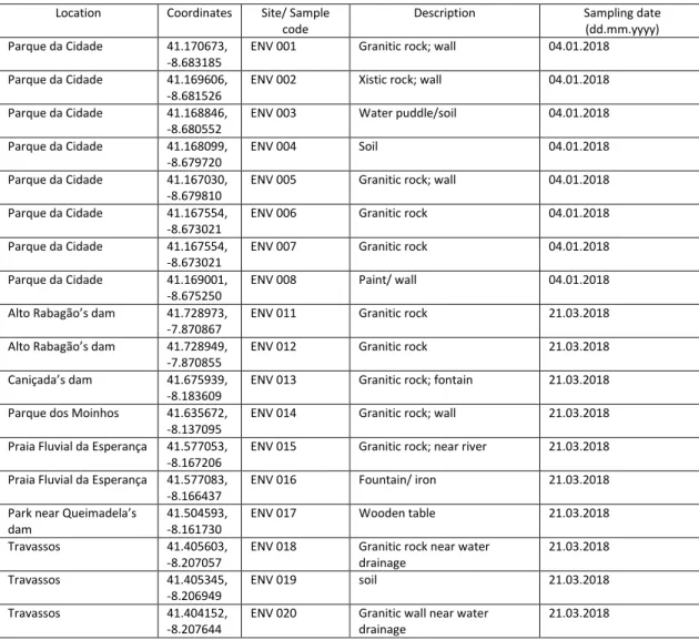
The samples were collected from several types of surfaces and ranged from several types of microbial communities, such as lithophilous communities inhabiting in rocks and soil (wet or near puddles) or inhabiting in cement or paint from man-made infrastructures, epixylous communities in wooden tables in a park, corticolous communities on the bark of a tree and epimetallous communities on the top of an iron faucet with continuously running water near a fountain, but the majority were lithophilous communities collected from granitic rocks either in nature or used in man-made infrastructures, and according to their relative humidity they can be classified as mesic, but mostly during winter because of higher rates of precipitation, nevertheless the samples collected were well wet and had a moist texture (with the exception of samples ENV001 and ENV002 that had a drier texture than the rest). The table 2 describes the characteristics of each sampling location. Some of those locations were chosen as sampling locations because they are frequently used by people. Indeed, the sampling sites include parks and freshwater beaches (for example, the park tables and a water fountain have been sampled).
Table 2. Description and coordinates of sampling locations. Location Coordinates Site/ Sample
code
Description Sampling date (dd.mm.yyyy) Parque da Cidade 41.170673,
-8.683185
ENV 001 Granitic rock; wall 04.01.2018 Parque da Cidade 41.169606,
-8.681526
ENV 002 Xistic rock; wall 04.01.2018 Parque da Cidade 41.168846,
-8.680552
ENV 003 Water puddle/soil 04.01.2018 Parque da Cidade 41.168099,
-8.679720
ENV 004 Soil 04.01.2018
Parque da Cidade 41.167030, -8.679810
ENV 005 Granitic rock; wall 04.01.2018 Parque da Cidade 41.167554,
-8.673021
ENV 006 Granitic rock 04.01.2018 Parque da Cidade 41.167554,
-8.673021
ENV 007 Granitic rock 04.01.2018 Parque da Cidade 41.169001,
-8.675250
ENV 008 Paint/ wall 04.01.2018 Alto Rabagão’s dam 41.728973,
-7.870867
ENV 011 Granitic rock 21.03.2018 Alto Rabagão’s dam 41.728949,
-7.870855
ENV 012 Granitic rock 21.03.2018 Caniçada’s dam 41.675939,
-8.183609
ENV 013 Granitic rock; fontain 21.03.2018 Parque dos Moinhos 41.635672,
-8.137095
ENV 014 Granitic rock; wall 21.03.2018 Praia Fluvial da Esperança 41.577053,
-8.167206
ENV 015 Granitic rock; near river 21.03.2018 Praia Fluvial da Esperança 41.577083,
-8.166437
ENV 016 Fountain/ iron 21.03.2018 Park near Queimadela’s
dam
41.504593, -8.161730
ENV 017 Wooden table 21.03.2018 Travassos 41.405603,
-8.207057
ENV 018 Granitic rock near water drainage 21.03.2018 Travassos 41.405345, -8.206949 ENV 019 soil 21.03.2018 Travassos 41.404152, -8.207644
ENV 020 Granitic wall near water drainage
Park near Queimadela’s dam 41.504693, -8.161734 ENV 035 soil 21.03.2018 Barcelos 41.527236, -8.622714
ENV 049 Granitic rock; very close to river 04.04.2018 Barcelos 41.527064,
-8.622775
ENV 050 Granitic rock; stairs 04.04.2018 Penide’s dam 41.550096,
-8.538160
ENV 051 Granitic rock; wall 04.04.2018 Praia Fluvial de Merelim 41.593770,
-8.464680
ENV 052 Tree (Quercus) 04.04.2018 Praia Fluvial dos Moinhos 41.654854,
-8.399625
ENV 053 Wall; cement 04.04.2018 Praia Fluvial da Navarra 41.613448,
-8.385260
ENV 054 Granitic rock; inside windmill 04.04.2018 Praia Fluvial da Navarra 41.613448,
-8.385260
ENV 055 Granitic rock; wall inside windmill
04.04.2018 Park near river Ave 41.529815,
-8.273271
ENV 056 Granitic rock; tank/fountain 04.04.2018 Santo Tirso’s park 41.355048,
-8.457896
ENV 057 Cement; ground 04.04.2018
2.1.3. Sample collection process
The itinerary plan was made in Google Maps and GPS coordinates were used to reach each chosen sampling location.
The samples were collected with a stainless steel spatula or knife which was used to scrape the cyanobacterial mats off of rocks into a 50mL Falcon tube, and whenever necessary a Pasteur pipette was used to help collect the mats from more humid surfaces that would sometimes be partially submerged in small puddles by collecting small cyanobacterial mat portions that were scraped off into the more wet parts of the surface. After collecting a sample, the Falcon tube was properly labeled with a code which served to identify the location from where each sample came from and a correspondence was made between each code and sample location characteristics, then the falcon tube was temporarily stored in a thermal box for transportation to the lab.
During the sample collection procedure, disposable nitrile gloves were used and the collection materials (spatula and knife) were always sterilized with alcohol (ethanol at 70%) and cleaned before and after each sample collection, all in an effort to avoid cross-contamination between samples.
One of the samples was lyophilized, after it was observed in the microscope it had a big community of cyanobacteria, to allow in the future to look for substances with potential biotechnology uses. That particular sample (ENV55) was collected inside an abandoned water mill, on the margin of river Cávado.
2.2. Sample processing: culturing and isolation
At the laboratory, the sample processing procedure was performed in aseptic conditions. It consisted in distributing a small portion of biomass from each collected
environmental sample in 4 types of media (solid Z8,solid BG110, liquid Z8 and BG110)
and in two 1,5µL Eppendorf tubes, one for the microscopic observation of the environmental samples and the other for extracting the sample environmental DNA (eDNA) which was used for molecular screening. The rest of the environmental biomass was left inside the falcon tube and stored at a temperature of -20C° as backup.
The raw environmental samples were observed through light microscopy using a Leica DMLB microscope (Wetzlar, Germany) to check for cyanobacterial presence and their predominance in each sample. Each sample was cultured in two liquid enrichment media, BG110 (Andersen, 2005) and Z8 (Kotai, 1972), and were allowed to grow freely,
some of them were attempted to isolate via micromanipulation at a later date. The solid BG110 and Z8 mediums in petri dish agar plates, with an agarose concentration of 1.2%,
were the primary method used to isolate the environmental samples. All themedia for the raw environmental samples were prepared with cycloheximide, at a concentration of 0.025%, to prevent the growth of eukaryotic microorganisms.
To isolate in solid media a bit of biomass from the environmental sample were placed and spread along an agar plate using a streaking technique, that consisted in making a series of strokes in the agar plate in which the last series of strokes had a more diluted concentration of microbial biomass than the first series. Strokes were made with the help of an inoculation loop that was sterilized in an infrared loop sterilizer after each series of strokes were made. Eventually, isolated colonies appeared in the more diluted strokes and were picked up with the help of a disposable surgeon’s blade or an inoculation loop to be placed in a new agar plate containing the same medium they were originally picked up from, and the process was repeated if deemed necessary. When two isolated colonies with a different macroscopic appearance appeared on the same agar plate, they were picked up and placed into two new separate agar plates. Colonies though to be isolated were picked up from the agar plate and inoculated in liquid media inside an Erlenmeyer flask where they were allowed to grow, after further confirmation of a monoculture in the flask, an aliquot was collected for extracting the genomic DNA (gDNA) of the isolated strain. All cultures were kept in LEGE’s isolation room at a temperature of 20°C, under artificial light with a period of 14h light/10h dark and a light intensity of (12 mol photons m−2 s −1). Strain isolates will be deposited at LEGE Culture Collection at CIIMAR (Porto, Portugal).
2.3. Morphological observation
After isolation, the strains were observed and characterized according to their morphotypes using a Leica DMLB microscope (Leica Microsystems GmbH, Wetzlar, Germany), and their microphotographs were captured with a Leica ICCA Camera
System at magnifications of 400x and 1000x, using the Qwin Leica software (Leica Microsystems GmbH) the microphotographs were properly processed. Morphometric characteristics of each strain were measured directly of the microscopic preparation (using an aliquot from the liquid culture medium) at a magnification of 1000x, length and width or diameter were the characters measured 20 times in different individuals of each strain.
2.4. Molecular methods 2.4.1. DNA extraction
Two methodologies were used to extract DNA. For eDNA extraction of raw environmental samples retrieved directly from the field, the DNeasy powersoil kit (QIAGEN, Netherlands) commercial kit was used due to being more suitable for the DNA extraction of mucilaginous subaerial cyanobacteria samples which are soil-like samples that may contain a lot of sediment and various kinds of debris. The extraction procedure was followed accordingly to the protocol provided by the manufacturer of the kit. For instance, the Vortex Genie 2 (MoBio laboratories, USA) was used and set at maximum vortex speed for a period of 10-15 minutes to properly mix and prepare the environmental samples for extraction.
For the gDNA extraction of both the isolated strains and the strains from LEGEcc (serving as positive controls for the molecular screenings), their respective cyanobacterial biomass, collected from the cultures was harvested by centrifugation. The biomass would be centrifuged at 10000 X g for 10 minutes and if after that a pellet was not observed the G-force would be increased to 16000 X g and the biomass was centrifuged again 10 minutes. After a pellet formed in the 1.5µL Eppendorf tube the liquid medium present in it would be removed and discarded with the aid of a micropipette, replacing it with ddH20 water. The biomass was then stored at -20°C for the DNA
extraction to be performed at a later date. After the cells were harvested, the DNA extraction of the isolated strains and of the positive controls strains was performed using the Purelink Genomic DNA Mini Kit (Invitrogen, USA), and the protocol for extracting DNA from gram-negative bacterial cell was followed according to the manufacturer instructions.
Finally, all the extracted DNA was stored in 1,5µL Eppendorf tubes in a freezer at a temperature of -20°C.
After completing the DNA extraction procedure an electrophoresis was always performed afterward to confirm that the DNA was indeed successfully extracted. Agarose gel (Ultrapuretm Agarose, Invitrogen, USA) at 1% concentration was prepared using a
Tris-Acetate EDTA buffer solution (TAE Ultrapuretm, Invitrogen, USA) at 1x (40mM
Tris-acetate and 1mM EDTA), to stain the gel 2µL of SYBRsafe (Invitrogen, USA) was used. Five microliters of extracted DNA mixed with 0.5µL of loading buffer was loaded into the gel, 1µL of molecular marker (1Kb Plus DNA Ladder) was loaded. The gel ran at a voltage of 90V during 45 minutes and was visualized and photographed in the transilluminator Molecular Imager® GEL DOCTM with the software Image LabTM(USA).
2.4.2. PCR screening
a) Primers:
To check if the isolated strains and environmental samples had cyanobacteria with the potential to produce secondary metabolites, several genes were targeted and amplified using specific primers through the method of Polymerase Chain Reaction (PCR) with the aim to screen for their presence. Most of the genes targeted belong to gene clusters related to the production of cyanotoxins (toxicity potential screening), and others were genes from the NRPS and PKS gene clusters that are responsible for encoding non-ribosomal peptides and polyketide peptides that are able to produce potential bioactive metabolites (biotechnological potential screening). Prior to all other screenings, the isolates and environmental samples were also screened for the presence of the 16S gene by using the cyanobacterial group-specific primer set CYA106F/CYA781R (Nübel et al., 1996). This amplification was used to check if there was any cyanobacterial DNA in the environmental samples and isolates in order to validate the extraction.
The primers used are listed in table 3 along with their target genes and their target groups. The primers used for the toxicity screening were the PKDF/PKDR (Ouahid et al. 2005), the HEPF/HEPR primers which were used to target a domain that is located in both the mcyE and ndaF genes to detect potential microcystin and nodularin producing strains (Jungblut and Neilan, 2006), the SxtI682F/sxtI877R (Lopes et al., 2012), the anaC-genF/anaC-genR (Rantala-Ylien, 2011), the cylnamR/ cylnamR (Mihali et al. 2008) and the CYTLATF/CYTLATR primers which were used to target the amidinotransferase (AMT) gene whose presence in a cyanotoxin gene cluster is unique to Cylindrospermopsin producing cyanobacteria (Kellman et al., 2006). The primers used for the assessment of biotechnological potential were the DKF/DKR and MTF2/MTR targeting the PKS and NRPS genes, respectively (Moffit et al. 2001; Neilan et al. 1999).
Table 3. Target genes and their respective primers, target groups, primer sequences, amplified fragment size and positive controls.
Target gene
Primer pair Target group Primer sequence (5’_3’) size (bp)
Positive control References
mcyD PKDF1; PKDR1 microcystin producers GACGCTCAAATGATGAAAC GCAACCGATAAAAACTCCC 657 Microcystis aeruginosa LEGE 91339 Ouahid et al. 2005 mcyE / ndaF HEPF; HEPR Microcystin and nodularin producers TTTGGGGTTAACTTTTTTGGGCATAGTC AATTCTTGAGGCTGTAAATCGGGTTT 472 Microcystis aeruginosa LEGE 91339 Jungblut and Neilan, 2006 sxtI SxtI682F; sxtI877R
Saxitoxin producers GGATCTCAAAGAAGATGGCA GCCAAACGCAGTACCACTT 195 Aphanizomenon gracile LMECYA40 Lopes et al., 2012 anaC anaC-genF; anaC-genR
Anatoxin producers TCTGGTATTCAGTCCCCTCTAT CCCAATAGCCTGTCATCAA 366 Anabaena sp. LEGE X-002 Rantala-Ylien, 2011 cyrJ cynsulF; cylnamR Cylindrospermopsin producers ACTTCTCTCCTTTCCCTATC GAGTGAAAATGCGTAGAACTTG 586 Cylindrospermopsis raciborskii LEGE 97047 Mihali et al. 2008 AMT CYTLATF; CYTLATR Cylindrospermopsin producers ATTGTAAATAGCTGGAATGAGTGG TTAGGGAAGTAATCTTCACAG 1105 Cylindrospermopsis raciborskii LEGE 97047 Kellman et al., 2006 PKS DKF; DKR Polyketide producers GTGCCGGTNCC(A/G)TGNG(T/C)(T/C)TC GCGATGGA(T/C)CCNCA(A/G)CA(A/G)(C/A)G 650-700 Microcystis aeruginosa LEGE 91339 Moffit et al. 2001 NRPS MTF2; MTR Non-ribosomal peptide producers GCNGG(C/T)GG(C/T)GCNTA(C/T)GTNCC CCNCG(AGT)AT(TC)TTNAC(T/C)TG ~1000 Microcystis aeruginosa LEGE 91339 Neilan et al. 1999 16S CYA106F; CYA781R Cyanobacteria, plastids
CGG ACG GGT GAG TAA CGC GTG A GAC TAC TGG GGT ATC TAA TCC CAT T
675 Microcystis aeruginosa LEGE 91339 Nübel et al. 1996 b) PCR amplification:
Each pair of primers listed in table 3 were used in the PCR reactions, the components and concentrations per reaction of 20 µL were: 1x GoTaq buffer, 2.5 mM for MgCl2, 1 µM for each primer, 0.5 mM for the dNTP mix, 0.5 U for GoTaqR Flexi DNA
polymerase, the final volume of each reaction was 20 µL and 1 µL of that volume was the DNA template. The volumes per reaction are listed in table 4.
Table 4. Mastermix preparation volumes for each component of the PCR reaction.
Components Volume per reaction
(1x) Molecular biology water 7.9
5x Buffer 4 µL MgCl2 2 µL Forward primer 2 µL Reverse primer 2 µL Deoxynucleotides (dNTP’s) 2 µL Taq Polymerase 0.1 DNA template 1 µL Total: 20 µL
The PCR reactions were made using a Biometra T-Professional Standard Gradient Thermocycler (Germany) and the PCR conditions for each pair of primers are described in table 5.
Table 5. PCR conditions for each primer set.
Primer pair PCR reaction References
Initial Denaturation PCR cycles Final extension
PKDF1; PKDR1 94 °C 5 min 35 cycles 72 °C 7 min Ouahid et al. 2005 95 °C 60 s 54 °C 30 s 72 °C 1 min HEPF; HEPR 92 °C 2 min 35 cycles 72 °C 5 min
Jungblut and Neilan, 2006 92 °C 20 s 52 °C 30 s 72 °C 1 min SxtI682F; sxtI877R 94 °C 3 min 35 cycles 72 °C 7 min Lopes et al., 2012 94 °C 10 s 52 °C 20 s 72 °C 1 min anaC-genF; anaC-genR 94 °C 2 min 25 cycles 72 °C 5 min Rantala-Ylien 2011 94 °C 30 s 50-60°C 30 s 72 °C 30 s cynsulF; cylnamR 94 °C 3 min 30 cycles 72 °C 7 min Mihali et al. 2008 94 °C 10 s 55-65°C 20 s 72 °C 1-3min CYTLATF; CYTLATR 94 °C 3 min 30 cycles 72 °C 7 min Kellman et al., 2006 94 °C 10 s 50-55°C 20 s 72 °C 1 min DKF; DKR 94 °C 2 min 30 cycles 72 °C 7 min Moffit et al. 2001 94 °C 5 s 65 °C 10 s 72 °C 20 s MTF2; MTR 94 °C 2 min 35 cycles 72 °C 7 min Neilan et al. 1999 93 °C 10 s 51 °C 20 s 72 °C 1 min CYA106F; CYA781R 94 °C 5 min 35 cycles 72 °C 7 min Nübel et al. 1996 94 °C 1 min 60 °C 1 min 72 °C 1 min
After PCR amplification, all PCR products would be analyzed and visualized via electrophoresis in an agarose gel ((Ultrapuretm Agarose, Invitrogen, USA) at a
concentration of 1.5% which was stained with 2µL of SYBRsafe (Invitrogen, USA). The selected voltage and running time was 90V and 45 minutes. The agarose gel was visualized and photographed in the transilluminator Molecular Imager® GEL DOCTM with
the software Image LabTM(USA), and the presence or absence of the amplified target
gene was observed for each environmental sample and for each isolate by looking at the position of the positive controls in the gel and by using the molecular markers to check if a fragment was present (or absent) in the expected position according to its amplified molecular size.
The strains used as positive controls for the molecular screenings were retrieved from the LEGE Culture Collection. They have been previously confirmed by sequencing to have the target genes, making them suitable to be used as positive controls.
2.4.3. Sequencing
Environmental samples that exhibited positive PCR results (i.e presence of the gene) for each cyanotoxin target gene were selected and sequenced.
The 16S rRNA gene was amplified and sequenced for all the isolates obtained, in order to identify the cyanobacteria and thus assess the diversity of the samples.
In this case, for sequencing purposes, the PCR preparation involved triplicating the number of reactions for each environmental sample (or isolate), in order to have enough amplified product to enable sequencing. So the final volume of PCR product per sample (and isolate) to be loaded into the 1.5% agarose gel was 60 µL. The primers and PCR conditions used were the same used for the screening and are listed in table 5. The electrophoresis ran at 90V for 60 minutes, then the amplified fragments were observed in the CSMICRODOC system (Cleaver scientific, UK) transilluminator coupled with a Canon PowerShot G9 camera. Then, bands with the expected size were excised from the gel and collected to be purified using the Nztech - genes & enzymes (NZYGelpure, Portugal) purification kit, following the manufacturer’s instructions. The check the efficacy of the DNA purification, an electrophoresis (90V; 45 minutes) ran in a 1% agarose gel, and the purified DNA was mixed with loading buffer, corresponding to a tenth of the total loaded DNA, with the aid of a micropipette. All the purified PCR products and the respetive pair of primers were sent to GATC Biotech (Germany) to be sequenced. 2.5. Sequence analysis
The forward and reverse sequences (i.e. 5’ and 3’) obtained from the same PCR product were examined in the bioinformatic software Geneious (v.8) and were assembled together (de novo assembly), their chromatograms were analyzed to check the quality of the sequences and to determine if further sequences were needed to form a consensus sequence (i.g. if the quality was bad on either one of the forward or reverse sequences). The sequences were usually trimmed at the extremities due to bad quality. Then, the consensus sequences of each strain isolate were compared with the sequences in the GenBank® database using the BLAST®n (Basic Local Alignment
Search Tool for nucleotides) tool available in the NCBI (National Center for Biotechnology Information) and compared with other cyanobacterial sequences in the GenBank® database to check for similarities and to help in their identification.
2.6. Phylogenetic analyses
To assess the relative position of our isolated strains relatively to each other and other reference strains a phylogenetic 16S rRNA gene-based tree was built based on the Maximum Likelihood method using the software MEGA7 (Molecular Evolutionary
Genetics Analysis Version 7.0). First, all the sequences were aligned using the algorithm ClustalW (Kumar et al, 2016) and then visually inspected. This multiple sequences alignment consisted of (1) the 16S rRNA consensus sequences of each strain isolate obtained in this study; (2) the best hit sequences of each isolates’ sequence obtained in this study (the isolates sequences were previously compared with the sequences in the GenBank® database using the Blast® tool; if the best hit was an unidentified cyanobacterium then a second sequence belonging to the closest identified cyanobacteria was also retrieved and used in the construction of the tree); and (3) 11 reference strains collected from LEGE’s Cyanotype database and from GenBank, in order to obtain a reliable representation of the diversity of the cyanobacteria. The model of substitution has been chosen according to the AICc criteria. Thus, the phylogenetic tree was built using the model GTR+G+I and with all the positions with a coverage of less than 97% site coverage removed. The labels in the phylogenetic tree were edited using the Inkscape software (V. 0.92; free software).
3. Results and Discussion
Although isolations attempts through micromanipulation were made using the liquid culture mediums containing the environmental samples those did not yield any success due to the fact that the strains exhibited poor growth in the liquid enrichment. Due to time constraints and deadlines the idea of any further attempts at micromanipulation was abandoned. So both the Z8 and BG110 liquid mediums holding
each sample served only as a sort of a backup in laboratory and the raw sample was allowed to grow, this most likely changed the community and the relative amount of certain strains of cyanobacteria in the sample because some strains are more capable of growing in those mediums than others, such is the case of opportunistic strains that can easily dominate in these mediums. Instead, all of the strains were isolated through solid media on agar plates.
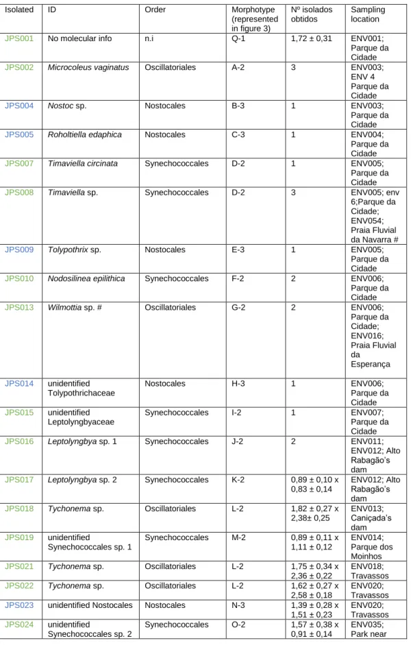
Following Komarek and co-authors’ (2014) criteria for identification of cyanobacteria, all isolates were properly identified using a polyphasic approach based mainly on molecular methods (amplification and sequencing of the 16S rRNA gene) combined with the strain morphological characteristics, 7 isolates could be identified to the taxonomic level of species, 13 isolated were identified to their genus and 5 were identified to their order. A few exceptions happened:
• For the isolate JPS1 no molecular data has been obtained due to the lack of biomass available in the liquid culture to properly perform a DNA extraction, so
that isolate was not identified. Only its morphological characteristics were able to be determined.
• For the isolates JPS13 and JPS26, it was only possible to obtain 1 sequence out of each one, corresponding for both strains to the reverse sequence (primer CYA785R). Blasting those sequences and comparing them with those in GenBank database along with their microscopical observation and characterization would allow us to conclude that they most likely belong to the genera Wilmottia and Timaviella, respectively. For all the other isolated strains I have obtained 2 or more sequences that were assembled together to form a consensus sequence that was used to build the phylogenetic tree. Since there was only 1 sequence for the isolates JPS13 and JPS26 it was not possible to produce a consensus sequence, consequently they were not included in the phylogenetic tree.
3.1. Morphological characterization and strain identification
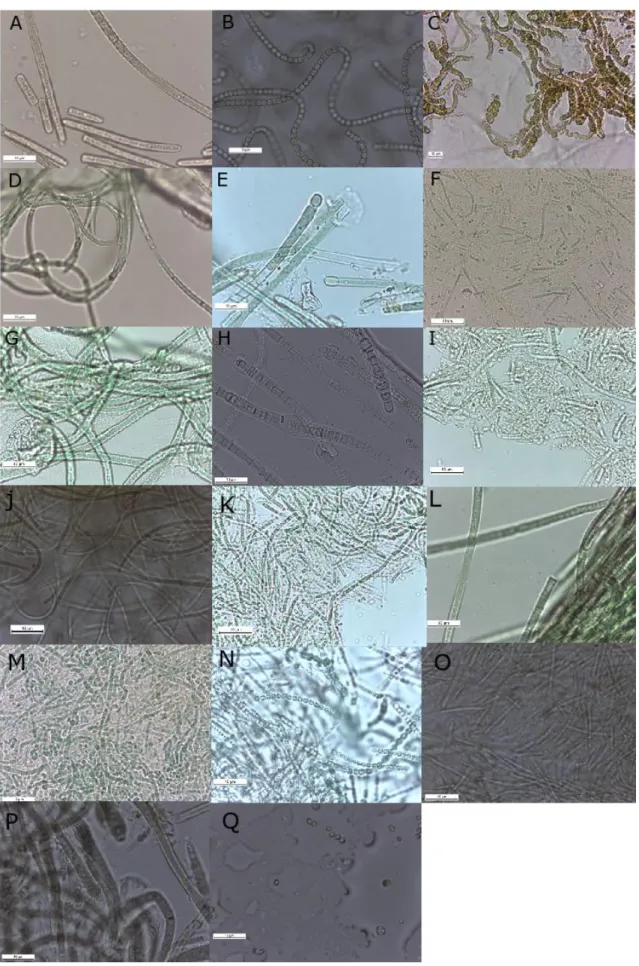
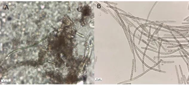
A total of 26 strains were isolated, characterized according to their morphological features (morphotypes) and their morphometric characteristics (i.e cell diameter or cell length and width) were measured under light microscopy. Morphological observations revealed 17 different cyanobacterial morphotypes (figure 3) and three main types of morphologies were distinguished as well, the filamentous cyanobacteria with heterocysts, filamentous cyanobacteria without heterocysts (non-heterocytous) and unicellular cyanobacteria (table 6).
Figure 3. Diversity of different morphotypes among strains; (image C at 400x magnification, all others at 1000x); A-
Microcoleus sp.; B- Nostoc sp.; C- Roholtiella edaphica; D- Timaviella sp.; E- Tolypothrix sp.; F- Nodosilinea epilithica;
G- Wilmottia sp.; H- Tolypothrichaceae; I- Leptolyngbyaceae; J- Leptolyngbya sp. 1; K- Leptolyngbya sp. 2; L-
; Q- unidentified unicellular strain; Scale Bar= 10 µm.
Table 6. Isolated strains, their respective identification (Komarek et al., 2014) with each correspondent morphotypes represented in figure 3 and their classification according to: 1- Unicellular, 2- heterocytous filamentous 3- non-heterocytous filamentous; the colors indicate: green- strains isolated using Z8 medium; blue- Strains isolated using BG110 medium.
Isolated ID Order Morphotype (represented in figure 3) Nº isolados obtidos Sampling location
JPS001 No molecular info n.i Q-1 1,72 ± 0,31 ENV001; Parque da Cidade
JPS002 Microcoleus vaginatus Oscillatoriales A-2 3 ENV003; ENV 4 Parque da Cidade
JPS004 Nostoc sp. Nostocales B-3 1 ENV003;
Parque da Cidade
JPS005 Roholtiella edaphica Nostocales C-3 1 ENV004;
Parque da Cidade
JPS007 Timaviella circinata Synechococcales D-2 1 ENV005; Parque da Cidade
JPS008 Timaviella sp. Synechococcales D-2 3 ENV005; env 6;Parque da Cidade; ENV054; Praia Fluvial da Navarra #
JPS009 Tolypothrix sp. Nostocales E-3 1 ENV005;
Parque da Cidade
JPS010 Nodosilinea epilithica Synechococcales F-2 2 ENV006; Parque da Cidade
JPS013 Wilmottia sp. # Oscillatoriales G-2 2 ENV006; Parque da Cidade; ENV016; Praia Fluvial da Esperança JPS014 unidentified Tolypothrichaceae Nostocales H-3 1 ENV006; Parque da Cidade JPS015 unidentified Leptolyngbyaceae
Synechococcales I-2 1 ENV007; Parque da Cidade
JPS016 Leptolyngbya sp. 1 Synechococcales J-2 2 ENV011; ENV012; Alto Rabagão’s dam JPS017 Leptolyngbya sp. 2 Synechococcales K-2 0,89 ± 0,10 x 0,83 ± 0,14 ENV012; Alto Rabagão’s dam JPS018 Tychonema sp. Oscillatoriales L-2 1,82 ± 0,27 x 2,38± 0,25 ENV013; Caniçada’s dam JPS019 unidentified Synechococcales sp. 1 Synechococcales M-2 0,89 ± 0,11 x 1,11 ± 0,12 ENV014; Parque dos Moinhos JPS021 Tychonema sp. Oscillatoriales L-2 1,75 ± 0,34 x 2,36 ± 0,22 ENV018; Travassos JPS022 Tychonema sp. Oscillatoriales L-2 1,62 ± 0,27 x 2,58 ± 0,18 ENV020; Travassos
JPS023 unidentified Nostocales Nostocales N-3 1,39 ± 0,28 x
1,51 ± 0,23 ENV020; Travassos JPS024 unidentified Synechococcales sp. 2 Synechococcales O-2 1,57 ± 0,38 x 0,91 ± 0,14 ENV035; Park near
Queimadela’s dam JPS025 Macrochaete sp. Nostocales P-3 2,82 ± 0,41 x 2,29 ± 0,32 ENV049; Barcelos # based on a singleton only
It was observed during the identification process, when comparing the sequences of the isolates from this study with their more similar sequences from GenBank, that most of the obtained strains are similar to other terrestrial strains from other studies that examined cyanobacterial diversity in extreme habitats. For example, our JPS002 and JPS003 isolates, which are very similar to each other, both revealed to be have high similarity with an uncultured cyanobacterium (acc.nbr: KC463588) (figure 6. and Annex A) from soil crust from a study conducted in south of Africa (Dojani et al., 2015), JPS007 and JPS008 show a 99% similarity with an uncultured cyanobacterium (acc.nbr: HQ188993) in the dry valleys of the high Himalayas and Antarctica (Schmidt et al., 2011) 3.2. Isolation and diversity analysis
Figure 4. Number of strains isolated per -order level.
In total 26 strains were isolated from both the urban park “Parque da Cidade” and from the hydrographic region “RH2” both situated in the north of Portugal. From these, 15 of the isolates obtained have an origin in “Parque da Cidade” and 11 have an origin in the “RH2” region. The figure 4 shows the number of strains obtained per -order:
Eleven strains belong to the order Synechococcales, which is an order that can have over 70 genera with unicellular (including colonial forms) and filamentous types, and it is a group that is not defined as monophyletic (Komarek et al., 2014).
Synechococcales, 11 Oscillatoriales; 8 Nostocales; 6 No molecular Info, 1
Eight strains isolated belonged to the Oscillatoriales order, which is a group that has the morphological characteristics of not having true-branching, heterocysts or akinetes, and with cells shorter than wide.
Six strains belonged to the order Nostocales, which is an order of filamentous cyanobacteria that can have very diversified thalli and have specialized cells such as heterocysts and akinetes (Komarek et al., 2014). The heterocysts are responsible for fixating atmospheric nitrogen, which means that in environments deprived of nitrogen, for example in the BG110 medium, the cyanobacteria that possess those cells have an
advantage over other strains are not able to fix N2. In that sense, in such conditions
heterocytous cyanobacteria are able to outcompete them and outgrow other colonies that might have formed in other conditions (Pentecost & Whitton, 2012).
Of the 26 isolated strains, 21 were isolated using the Z8 solid medium while 5 were isolated using the BG110 solid medium. All of the strains isolated using the BG110
solid medium belonged to the Nostocales order, something that was to be expected because it is in accordance with the fact that they fix dinitrogen. Although there are some unicellular or non-heterocytous cyanobacteria that are also capable to fix N2 (Berrendero
et al., 2016), none was isolated in this work. The low number of strains successfully isolated by using the BG110 solid medium, in comparison with the number of strains
isolated from the Z8 solid medium, can be attributed to their slow growth and colony formation in that media, fact that was observed during the isolation process. Sometimes no growth would occur at all, or the growth would be so minimal that no isolated colonies would appear, thus not allowing for the isolation process to proceed for those particular plates. In conclusion, the strains in the Z8 medium grew faster and yielded more isolates (mostly from the Synechococcales and Oscillatoriales order, only 1 Nostocales strain) than in the BG110 medium. However, as said, with BG110 it was possible to obtain new
diversity that was not possible with Z8.
It is to note that while the most predominant strain of cyanobacteria present in the environmental sample – and that were observed under the microscope would possibly be isolated, this was not certain due to the ubiquity and opportunistic behavior of certain strains. This is a major point that possibly determined the diversity of the isolates obtained, which was performed following a culture-dependent approach. Still, in most cases the predominant strain in the environmental sample would be the one isolated. For example, two Microcoleus vaginatus strains were isolated from the sample ENV003, whichwas dominated by Microcoleus strains (figure 5), although during the isolation process two isolated colonies with different macroscopic characteristics appeared in the agar plate (at the time assumed to be different strains). They were
separated into two Petri dishes in order to be isolated: both yielded the same strain belonging to the genus that dominated the environmental sample.
Figure 5. A- ENV003 environmental sample showing Microcoleus spp. B- Microscopic preparation of biomass collected from the ENV003 agar plate, during the later stages of isolation, showing the Microcoleus vaginatus strain. Scale bar= 10 µm.
Almost all the isolated strains are filamentous cyanobacteria, which are capable of producing an exopolysaccharide (EPS) matrix that promotes the stabilization of the mats and helps maintain favorable conditions (preventing the loss of water) that allow colonization of other microorganisms (Mager & Thomas, 2011). Filamentous strains similar to Microcoleus traditionally recognized as EPS producers, are the group of organisms to first colonize a dry terrestrial-like environment (Garcia-Pichel & Wojciechowski, 2009). Only one non-filamentous unicellular strain strain was isolated (I was not able to identify it).
A higher number of non-heterocystous strains were isolated from the Z8 but this might be due to the faster growth occurring in this medium, which seemed to favor the growth of non-heterocystous strains. Still, 6 heterocystous strains were isolated, and although they were less prominent than the non-heterocystous strains they are known to play an important role as nitrogen contributors in terrestrial environments (e.g biocrusts) by fixing dinitrogen (Belnap, 2002).
The characteristics of a terrestrial surface (e.g texture, slope, chemical composition) may determine the cyanobacteria strain or groups that are able to attach on it, successfully colonize and dominate it (Pentecost & Whitton, 2012). The table 6 gives an insight into the type of surface and the strain isolated, and it is noted that all the strains belonging to the genus Timaviella (figure 3-D) were isolated from a biocrust on top of granitic surfaces, from two distanced locations correspondent to sites ENV005 and ENV006 (same location) and to ENV054.
3.3. Phylogenetic results
In order to verify the relative positioning of the isolates obtained, a Maximum likelihood (ML) tree was constructed with the sequences from the isolates, their Best Hits and some reference strains.
The phylogenetic tree consisted of 12 clusters (figure 6), which were defined by being monophyletic groups with a bootstrap value of 70% or higher and had to include at least 1 isolate strain or 1 reference strain. An exception was made to the Cluster D which had a bootstrap value lower than 50% but was defined as a cluster because it is a monophyletic group that includes the isolate strain JPS004 (Nostoc sp.) and a matching reference strain (Nostoc punctiforme), and also no other bootstrap values justified the definition of another clade, that would include either of these trains. For the most part, the species or genus attributed to the isolates match with the reference strains they are clustered with. However, some isolates are not clustered with any reference strain being only matched by another strain (Best hit) with a high bootstrap value support.
Clade A (Figure 6) comprises of 3 Tolypothrix strains and one Kryptousia strain, all belonging to the Tolypothrichaceae family, it includes: the isolate JPS009 that has been identified to the genus level only Tolypothrix sp. the Best hit strain Tolypothrix UAM 357 and 2 reference strains, Tolypothrix distorta ACOI 731 and the Kryptousia microlepis CENA343 strain, which is placed more distantly in relation the previous strains. The genus Kryptousia despite being morphologically similar to the Tolypothrix has been distinguished from it through molecular methods (Alvarenga et al., 2017). Still they are closely related, as it is shown in the phylogenetic tree (figure 6). The clade B includes the isolate obtained in this study (JPS014) – an unidentified Tolypothrichaceae and two Best Hit strains, an unidentified cyanobacteria (acc.nbr: KC463244) and the strain
Hassallia cf. pseudoramosissima ACSSI 158 (Tolypothrichaceae family), although no
reference strain is present. This clade is supported by a high bootstrap value of 99%. The clade C (figure 6) includes the isolated strain (JPS005) identified as Roholtiella
edaphica, its respective Best hit Roholtiella edaphica AR5 strain and the reference strain Roholtiella edaphica CCALA 1063. Clade D (figure 6) includes the isolated strain
(JPS004), the Best hit strains, Nostoc commune NTC and an unidentified cyanobacterium (acc.nbr: JX255093) and the reference strain Nostoc punctiforme PCC 73102. With a low bootstrap value (<50 %) this clade was only defined to illustrate the close proximity of the isolate JPS004 (Annex A) to the reference strain according to the topology of the tree, relatively to the other strains, and they have shown a 97% of similarity by blasting them. So, according to molecular data only we can say that they belong to the same genus (Kim et al., 2014; Yarza et al., 2014), but not the same species