Comparative Genomic Analysis of Two Novel
Sporadic Shiga Toxin-Producing
Escherichia
coli
O104:H4 Strains Isolated 2011 in
Germany
Erhard Tietze1*, Piotr Wojciech Dabrowski2, Rita Prager1, Aleksandar Radonic2, Angelika Fruth1, Philipp Auraß1, Andreas Nitsche2, Martin Mielke3, Antje Flieger1
1Department of Infectious Diseases, Division of Enteropathogenic Bacteria andLegionella, National Reference Centre forSalmonellaand other Bacterial Enteric Pathogens, Wernigerode Branch of Robert Koch-Institute, Berlin, Germany,2Centre for Biological Threats and Special Pathogens, Robert Koch-Institute, Berlin, Germany,3Department of Infectious Diseases, Robert Koch-Institute, Berlin, Germany
Abstract
A large outbreak of gastrointestinal disease occurred in 2011 in Germany which resulted in almost 4000 patients with acute gastroenteritis or hemorrhagic colitis, 855 cases of a hemo-lytic uremic syndrome and 53 deaths. The pathogen was an uncommon, multiresistant
Escherichia colistrain of serotype O104:H4 which expressed a Shiga toxin characteristic of
enterohemorrhagicE.coliand in addition virulence factors common to enteroaggregative E.coli. During post-epidemic surveillance of Shiga toxin-producingE.coli(STEC) all but
two of O104:H4 isolates were indistinguishable from the epidemic strain. Here we describe two novel STEC O104:H4 strains isolated in close spatiotemporal proximity to the outbreak which show a virulence gene panel, a Shiga toxin-mediated cytotoxicity towards Vero cells and aggregative adherence to Hep-2 cells comparable to the outbreak strain. They differ however both from the epidemic strain and from each other, by their antibiotic resistance phenotypes and some other features as determined by routine epidemiological subtyping methods. Whole genome sequencing of these two strains, of ten outbreak strain isolates originating from different time points of the outbreak and of one historical sporadic EHEC O104:H4 isolate was performed. Sequence analysis revealed a clear phylogenetic distance between the two variant strains and the outbreak strain finally identifying them as epidemio-logically unrelated isolates from sporadic cases. These findings add to the knowledge about this emerging pathogen, illustrating a certain diversity within the bacterial core genome as well as loss and gain of accessory elements. Our results do also support the view that dis-tinct new variants of STEC O104:H4 repeatedly might originate from yet unknown reser-voirs, rather than that there would be a continuous diversification of a single epidemic strain established and circulating in Germany after the large outbreak in 2011.
OPEN ACCESS
Citation:Tietze E, Dabrowski PW, Prager R, Radonic A, Fruth A, Auraß P, et al. (2015) Comparative Genomic Analysis of Two Novel Sporadic Shiga Toxin-ProducingEscherichia coli
O104:H4 Strains Isolated 2011 in Germany. PLoS ONE 10(4): e0122074. doi:10.1371/journal. pone.0122074
Academic Editor:Axel Cloeckaert, Institut National de la Recherche Agronomique, FRANCE
Received:November 27, 2014
Accepted:February 11, 2015
Published:April 2, 2015
Copyright:© 2015 Tietze et al. This is an open access article distributed under the terms of the
Creative Commons Attribution License, which permits unrestricted use, distribution, and reproduction in any medium, provided the original author and source are credited.
Introduction
During a large outbreak of gastrointestinal disease 2011 in Germany, almost 4000 people con-tracted acute gastroenteritis or hemorrhagic colitis, 855 cases developed a hemolytic uremic syndrome (HUS) and 53 patients died [1]. Simultaneously, a smaller outbreak occurred in France [2,3]. The pathogen in both of the outbreaks was an uncommon, multiresistant Shiga-toxinogenicEscherichia coli(STEC) strain of serotype O104:H4. PCR-based virulence gene analysis of the outbreak strain revealed a combined pattern of virulence properties inherent to two differentE.colipathovars [4–6]. The production of a Shiga toxin (Stx) is a signature prop-erty of enterohemorrhagicE.coli(EHEC). In addition this strain contained a plasmid which encodes the aggregative adherence-mediating fimbriae (AAF) characteristic of enteroaggrega-tiveE.coli(EAEC). Immediate extensive characterization of the outbreak strain by whole ge-nome sequencing confirmed its hybrid pathovar type [7–11]. Routine epidemiological typing at the German National Reference Center forSalmonellaand other Bacterial Enteric Pathogens (NRC) of hundreds of STEC O104:H4 isolates during the outbreak using pulsed-field gel elec-trophoresis (PFGE) after macrorestriction, plasmid profile analysis, virulence gene profiling and antibiotic susceptibility testing did not recognize any variation [12].
On July 26th2011 the German national public health authority, the Robert Koch Institute, declared the German outbreak to be over, dating the begin on May 5thand the end on July 4th with the peak on May 22nd[13]. During post-epidemic surveillance of STEC-caused disease by the German NRC, all but two of O104:H4 isolates remained indistinguishable from the epi-demic strain. Here we describe two novel post-epiepi-demic STEC O104:H4 strains isolated in close spatiotemporal proximity to the outbreak. These isolates differed both, from the epidemic strain and from each other by features as determined by the routine epidemiological typing methods to such an extent, that they were not considered to be isolates of the O104:H4 out-break strain but most likely isolates from sporadic cases. Whole genome sequencing of the two sporadic strains, of ten outbreak strain isolates originating from different cases and time points of the outbreak and of one historical sporadic EHEC O104:H4 isolate was performed. Analyses and comparison with other published sequences of EHEC O104:H4 outbreak and sporadic iso-lates clearly confirmed a phylogenetic relatedness but epidemiological distance between the sporadic strains and the outbreak strain.
Materials and Methods
Escherichia coli
O104:H4 strains
AllE.colistrains under investigation (Table 1) were isolated from enriched cultures of coliform bacteria and serotyped at the NRC. Isolates obtained during and shortly after the outbreak peri-od are from infections between May and August 2011. Strain 11–02027 is the index strain pro-vided for reference by the Robert Koch-Institute in May 2011. PCR-based virulence gene profiling revealed the strain Shiga toxin genestx1negative,stx2apositive, intimin geneeae
neg-ative, ABC-transporter protein geneaatApositive, master regulator gene of virulence-plasmid genesaggRpositive, secreted protein dispersin geneaappositive, AAF-fimbrial operon genes
aggA/type I andaggC/type I positive, enteroaggregativeE.coliheat-stable enterotoxin gene
astAnegative [5,12]. Nine more typical outbreak isolates from clinical cases (Table 1) which were indistinguishable from 11–02027 by routine epidemiological typing methods such as viru-lence gene profiling, plasmid profile analysis, antibiotic susceptibility testing and PFGE after macrorestriction were selected for whole genome sequencing. Isolates 11–06681 and 11–07153 attracted attention during post epidemic routine surveillance as they share the virulence gene profile with the outbreak strain but differed from the outbreak strain and from each other with
(11-06837), SAMN03168472 (11-07153), SAMN03174138 (01-09591)].
Funding:The funder provided support in the form of salaries for authors Erhard Tietze, Piotr Wojciech Dabrowski, Rita Prager, Aleksandar Radonic, Angelika Fruth, Philipp Auraß, Andreas Nitsche, Martin Mielke and Antje Flieger, but did not have any additional role in the study design, data collection and analysis, decision to publish, or preparation of the manuscript. The specific roles of these authors are articulated in the‘author contributions’section.
respect to the antibiogram (Table 1). The historical O104:H4 isolate 01–09591 originates from the same patient sample as the well characterized EHEC strain HUSEC041 [7,14]. Isolated in 2001, it is considered epidemiologically unrelated to the 2011 outbreak strain and was therefore included in our investigations for comparison.
Standard diagnostic procedures
Conventional serotyping, a broth microdilution method for testing susceptibility against anti-biotics, PCR for virulence gene profiling, macrorestriction analysis using enzyme XbaI, pulsed-field gel electrophoresis (PFGE), plasmid profiling and multi locus sequence typing (MLST) assigning alleles and sequence type in accordance with theE.coliMLST database [16] were car-ried out as described elsewhere [5,15,17].
Cytotoxicity Assay. Toxicity towards Vero cells was determined as described previously [17]. Briefly, strains were grown to exponential phase in TSB (Difco), then diluted 1:100 in 5ml TSB and incubated for 20h at 37°C with agitation (180rpm). Next, 100ul of 8-fold to 512-fold DMEM (GE Healthcare) diluted cell free culture supernatants of the TSB-grown strains were added to washed confluent Vero cell monolayers in 100ul DMEM/10% FCS in 96 well plates in triplicates. For each experiment fresh culture supernatants were produced and equal growth of the bacterial cultures was confirmed by OD600 readings. After 48h of incubation at 37°C, su-pernatants were analyzed for LDH release by means of the CytoTox96 Non-Radioactive Cyto-toxicity Assay (Promega) according to the manufacturer’s protocol.
Adherence to Hep-2 cells. The Hep-2 cell adherence assay was performed as previously described [17]. Briefly, bacteria were grown to exponential growth phase in TSB (Difco), then inoculated 1:100 in 5ml TSB containing 1% D-mannose and incubated for 20h statically at 37°C. Equal growth of the cultures was confirmed by reading OD600. Hep-2 cells, grown to 70 to 90% optical confluence in 24 well plates (in DMEM/10% FCS, GE Healthcare), were washed with PBS nd the medium was replaced with DMEM containing 1% D-mannose. Subsequently, 40ul of the bacterial cultures were added per well. After 3h of incubation, cells were washed three times with PBS, followed by fixation in ice-cold 70% ethanol on ice for 15min. Next, sam-ples were stained with Giemsa staining solution (1/20 diluted 0.4% stock solution, diluted in Table 1. Origin of the STEC O104:H4 isolates under investigation and characteristics as determined by standard subtyping methods.
Strainsa) Antibiotic
resistance profile
b)
PFGE profilec)
Plasmid profiled) [kilo basepairs]
date of isolation / clinical background / note
11–02027 Tc Sm Sxt Ap Nal
Ceph type I 88; 75; 1.5 May 19
th, 2011 / bloody diarrhoea, HUS / outbreak
index isolate 11–02058, 11–02135, 11–03424, 11–
03944, 11–04083, 11–06601, 11–06782, 11–06811, 11–06837
Tc Sm Sxt Ap Nal
Ceph type I 88; 75; 1.5 outbreak isolates obtained between May 20
thand
August 12th, 2011 / bloody diarrhoea, HUS,
post-diarrhoeal shedding
11–06681 Tc Sm Sxt Ap Nal type II 95; 75; 53; 36 August 1st, 2011 / asymptomatic / sporadic isolate
11–07153 Tc Sm Sxt Ap Nal
Cm type III 75; 60 August 22
nd, 2011 / bloody diarrhoea / sporadic
isolate
a)All isolates share the same virulence gene PCR pro
file:stx1negative,stx2apositive,eaeAnegative,aatApositive,aggRpositive,aappositive,aggA/ type I andaggC/type I positive,astAnegative
b)Antibiotic resistance phenotypes: Tc—tetracyclines, Sm—streptomycin, Sxt—trimethoprim/ sulphonamides, Ap—ampicillin, Nal—nalidixic acid: Nal,
Ceph—cephalosporins, Cm—chloramphenicol.
c)seeFig 1 d)seeFig 2
PBS) for 20min at room temperature. Samples were then rinsed with water, air dried, and mounted for microscopy at 600-fold magnification on a Nikon Eclipse inverted microscope.
Library preparation and genome sequencing. Genomic DNA was prepared using the Qia-gen DNeasy Blood & Tissue-Kit according to the instructions of the supplier. Oneμg of DNA as determined with the Qubit-dsDNA BR assay and instrument (Invitrogen) was fragmented using a Covaris S2 instrument (Covaris Ltd., Woodingdean Brighton, UK). The fragmented DNA was used to generate libraries for 454 sequencing utilising the GS Rapid Library Prep Kit (Roche Diagnostics, Mannheim, Germany) following the manufacturer’s instructions. All librar-ies contained the genomic sequences for amplification, the sequencing primer binding sequence and multiplex identifier sequences for multiplexing. The libraries were amplified utilising the GS Titanium MV emPCR v2 Kit (Roche Diagnostics, Mannheim, Germany). Sequencing was based on the 454-pyrosequencing chemistry from Roche. The Roche FLX+ instrument was used in combination with the GS FLX Titanium Sequencing Kit XL+ chemistry (Roche Diagnostics, Mannheim, Germany). Base calling was performed by the instrument’s software.
The raw data are deposited in the single read archive at GeneBank and available under the project numbers SAMN03168461 (11–02027), SAMN03174137 (11–02058), SAMN03168462 (11–02135), SAMN03168463 (11–03424), SAMN03168465 (11–03944), SAMN03168466 (11–04083), SAMN03168467 (11–06601), SAMN03168471 (11–06681), SAMN03168468 (11–06782), SAMN03168469 (11–06811), SAMN03168470 (11–06837), SAMN03168472 (11–07153), SAMN03174138 (01–09591).
Genome sequence analysis, single nucleotide polymorphism prediction and phylogenet-ic analysis. The reads for each of the 13 isolates were mapped to the genome of theE.coli
O104:H4 strain TY2482 (accession number NZ_AFVR01000000) [8,9] using Bowtie2 [18]. The mappings of the reads from the outbreak index isolate 11–02027 and those from the two variant isolates 11–06681 and 11–07153 to TY2482 were further used for a basic analysis of the coverage of chromosomal coding sequences. The number of bases covered in each of the (puta-tive) coding sequences annotated for the TY2482 chromosome [19] was recorded for each of these strains (S2 Table).
remaining 224 positions was then created (filesS1 Datasetfor SNP positions andS2 Dataset for sequences) and a phylogenetic tree was calculated using MrBayes [24] with the HKY85 sub-stitution model and a chain length of 1,100,000. The historical isolate 01–09591 was used as outgroup.
All reads from the samples 11–02027, 11–06681 and 11–07153 were also mapped against a total of 11 reference sequences of plasmids present in strains TY2482 (three plasmids) [8,9], 2011C-3493 (three plasmids), 2009EL-2050 (three plasmids) and 2009EL-2071 (two plasmids) [21]. Mapping was performed using bowtie2 [18]. The percentage of coverage of the respective reference sequences is recorded in supplementary fileS3 Table.
From the single reads, contigs for use in homology analyses were generated for each of our isolates. Reads were both mapped to the TY-2482 genome using GS Reference Mapper 2.4 (Roche) and assembled de novo using MIRA 3.2 [25,26]. The contigs resulting from both ap-proaches were then assembled together using Geneious 7.1 [27] to create a set of longer contigs for each strain. The tools provided by the Geneious software were also used for phylogenetic analysis and further DNA manipulations such as extraction of DNA sequences for homology analyses utilizing the bl2seq facility [28] at the NCBI BLAST homepage [29].
Results
Conventional subtyping
Identification of two novel isolates of STEC O104:H4 from sporadic cases. Several hun-dred STEC O104:H4 isolates obtained in Germany from May to end of August 2011 were fur-ther subtyped at the German NRC [13]. Typing by conventional methods such as PFGE after macrorestriction, virulence gene profiling, plasmid profile analysis and antibiotic susceptibility testing revealed the vast majority of these isolates indistinguishable from the prototypic out-break isolate 11–02027 (Table 1). However, during post-epidemic surveillance, two STEC O104:H4 isolates were detected which shared the virulence gene profile and the MLST se-quence type ST678 with the epidemic strain but differed by several features as determined by the routine typing methods (see 11–06681 and 11–07153 inTable 1). The antibiograms of these two strains differed from that of the epidemic strain by a lack of the cephalosporin resis-tance phenotype. In addition, strain 11–07153 showed a chloramphenicol resistance phenotype while neither the typical outbreak strain isolates nor isolate 11–06681 did so (Table 1). With re-gard to theirXbaI-PFGE patterns, strains 11–06681 and 11–07153 differed by several bands from the epidemic strain and by at least one band from each other while the historical isolate 01–09591 (seeMaterial and Methodssection) was clearly distinct (Fig 1). Plasmid profile anal-ysis showed obvious differences between the strains (Fig 2). A plasmid of about 75kbp present in 11–02027 was identified as the AAF encoding plasmid of the epidemic strain [17]. A plasmid of same size is also present in 11–06681 and 11–07153. Three more large plasmids present in 11–06681 and one more plasmid present in 11–07153 distinguished these strains from both the epidemic strain and the historical isolate 01–09591 (Fig 2). Overall, according to customary epidemiological typing results, strains 11–06681 and 11–07153 would be considered phyloge-netically related to the outbreak strain but not as outbreak strain isolates. Rather they would be regarded as independent isolates from sporadic cases.
brick-like aggregative adherence pattern type as the outbreak strain which is characteristic for EAEC (Fig 4).
Genomic analysis
Comparative whole genome sequence analysis of epidemic and sporadic STEC O104:H4 isolates. In order to verify the conclusions from routine epidemiological typing and to ana-lyze their phylogenetic relatedness, comparative whole genome sequencing was performed for the two variant STEC O104:H4 strains and for the outbreak index isolate 11–02027. In addition nine independent outbreak-strain isolates and a historical STEC O104:H4 isolate from 2001 were sequenced (Table 1). For the outbreak isolate 11–02027, de novo assembly of the reads re-sulted in 33 contigs and for the two sporadic isolates 11–06681 and 11–07153 in 52 and 53 con-tigs, respectively. Mapping of the reads obtained for these strains onto the O104:H4 outbreak strain TY2482 genome covered 99.22% of this reference chromosome in the case of outbreak isolate 11–02027 and 98.21% and 98.18% with the variant strains 11–06681 and 11–07153, re-spectively (S1 Table). Moreover, a preliminary assessment of coverage of the chromosomal Fig 1. TheXbaI macrorestriction patterns of the two sporadic STEC O104:H4 strains 11–06681 and 11–07153 are distinct from outbreak strain isolates and from the historical strain.Lanes 1, 6, 9: molecular weight standardSalmonellaserotype Braenderup strain H9812; Lane 2: historical O104:H4 isolate 01–09591; Lanes 3, 4, 5: independent isolates of the epidemic strain (11–02027, 11–02135, 11–06811); Lanes 7, 8: variant isolates 11–07153 and 11–06681.
(putative) coding sequences annotated for TY2482 [19] was performed from these mappings (S2 Table). This approach revealed about 96% of the (putative) genes annotated for TY2482 present in our outbreak isolate 11–02027 genome sequence. Of these genes 2.98% were not de-tected in 11–06681, 2.87% were not detected in 11–07153 and 1.42% remained undetected in both variant isolates.
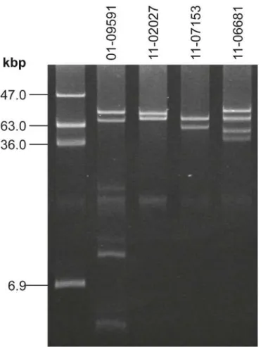
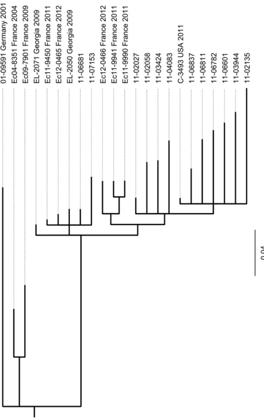
Phylogenetic analysis. In order to establish the phylogenetic relatedness of STEC O104: H4 strains, we included other published high quality genome sequences of STEC O104:H4 iso-lates in our SNP-based phylogenetic analysis. Among these are three finished sequences pub-lished in 2012 by Ahmed et al. [21], one from another outbreak isolate and two others from independent isolates discovered in Georgia in 2009. In addition the sequences of two other his-torical STEC O104:H4 isolates from France and of five more recent STEC O104:H4 isolates from sporadic cases in France as published by Grad et al. [22,23] were also included in the analysis. SNPs were identified comparing these sequences and the genome sequences of our 13 STEC O104:H4 isolates and a phylogenetic tree was calculated as described in the Materials and Methods section. Using a set of 224 selected SNPs gives rise to the tree shown inFig 5. Obviously, all epidemic strain isolates including the US-isolate C-3493 which originated from Fig 2. The plasmid profiles of the two sporadic STEC O104:H4 strains 11–06681 and 11–07153 are distinct from outbreak strain isolates and from the historical strain as well as from each other.Lane 1: molecular weight referenceE.coli39R862; Lane 2: historical O104:H4 isolate 01–09591; Lane 3: index isolate of the epidemic strain 11–02027—another plasmid of 1.5kbp is not visible here; Lanes 4, 5: variant isolates 11–07153 and 11–06681. A small plasmid of 1.5kbp present in 11–02027 as well as in 11–07153 and 11–06681 is not visible here (but seeS3 Table).
a person who travelled to Germany during the epidemic in 2011, cluster into one clade. The pairwise deviation among them with respect to numbers of SNPs ranges from three to thirty-six (S4 Table). The two German sporadic isolates 11–06681 and 11–07153 which differ from one another by only 10 SNPs do cluster separately from the epidemic strain isolates together with several recent sporadic isolates from France and with historical isolates from Georgia. Moreover, the two historical strains isolated in France in 2004 and 2009, respectively, belong to Fig 3. The two sporadic STEC O104:H4 strains 11–06681 and 11–07153 and the outbreak strain isolate 11–02027 show comparable levels of toxicity towards Vero cells.EHEC EDL933 served as a positive control andE.coliK12 C600 as a negative control. Toxicity of strain EDL933 as a quantitative reference was set to 100%. Shown are mean values of three independent experiments, each performed in triplicates. Bars represent means and standard deviation of three experiments. No significant difference in cytotoxicity of the STEC strains was observed.
doi:10.1371/journal.pone.0122074.g003
Fig 4. The two sporadic STEC O104:H4 strains 11–06681 and 11–07153 and the outbreak strain isolate 11–02027 show aggregative adherence as characteristic for EAEC.Assay of mannose-resistant adherence to Hep-2 cells included the EHEC O157:H7 strain EDL933 for comparison. Images were taken at 600-fold magnification.
an even more distant clade. Furthermore, the German historical STEC O104:H4 strain 01–09591 isolated in 2001 is clearly separated from all other isolates.
In the studies by Grad et al. and Guy et al. [22,30,31] comparing isolates from the German and the French STEC O104:H4 outbreaks in 2011, a number of SNPs were identified and have been mapped onto the TY2482 chromosome. Accordingly, there are two SNPs that define the German outbreak clade. We extracted the respective positions from the sequence of TY2482 together with surrounding 100 bases upstream and downstream and used the resulting 201-nucleotide sequences to examine the corresponding regions in our genome sequences using the bl2seq-facility at the NCBI BLAST homepage. The two SNPs typical of the German outbreak strain do also occur in all of our outbreak isolates. They are not present, however, in our two sporadic isolates and in the historical strain, which all have a G instead of an A at the position corresponding to 1568661 in TY2482 and a G instead of an A at the position corre-sponding to 2252380 in TY2482. The same way we could confirm that two SNPs identified as unique to TY2482 by Guy et al. [30] are not present neither in any of our outbreak isolates nor in the two sporadic isolates or in the historical strain. In the analyses performed by Grad et al. and Guy et al. [22,30] there is only one (from a Swedish who travelled to Germany) among 22 German isolates that was assigned to a clade different from the one where all other German iso-lates accumulate. For this isolate three SNPs (at positions 1262666, 2564789 and 3089339) were identified. Our two sporadic isolates which would also not fall into the German outbreak clade do not have these SNPs, indicating an independent ancestry of these strains. Moreover, they do not belong to any of the other lineages identified in the French outbreak because none of the 18 SNPs detected in the 11 French outbreak strains studied by Grad et al. [22,31] is present.
Identification and homology analysis of selected features of the sporadic STEC O104:H4 isolates 11–06681 and 11–07153. Based on the contigs assembled for each of the strains, se-lected features were compared using the BLAST facilities at NCBI. As a quality check of our ge-nome sequences, we analysed the alleles of those housekeeping genes targeted by classical MLST [16,32] in silico by BLAST analysis of the assembled contigs. Using the sequences deter-mined from the respective PCR products, there was perfect homology confirming the MLST sequence type ST678 (adk6,fumC6,gyrB5,icd136,mdh9,purA7,recA7) in all of our isolates. The Shiga toxin-encodingE.coliphage P13374 was induced from a German STEC O104: H4 outbreak isolate and sequenced to completion by Beutin et al. [33]. We extracted from the complete prophage genome (accession number HE664024) thestx-operon including 200 nu-cleotides adjacent to each end to give a sequence 1641 nunu-cleotides in length. This sequence was used for BLAST analyses of the contigs assembled for the outbreak isolate 11–02027, for the two sporadic STEC O104:H4 isolates 11–06681 and 11–07153 and for the historical isolate 01–09591 and was found completely present in one of the contigs of each genome sequence, re-spectively. For 11–02027 and for 01–09591, a sequence identical toE.coliphage P13374 was observed. Strains 11–06681 and 11–07153, however, show a difference of two nucleotides, one silent T to A mutation at position 909 in the open reading frame of thestxA2asubunit gene and
another G to A exchange 133 nucleotides downstream of the stop codon of thestxB2asubunit
gene resulting in an exchange of serine to leucine in a predicted hypothetical protein. Compar-ing the correspondCompar-ingstx2-operon containing DNA segment of 11–07153 with the two
sequences for Ec04-8351 and Ec09-7901, two historical strains from France and the sequences of Ec11-9450, Ec11-9941, Ec11-9990, Ec12-0456, Ec12-0466, five more recent sporadic isolates from France were published by Grad et al [22,23]. The selected SNPs used for tree calculation are given in supplementary files S1 Dataset(SNP positions) andS2 Dataset(sequences).
Georgian STEC O104:H4 isolates from 2009 as sequenced by Ahmed et al. [21] revealed identi-ty with strain 2009EL-2050. In contrast, the other strain 2009EL-2071 only showed the G to A exchange 133 nucleotides downstream of the stop codon of thestxB2asubunit gene whereas
the silent mutation in the open reading frame of thestxA2asubunit gene was not present.
The respectivestx-operon-comprising contig from the sporadic strain 11–07153 genome se-quence aligns to the phage P13374 sese-quence continuously along about half of the phage ge-nome to 96.1% pairwise identity. The equivalent contig of 11–06681 contains the left end of the phage and aligns continuously along about 40% of the phage sequence with 95.5% pairwise identity. An appropriate BLAST-based approach, using each of the ends of the prophage and 1000 nucleotides of the adjacent DNA sequence from the TY2482 chromosome, mapped the phage integration site in the genome of isolates 11–07153 and 11–06681 to exactly the same po-sition as in the outbreak strain 11–02027 inside the flavoprotein genewrbA, that is altered due to the integration [33].
Grad et al. [23] discussed thegyrAmutations responsible for the nalidixic acid resistance of their O104:H4 strains. They found an amino acid exchange S83A in the GyrA protein sequence of the 2011 epidemic O104:H4 strain as well as in all sporadic isolates from 2011 but a GyrA S83L exchange in HUSEC041, the historical EHEC O104:H4 strain from the HUSEC collection [14] and in their two historical French isolates from 2004 and 2009, respectively. Analysing the genome sequences of the epidemic isolate 11–02027, of the two sporadic isolates 11–06681 and 11–07153 and of the historical strain 01–09591 we found a completegyrAgene assembled into one of the contigs of each of the genome sequences. The predicted GyrA protein sequences of all but strain 01–09591 were identical with an alanine at position 83 of the translated protein sequence revealing the S83A mutation responsible for the Nal phenotype (Table 1). The Ger-man historical strain 01–09591 revealed a GyrA S83L exchange as seen in HUSEC041.
In order to identify the chloramphenicol resistance determinant of strain 11–07153 (Table 1), a BLAST analysis in our genome sequence assembly was performed using the open reading frames of common chloramphenicol resistance genes such ascat(extracted from BX664015),cmlA(AY509004) andfloR(AF231986) as a query. Not any of these genes was found assembled into a contig of 11–07153. However, mapping of all sequence reads of this ge-nome onto the open reading frames of the chloramphenicol resistance genes identifiedfloRbut notcatorcmlApresent in 11–07153. For the 11–06681 genome, no homology with any of the chloramphenicol resistance genes was detected, what is in agreement with the chloramphenicol sensitive phenotype of this strain (Table 1). ThefloRgene of 11–07153 differs from the most similarfloR-allele (accession number AB591424) on aSalmonellaplasmid by one nucleotide (transversion of A to C) at position 93 in the open reading frame, resulting in an amino acid ex-change of leucine for isoleucine.
Analysis of the plasmids of the sporadic STEC O104:H4 isolates 11–06681 and 11–
analysis (Fig 5) and epidemiological evidence of 11–02027 being an outbreak strain isolate it is reasonable to conclude that the 100% coverage actually indicates presence of the plasmids. By a similar line of arguments one might conclude that the 100% coverage of the EAEC-type adherence-encoding plasmids and of a small cryptic plasmid common to all of the reference ge-nomes in the genome sequences of the two sporadic isolates 11–06681 and 11–07153 does indi-cate the presence of those plasmids in these strains. The coverage of the ESBL-encoding epidemic reference strain plasmid pTY-1 or pESBL-EA11 in the genome sequences of the two sporadic isolates 11–06681 and 11–07153 is only around 50% (S3 Table). Therefore, one can conclude that such a plasmid is not present in the sporadic isolates what is in agreement with their plasmid profiles (Fig 2) and phenotypes (Table 1). A coverage level of 50% might indicate that the sporadic isolates contained related plasmids (e.g. of the same incompatibility group) which share some plasmid specific sequences encoding similar replication and conjugative transfer systems with pTY-1 and pESBL-EA11. Alternatively there could be some mobile ge-netic elements (e.g. transposons, IS elements) present somewhere in the genome of the sporad-ic strains whsporad-ich are also present as accessory genetsporad-ic elements on pTY-1 and pESBL-EA11. Finally, only a coverage of less than 5% for the large IncF plasmid unique to strain 2009EL-2050 was detected, indicating that there is no similar plasmid present in the German sporadic STEC O104:H4 strains 11–06681 and 11–07153.
Discussion
Infections with pathogenicE.coliof the serovar O104:H4 only rarely were reported before the year 2011. An enteroaggregativeE.coliisolated in the 1990s in Africa [34] and a Shiga toxin-producing isolate obtained in 2001 in Germany, HUSEC041 [14], which is identical to the strain 01–09591 included in our investigations, have been the best studied strains of this sero-var for many years. After the 2011 outbreaks with a Shiga toxin-producing, enteroaggregative
E.coliO104:H4 in Germany [1] and France [2], substantial effort has been made in studying this emerging pathogen. A few more historical isolates were extensively characterized in com-parison to the more recent outbreak strains [21–23,35,36]. Whole genome sequencing ap-proaches in addition to conventional typing methods, rapidly uncovered the particular pathogenetic background of this uncommon pathogen [5–11].
Besides the ten outbreak-related STEC O104:H4 isolates, we investigated two distinct STEC O104:H4 strains, 11–06681 and 11–07153, which were isolated in 2011 in Germany and resem-ble the outbreak strain not only regarding the MLST and the serotype, but also with respect to the virulence gene profile, thestx2operon-containing prophage, the expression of Shiga toxin,
the degree of cytotoxicity (Fig 3) and the aggregative adherence pattern (Fig 4). However, they differ from the outbreak strain and from each other as well as from the historical 2001 isolate with respect to the antibiograms (Table 1), the PFGE patterns (Fig 1) and the plasmid profiles (Fig 2,S3 Table). Moreover, although isolated in close spatiotemporal proximity to the out-break in 2011, according to the SNP-based phylogenetic analysis they are clearly separated from outbreak strain isolates (Fig 5). In particular, at the two positions corresponding to 2252380 and 1568661, respectively, in TY2482, which define the clade comprising the German outbreak isolates in the study of Grad et al. [22,31], they differ from all of the outbreak isolates. Moreover, none of the SNPs identified by Grad et al. [22,31] and Guy et al. [30] in German or French outbreak isolates is present in neither 11–06681 nor 11–07153 indicating that there is no direct ancestry. Concluding from SNP analyses, the two German sporadic STEC O104:H4 isolates from 2011 are closely related to each other and in terms of phylogeny less distant to the two historical Georgian isolates described by Ahmed et al. [21] than to the historical German isolate from 2001 or to the two historical isolates from France (Fig 5). Moreover, the two spo-radic German STEC O104:H4 isolates from 2011 fall into one clade with more recent isolates obtained from sporadic cases in France in 2011 and 2012, respectively (Fig 5).
It is interesting to note, that despite beeing phylogenetically closely related (Fig 5,S4 Table) the two German sporadic STEC O104:H4 isolates considerably differ from each other with re-spect to their plasmid content. Strains 11–06681 and 11–07153 both do contain a plasmid, identical in size to the EAEC-type adherence-encoding plasmid in the epidemic STEC O104: H4 strain (Fig 2). Most likely, these plasmids are genetically identical or very similar, since the entire DNA sequence of the EAEC-type adherence-encoding plasmid is also present in the ge-nome sequences of strains 11–06681 and 11–07153 (S3 Table). However, there are several more unknown large plasmids in strain 11–07153 and 11–06681 (Fig 2).
Grad et al. [23] concluded from their comprehensive genome comparison analyses that there were two lineages among STEC O104:H4 strains, one comprising the two French histori-cal isolates from 2004 and 2009 and another comprising the 2011 epidemic strain and several French sporadic isolates from 2011 and 2012. These two lineages separated before indepen-dently acquiring distinctgyrAmutations resulting in resistance to nalidixic acid [23]. In agree-ment with this conclusion and the SNP-based tree shown inFig 5, our two German sporadic isolates belong to the lineage comprising the 2011 epidemic strain and the 2011–2012 French sporadic isolates in sharing their GyrA S83A genotype, whereas the German isolate from 2001 shares the GyrA S83L genotype with the French historical isolates. Overall, for the two German sporadic STEC O104:H4 isolates under investigation, the analysis of their whole genome se-quences confirms the conclusions drawn from conventional epidemiological subtyping. More-over, the results do not support a direct derivation of these isolates from the outbreak strain.
report of Jourdan-da Silva et al. with respect to the Turkish origin of their STEC O104:H4 iso-late it is worth mentioning, that the two German sporadic STEC O104:H4 strains 11–06681 and 11–07153 (Table 1) were isolated from patients with a travel history to Turkey [38]. How-ever, it is not completely without doubt whether the German patients were infected in Turkey because the onset of symptoms or date of isolation, respectively, was only late after returning from Turkey (11 and 18 days, respectively) in contrast to the French traveler, where there is strong epidemiological evidence that this infection was contracted in Turkey [37].
In conclusion, we here describe two additional EHEC/EAEC strains of the serovar O104:H4 which share virulence determinants with the 2011 outbreak strain and with several other spo-radic STEC O104:H4 isolates but are epidemiologically unrelated. The results of our investiga-tion of STEC O104:H4 strains add to the knowledge about this emerging pathogen, concerning a certain diversity within the bacterial core genome as well as loss and gain of accessory ele-ments such as plasmids. Our results do also support the view that distinct new variants of STEC O104:H4 might repeatedly originate from a yet unknown reservoir, rather than that there would be a continuous diversification of a single strain established and circulating in Germany after the large outbreak in 2011 [38,39,40].
Supporting Information
S1 Dataset. Positions in the TY2482 genome of the 224 SNPs selected for phylogenetic analysis.
(TXT)
S2 Dataset. Sequences in fasta format created by concatenating those nucleotides from each of the 23 genome sequences under investigation which correspond to the positions of the 224 SNPs selected for phylogenetic analysis (see alsoS1 Datasetfor SNP positions in the TY2482 genome).
(FASTA)
S1 Table. Sequencing statistics. (DOCX)
S2 Table. Coverage of chromosomal coding sequences identified for the reference genome of STEC O104:H4 strain TY2482.
(XLSX)
S3 Table. Coverage of reference plasmids from various STEC O104:H4 reference genomes. (XLSX)
S4 Table. Matrix giving the numbers of SNPs different between the STEC O104:H4 ge-nomes under investigation.
(XLSX)
Acknowledgments
We thank S. Dumschat, S. Karste, S. Schidlo, B. Leiste, G. Bartel, U. Siewert, U. Strutz, T. Eitze for excellent technical assistance. We are indebted to Ulrich Nübel for advice with respect to sequence analysis.
Author Contributions
reagents/materials/analysis tools: ET PWD RP AR A. Fruth PA AN MM A. Flieger. Wrote the paper: ET PWD RP AR A. Fruth PA AN MM A. Flieger.
References
1. Frank C, Werber D, Cramer JP, Askar M, Faber M, an der Heiden M, et al. Epidemic profile of Shiga-toxin-producingEscherichia coliO104:H4 outbreak in Germany. N Engl J Med. 2011; 365: 1771–1780. doi:10.1056/NEJMoa1106483PMID:21696328
2. Gault G, Weill FX, Mariani-Kurkdjian P, Jourdan-da Silva N, King L, Aldabe B, et al. Outbreak of haemo-lytic uraemic syndrome and bloody diarrhoea due toEscherichia coliO104:H4, south-west France, June 2011. Euro Surveill. 2011 Jun 30. 16(26). pii: 19905. PMID:21749817
3. Mariani-Kurkdjian P, Bingen E, Gault G, Jourdan-Da Silva N, Weill FX.Escherichia coliO104:H4 south-west France, June 2011. Lancet Infect Dis. 2011; 11: 732–733. doi:10.1016/S1473-3099(11)70266-3 PMID:21958580
4. Characteristics of the pathogen and information and assistance by the RKI in diagnosis of the currently circulating outbreak strain. Robert Koch-Institute, Germany, Berlin. 2011. Available:http://www.rki.de/ EN/Home/EHECO104.pdf?__blob = publicationFile. Accessed 2015 Feb 24.
5. Bielaszewska M, Mellmann A, Zhang W, Köck R, Fruth A, Bauwens A, et al. Characterisation of the
Escherichia colistrain associated with an outbreak of haemolytic uraemic syndrome in Germany, 2011: a microbiological study. Lancet Infect Dis. 2011; 11(9): 671–676. doi:10.1016/S1473-3099(11)70165-7 PMID:21703928
6. Scheutz F, Møller Nielsen E, Frimodt-Møller J, Boisen N, Morabito S, Tozzoli R, et al. Characteristics of the enteroaggregative Shiga toxin/verotoxin-producingEscherichia coliO104:H4 strain causing the outbreak of haemolytic uraemic syndrome in Germany, May to June 2011. Euro Surveill. 2011 Jun 16. 16(24). pii: 19889. PMID:21699770
7. Mellmann A, Harmsen D, Cummings CA, Zentz EB, Leopold SR, Rico A, et al. Prospective genomic characterization of the German enterohemorrhagicEscherichia coliO104:H4 outbreak by rapid next generation sequencing technology. PLoS One. 2011; 6: e22751. doi:10.1371/journal.pone.0022751 PMID:21799941
8. Rohde H, Qin J, Cui Y, Li D, Loman NJ, Hentschke M, et al. Open-source genomic analysis of Shiga-toxin-producingE.coliO104:H4. N Engl J Med. 2011; 365: 718–724. doi:10.1056/NEJMoa1107643 PMID:21793736
9. Li D, Xi F, Zhao M, Chen W, Cao S, et al. Genomic data fromEscherichia coliO104:H4 isolate TY-2482. BGI Shenzhen. 2011. Available: doi:10.5524/100001. Accessed 2015 Feb 24.
10. Rasko DA, Webster DR, Sahl JW, Bashir A, Boisen N, Scheutz F, et al. Origins of theE.coliStrain Causing an Outbreak of Hemolytic-Uremic Syndrome in Germany. N Engl J Med. 2011; 365: 709–717. doi:10.1056/NEJMoa1106920PMID:21793740
11. Brzuszkiewicz E, Thurmer A, Schuldes J, Leimbach A, Liesegang H, Meyer FD, et al. Genome se-quence analyses of two isolates from the recentEscherichia colioutbreak in Germany reveal the emer-gence of a new pathotype: Entero-Aggregative-HaemorrhagicEscherichia coli(EAHEC). Arch
Microbiol. 2011; 193: 883–891. doi:10.1007/s00203-011-0725-6PMID:21713444
12. Bakteriologische Untersuchungen im Rahmen des Ausbruchs mitE.coliO104:H4. Robert Koch-Institute, Germany, Berlin. 2011. Epid Bull. 35: 325–329. Available:https://www.rki.de/DE/Content/ Infekt/EpidBull/Archiv/2011/Ausgaben/35_11.pdf?__blob = publicationFile. Accessed 2015 Feb 24.
13. Report: Final presentation and evaluation of epidemiological findings in the EHEC O104:H4 outbreak, Robert Koch-Institute, Germany, Berlin. 2011. Available:http://www.rki.de/EN/Home/EHEC_final_ report.pdf?__blob = publicationFile. Accessed 2015 Feb 24.
14. Mellmann A, Bielaszewska M, Köck R, Friedrich AW, Fruth A, Middendorf B, et al. Analysis of a collec-tion of hemolytic uremic syndrome-associated enterohemorrhagicEscherichia coli. Emerg Infect Dis. 2008; 14: 1287–1290. doi:10.3201/eid1408.071082PMID:18680658
15. Prager R, Fruth A, Busch U, Tietze E. Comparative analysis of virulence genes, genetic diversity, and phylogeny of Shiga toxin 2 g and heat-stable enterotoxin STIa encodingEscherichia coliisolates from
humans, animals, and environmental sources. Int J Med Microbiol. 2011; 301: 181–191. doi:10.1016/j. ijmm.2010.06.003PMID:20728406
16. Escherichia coliMLST Database [Internet]. Available:http://mlst.warwick.ac.uk/mlst/dbs/Ecoli. Ac-cessed 2015 Feb 24.
18. Langmead B, Salzberg SL. Fast gapped-read alignment with Bowtie 2. Nat Methods. 2012; 9: 357–359. doi:10.1038/nmeth.1923PMID:22388286
19. automatic annotation of the third BGI assembly of theE.coliTY-2482 strain genome. Available:https:// github.com/ehec-outbreak-crowdsourced/BGI-data-analysis/wiki/Automatic-annotation-of-bgi-v3-assembly-of-e.-coli-ty-2482-genome. Accessed 2015 Feb 24.
20. McKenna A, Hanna M, Banks E, Sivachenko A, Cibulskis K, Kernytsky A, et al. The Genome Analysis Toolkit: a MapReduce framework for analyzing next-generation DNA sequencing data. Genome Res. 2010; 20:1297–1303. doi:10.1101/gr.107524.110PMID:20644199
21. Ahmed SA, Awosika J, Baldwin C, Bishop-Lilly KA, Biswas B, Broomall S, et al. Genomic Comparison ofEscherichia coliO104:H4 Isolates from 2009 and 2011 Reveals Plasmid, and Prophage Heterogene-ity, Including Shiga Toxin Encoding Phage stx2. PLoS One. 2012; 7: e48228. doi:10.1371/journal. pone.0048228PMID:23133618
22. Grad YH, Lipsitch M, Feldgarden M, Arachchi HM, Cerqueira GC, Fitzgerald M, et al. Genomic epidemi-ology of theEscherichia coliO104:H4 outbreaks in Europe, 2011. Proc Natl Acad Sci USA. 2012; 109: 3065–3070. doi:10.1073/pnas.1121491109PMID:22315421
23. Grad YH, Godfrey P, Cerquiera GC, Mariani-Kurkdjian P, Gouali M, Bingen E, et al. Comparative geno-mics of recent Shiga toxin-producingEscherichia coliO104:H4: short-term evolution of an emerging pathogen. mBio. 2013; 4: e00452–12. doi:10.1128/mBio.00452-12PMID:23341549
24. Huelsenbeck JP, Ronquist F. MRBAYES: Bayesian inference of phylogeny. Bioinformatics. 2001; 17: 754–755. PMID:11524383
25. Chevreux B. MIRA: An Automated Genome and EST Assembler [dissertation]. German Cancer Re-search Center Heidelberg; 2005. Available:http://www.chevreux.org/thesis/index.html. Accessed 2015 Feb 24.
26. MIRA web site [Internet]. Available:http://sourceforge.net/projects/mira-assembler. Accessed 2015 Feb 24.
27. Geneious 7.1 created by Biomatters. Available fromhttp://www.geneious.com. Accessed 2015 Feb 24.
28. Zhang Z, Schwartz S, Wagner L, Miller W (2000) A greedy algorithm for aligning DNA sequences. J Comput Biol 7: 203–214. PMID:10890397
29. Basic Local Alignment Search Tool [Internet]. National Center for Biotechnology Information. Available: http://blast.ncbi.nlm.nih.gov/Blast.cgi. Accessed 2015 Feb 24.
30. Guy L, Jernberg C, Ivarsson S, Hedenström I, Engstrand L, Andersson SG. Genomic diversity of the 2011 European outbreaks ofEscherichia coliO104:H4. Proc Natl Acad Sci USA. 2012; 109: E3627–E3628. doi:10.1073/pnas.1206246110PMID:23248326
31. Grad YH, Lipsitch M, Griggs AD, Haas BJ, Shea TP, McCowan C, et al. Reply to Guy et al.: Support for a bottleneck in the 2011Escherichia coliO104:H4 outbreak in Germany. Proc Natl Acad Sci USA. 2012; 109: E3629–E3630. PMID:23479789
32. Wirth T, Falush D, Lan R, Colles F, Mensa P, Wieler LH, et al. Sex and virulence inEscherichia coli: an evolutionary perspective. Mol Microbiol. 2006; 60: 1136–1151. PMID:16689791
33. Beutin L, Hammerl JA, Strauch E, Reetz J, Dieckmann R, Kelner-Burgos Y, et al. Spread of a distinct Stx2-encoding phage prototype amongE.coliO104:H4 strains from outbreaks in Germany, Norway and Georgia. J Virol. 2012; 86: 10444–10455. doi:10.1128/JVI.00986-12PMID:22811533
34. Mossoro C, Glaziou P, Yassibanda S, Lan NT, Bekondi C, Minssart P, et al. Chronic diarrhea, hemor-rhagic colitis, and hemolytic-uremic syndrome associated with HEp-2 adherentEscherichia coliin adults infected with human immunodeficiency virus in Bangui, Central African Republic. J Clin Micro-biol. 2002; 40: 3086–3088. PMID:12149388
35. Monecke S, Mariani-Kurkdjian P, Bingen E, Weill FC, Baliere C, Slickers P, et al. Presence of Enterohe-morrhagicEscherichia coliST678/O104:H4 in France Prior to 2011. Appl Environ Microbiol. 2011; 77: 8784–8786. doi:10.1128/AEM.06524-11PMID:22003010
36. Guy L, Jernberg C, Arvén Norling J, Ivarsson S, Hedenström I, Melefors Ö, et al. Adaptive Mutations and Replacements of Virulence Traits in theEscherichia coliO104:H4 Outbreak Population. PLoS One. 2013; 8(5): e63027. doi:10.1371/journal.pone.0063027PMID:23675451
37. Jourdan-da Silva N, Watrin M, Weill FX, King LA, Gouali M, Mailles A, et al. Outbreak of haemolytic uraemic syndrome due to Shiga toxin-producingEscherichia coliO104:H4 among French tourists returning from Turkey, September 2011. Euro Surveill. 2012 Jan 26. 17(4). pii: 20065. PMID: 22297137
38. Frank C, Milde-Busch A, Werber D. Results of surveillance for infections with Shiga toxin-producing
39. Abu Sin M, Takla A, Flieger A, Prager R, Fruth A, Tietze E, et al. Carrier prevalence, secondary house-hold transmission and long-term shedding in two districts during theEscherichia coliO104:H4 outbreak in Germany, 2011. J Infect Dis. 2013; 207:432–438. doi:10.1093/infdis/jis702PMID:23175763