S
TUDIES ON THE MODE OF ACTION OF AUXIN IN PLANT DEVELOPMENT–
ANALYSIS OF THEPIN6
GENE IN MERISTEM FUNCTION AND CELL DIFFERENTIATIONVIOLANTE DE FARIA E MAIA PACHECO DE MEDEIROS
DOUTORAMENTO EM BIOLOGIA BIOLOGIA MOLECULAR
S
TUDIES ON THE MODE OF ACTION OF AUXIN IN PLANT DEVELOPMENT–
ANALYSIS OF THEPIN6
GENE IN MERISTEM FUNCTION AND CELL DIFFERENTIATION.
VIOLANTE DE FARIA E MAIA PACHECO DE MEDEIROS
Tese orientada pela Professora Doutora Maria Salomé Soares Pais
Professora Catedrática da Faculdade de Ciências da Universidade de Lisboa, Portugal
e co-orientada pelo Professor Doutor Klaus Palme
Professor da Universidade Albert-Ludwig, Freiburg, Alemanha
DOUTORAMENTO EM BIOLOGIA BIOLOGIA MOLECULAR
Medeiros,V., Ditengou, F., Pais, M., Palme, K. Characterization of PIN6, a member of the PIN gene family of auxin efflux regulators. (publicação em poster)
Baumann, M., Ditengou, F., Dovzhenko, A., Krieghoff, N., Li, X., Medeiros, V., Paponov, IA., Paponov, M., Pasternak. T., Santos, F., Teale, WD, Tietz, O., Wang, B., Palme, K. Role of auxin in cell
differentiation and organ formation in A. thaliana. (publicação em poster)
Violante Medeiros, Franck Ditengou, Hugues Nziengui, Xugang Li, Maria S. Pais, Klaus Palme. PIN6 is a negative regulator of new organ development in meristems. (publicação em preparação)
Para efeitos do disposto no nº 2, art. 8, Dec. -Lei 388/70, a autora da dissertação declara que interveio na concepção e execução do trabalho experimental, na interpretação dos resultados e na redacção do manuscrito enviado para publicação.
The study hereby presented was developed under supervision of Prof. Dr. Maria Salomé Pais, from the Unit for Molecular Biology and Plant Biotechnology at the Institute for Applied Science and Technology of the Faculty of Sciences, University of Lisbon, and supervision of Prof. Dr. Klaus Palme, from the Institute Biology II at the Albert-Ludwig University in Freiburg, Germany, where the majority of the work was carried out.
My sincere gratitude goes to Prof. Dr. Maria Salomé Pais, for accepting me under her supervision, for allowing me to develop this work, for all the regular scientific discussions and for long-distance encouragement.
I am very grateful to Prof. Klaus Palme for taking the supervision of this work and welcoming me in his lab, for allowing me to participate in other projects of his thus allowing me to expand my technical and scientific knowledge. To my close supervisor Dr. Franck Ditengou, a very special thank you for adopting me and my project, for his endless ideas and the fruitful discussions we had, and for being an example of neverending motivation.
To Dr. Stefan Kirscher and Dr. Roman Ulm (Albert-Ludwig University Freiburg, Germany), my gratitude for allowing me to perform light quality experiments in their laboratories, as well as for critical advice and analysis on the subject.
I am also very grateful to Dr. Franck Ditengou, Dr. Filipa Santos, Dr. Olaf Tietz, Óscar Oliveira and Nicola Krieghoff for critical reading of this manuscript and additional help with formatting and proofreading.
A hearted thank you goes to all the colleagues in AG Palme for these years of frienly collaborations. My special gratitude goes to: Dr. Franck Ditengou, for his guidance throughout this work, Dr. Benedetto Ruperti and Dr. Hugues Nziengui, for close collaboration and helpful discussions, Irina Petrovà, Karl-Andreas Flittner and Philip Kochersperger, for technical assistance,, Dr. Xugang Li, Dr. Olaf Tietz and Dr. Ivan Paponov, for providing the anti-PIN1 and anti-PIN6 antibodies, Claudia Gilles, for technical assistance, and Dr. William Teale and Dr. Filipa Santos for many helpful scientific discussions. A special thank you to my supportive fellow PhD student colleagues: Claude, Filipa, Francesco, Irina, Manoj, Nicola, Óscar and Tatyana.
To all my colleagues in ICAT I wish to acknowledge their helpfulness and good working atmosphere; the long-distance support and constant warm welcome whenever I was in the lab. A special thank you to Dr. Ana Margarida Fortes, Dr. Mónica Sebastiana, Andreia Figueiredo, Cláudia Rato, Eva Sousa and Silvia Ferreira for helpful ideas and support.
A special thank you goes to my azorean “bio” friend Dr. Luisa Mota Vieira, for many friendly discussions about carrier and prospects, but mainly for taking an interest and for all the good advice throughout the years.
To those friends outside the lab who followed similar paths as mine and so many times listened and supported me: Anabela Maia, Catarina Vinagre, Cláudia Rato, Joana Osório, Leena Luis, Luisa Arruda and João Pereira.
A very special thank you goes to the new friends met through this PhD experience: Isabell, whose non-biology-related friendship was a pillar to stand on, to m “not-only-in-the-lab-buddies” Benedetto, Francesco, Ines, Nico, Olaf, Óscar, Ulrike, Yamuna, for everything else, and specially the Madrinha Filipa, for many years of listening to me, for “mentoring” me, and for always looking for (and finding!) a silver lining. Also to the big family of the Quasilusos, for dreaming, for the big adventures lived, the endless marathons survived and the full-proof friendships made.
Last, but not least, a very big OBRIGADA to my whole family. Ao lado “Pê de Mê”, pelo interesse demonstrado e o recarregar de baterias nas minhas estadas em Lisboa; à Godmother, pelas distracções e apoio em momentos críticos e ao Primo, pelos pequenos gestos e a constante parceria com a sua “irmã mais velha”. E, finalmente, aos meus pais, Tonecas e Graça, e à minha avó Lú, por aceitarem as minhas escolhas, por me terem sempre incentivado a fazer mais e melhor, por me terem sempre apoiado nos momentos mais difíceis e sabido aceitar as mudanças impostas.. enfim, obrigada, simplesmente, por acreditarem!
A bolsa de Doutoramento com a referência SFRH/BD/10891/2002 foi atribuída pela Fundação para a Ciência e Tecnologia (FCT), financiada pelo Programa Operacional Ciência e Inovação (POCI) 2010- Formação Avançada para a Ciência, no âmbito do III Quadro Comunitário de Apoio, comparticipado pelo Fundo Social Europeu (FSE) e por fundos nacionais do Ministério da Ciência e Ensino Superior (MCES). A publicação da dissertação foi financiada pela mesma instituição.
União Europeia Fundo Social Europeu União Europeia Fundo Social Europeu
“Philosophy (nature) is written in that great book which ever lies before our eyes. I mean the universe, but we cannot understand it if we do not first learn the language and grasp the symbols in which it is written. The book is written in the mathematical (scientifical) language... without whose help it is humanly impossible to comprehend a single word of it, and without which one wanders in vain through a dark labyrinth.”
Galileo Galilei
Aos meus pais
e à avó Lú
A
CKNOWLEDGMENTSiii
FIGURE INDEX
x
TABLE INDEX xi RESUMO xii ABSTRACT xv ABBREVIATIONS AND SYMBOLS xvi
1 INTRODUCTION
1.1
AUXIN IN PLANT DEVELOPMENT1
1.2
AUXIN MODE OF ACTION:
PERCEPTION AND SIGNALLING3
1.3
AUXIN TRANSPORT5
1.3.1 INFLUX AND EFFLUX TRANSPORTERS
7
1.4 ATPIN FAMILY OF AUXIN EFFLUX CARRIERS
7
1.4.1 PINS IN PLANT DEVELOPMENT 10
1.4.1.1 PINs in leaf development and phyllotaxis 10
1.4.1.2 PINs in vascular patterning 14
1.4.1.3 PINs in root growth 15
1.4.1.4 PINs in transition from vegetative to reproductive development 17
1.4.1.5 PINs and lateral root formation 18
1.4.1.6 PINs in gravity perception 21
1.4.2 REGULATION OF PIN EXPRESSION 23
1.4.3 PIN PROTEIN DYNAMICS 25
1.5 AIM OF THE THESIS 26
2 MATERIALS
AND
METHODS
2.1
MATERIALS 27 2.1.1 PLANTS 27 2.1.2 BACTERIA 27 2.1.3 PLASMID VECTORS 27 2.1.4 SYNTHETIC OLIGONUCLEOTIDES 28 2.1.5 ANTIBODIES 29 2.1.6 ENZYMES 302.1.10 BATERIA MEDIA 31
2.1.11 MICROSCOPES 31
2.1.12 COMPUTER PROGRAMS 31
2.1.13 DATABASES 32
2.2
METHODS 322.2.1 METHODS FOR THE CULTIVATION AND TRANSFORMATION OF ARABIDOPSIS 32
2.2.1.1 Arabidopsis seed sterilization 32
2.2.1.2 In vitro germination of Arabidopsis seeds 32
2.2.1.3 Arabidopsis growth on soil 33
2.2.1.4 Transformation of Arabidopsis WT plants 33
2.2.1.5 In vitro selection of transformed WT plants 34
2.2.1.6 Preparation of seedlings for microscopy 34
2.2.1.6 Measurements of root length, lateral root number and root curvature 34 2.2.2 METHODS FOR THE CULTIVATION AND TRANSFORMATION OF BACTERIA 34
2.2.2.1 Preparation of electrocompetent Escherichia coli cells 34
2.2.2.2 Electroporation of Escherichia coli cells 34
2.2.2.3 Preparation of electrocompetent Agrobacterium tumefaciens cells 34 2.2.2.4 Electroporation of Agrobacterium tumefaciens cells 35
2.2.3 CLONING STRATEGIES 35
2.2.4 OLIGONUCLEOTIDE DESIGN 36
2.2.5 METHODS FOR NUCLEIC ACID ANALYSIS 36
2.2.5.1 Small scale plasmid isolation from Escherichia coli and Agrobacterium tumefaciens
36
2.2.5.2 Isolation of genomic DNA from Arabidopsis – crude protocol, for genotyping (adapted from Edwards et al., 1991)
36
2.2.5.3 Isolation of genomic DNA from Arabidopsis – CTAB method 36 2.2.5.4 Separation of DNA fragments on agarose gel electrophoresis 37
2.2.5.5 Polymerase Chain Reaction (PCR) 37
2.2.5.6 Reverse Transcriptase Polymerase Chain Reaction (R.T.-PCR) 38
2.2.5.7 Purification of PCR products 38
2.2.5.7.1. PEG Clean-Up 38
2.2.5.7.2. Purification of plasmids or PCR products from an agarose gel 38 2.2.5.8 Purification of DNA by phenol/chloroform extraction 38
2.2.5.9 Precipitation of DNA 39
2.2.5.10 Recombination of DNA fragments 39
2.2.5.11 Southern Blot 39
2.2.5.12 Isolation of total RNA from Arabidopsis – Trizol method 40
2.2.5.13 RNA quantification 41
2.2.5.14 In vitro transcription of the RNA probe 41
2.2.6 METHODS FOR PROTEIN ANALYSIS 41
2.2.6.1 Determination of protein concentration 41
2.2.6.2 Separation of proteins by SDS polyacrylamide gel electrophoresis (SDS-PAGE) 41
2.2.6.3 Coomassie blue staining of SDS-PAGE gels 42
2.2.6.4 Western Blot 42
2.2.6.5 Ponceau staining 42
2.2.6.6 Immunostaining of Western Blots 42
2.2.6.7 Epitope expression for antibody generation and purification 42
2.2.6.8 Affinity purification of antibodies 43
2.2.7 METHODS FOR HISTOCHEMISTRY ASSAYS 43
2.2.7.1 Lugol staining 43
2.2.7.2 Histochemical detection of reporter gene activity (GUS staining) 43
2.2.7.3 Embedding plant tissue for sectioning 44
2.2.7.4 In situ hybridization on sections 44
2.2.7.5 Whole-mount in situ hybridization 44
2.2.7.6 Immunolocalization on sections 45
3 RESULTS
3.1
IN SILICO ANALYSIS OFPIN6 46
3.1.1 PREDICTION OF PROTEIN PROPERTIES 46
3.1.2 PROMOTER SEQUENCE ANALYSIS 49
3.1.3 EXPRESSION PATTERN ANALYSIS 52
3.1.4 PIN6 REGULATION 52
3.2 PIN6
LOCALIZATION54
3.2.1 GENE EXPRESSION 54
3.2.2 PROTEIN LOCALIZATION 60
3.3
SCREENING FOR PIN6
NULL MUTANTS 613.3.1 GENOTYPING 62
3.3.2 PIN6 EXPRESSION LEVELS IN pin6 SEEDLINGS 63
3.4
GENERATING PIN6 OVEREXPRESSING PLANTS AND RNAILOSS-OF-FUNCTION TRANSGENIC LINES
64
PLANTS
3.5.3 LIGHT QUALITY EFFECT ON pin6 GROWTH 73
3.5.4 LATERAL ROOT DEVELOPMENT IN pin6 SEEDLINGS 77
3.5.5 GRAVITY PERCEPTION OF pin6 SEEDLINGS 77
3.5.6 AUXIN SENSITIVITY OF pin6 SEEDLINGS 80
3.5.7 ETHYLENE EFFECT ON pin6 SEEDLINGS 84
4 DISCUSSION
4.1
CHARACTERIZATION OF A NOVEL PIN PROTEIN:
PIN6 87
4.1.1 USING GENE KNOCKOUT AND –DOWN STRATEGIES AS TOOLS FOR
ANALYZING PIN6 FUNCTION
88
4.2 PIN6 AND AUXIN TRANSPORT MODULATION 90
4.3 PIN6 AND OTHER HORMONES 91
4.4 PIN6 IN MERISTEM DEVELOPMENT 93
4.4.1 GERMINATION 93
4.4.2 LEAF PRIMORDIA EMERGENCE 94
4.4.3 MERISTEM TRANSITION TO THE REPRODUCTIVE PHASE 95
4.4.4 RESPONSE TO LIGHT AND PHOTOPERIOD 98
4.4.5 LATERAL ROOT DEVELOPMENT 101
4.5 PIN6
AND GRAVITROPIC RESPONSE103
4.6
OTHER LEVELS OFPIN6
REGULATION104
4.7 PIN6 AND REDUNDANCY AMONG THE PIN FAMILY 104
4.8
CONCLUDING REMARKS 1054.9
FUTURE PERSPECTIVES 106Page Nr.
Fig 1.1. Chemiosmotic hypothesis of polar, cell-to-cell, auxin transport. 6
Fig 1.2. Typical PIN protein topology. 8
Fig 1.3. Changes in gravity vector orientation induce asymmetric DR5-GFP expression in
lateral root cap cells. 22
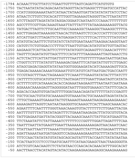
Fig 3.1. Full-length genomic DNA sequence of PIN6 (At1g77110). 47
Fig 3.2. Tree view of PIN genes sequence alignment. 47
Fig 3.3. PIN6 amino acid sequence. 47
Fig 3.4. Exon-intron structure of PIN1, PIN2 and PIN6 gene sequences. 48
Fig 3.5. Sequence alignment of PIN proteins. 48
Fig 3.6. Promoter sequence of PIN6 used previously for the pPIN6::GUS construct. 49
Fig 3.7. PIN6 and DR5 expression in Arabidopsis roots. 56
Fig 3.8. PIN6 and DR5 expression in the Arabidopsis shoot apical meristem. 57
Fig 3.9. PIN6 expression in the Arabidopsis floral meristem. 57
Fig 3.10. In situ hybridization of PIN6 mRNA transcripts in the floral meristem. 58 Fig 3.11. pPIN6::GUS expression in 7-days old seedlings grown in AM medium under long-days
condition.
59 Fig 3.12. PIN6 mRNA expression in 7-days old seedlings grown in AM medium under long-days
condition. 60
Fig 3.13. Visualization of PIN expression in Arabidopsis. 61
Fig 3.14. Southern Blot analysis of the T-DNA insertion lines. 63
Fig 3.15. R.T.-PCR analysis of the T-DNA insertion lines. 64
Fig 3.16. R.T.-PCR analysis of the generated overexpressor and RNAi lines. 65
Fig 3.17. Main root length of 2- to 10-days old WT and pin6 seedlings grown under continuous
light. 66
Fig 3.18. Main root length of 2- to 10-days old WT and pin6 knockdown lines grown under continuous light.
67 Fig 3.19. Main root length of 2- to 10-days old WT and pin6 seedlings grown under long-days
condition.
67 Fig 3.20. Main root length of 2- to 10-days old WT and pin6 knockdown lines grown under
long-days condition. 68
Fig 3.21. Hypocotyl length of 6-days old WT and pin6 seedlings grown under continuous light, long-days, short-days and darkness.
69 Fig 3.22. PIN6 expression in 6-days old pPIN6::GUS seedlings grown under different
photoperiodic regimes.
70 Fig 3.23. Flowering status of 4-weeks old seedlings grown under long-days condition. 71
Fig 3.24. Numbers of leaves developed prior to the transition to flowering. 72
Fig 3.25. Effect of different light qualities on hypocotyl growth of WT and pin6 seedlings. 75
Fig 3.26. Effect of different light qualities on PIN6 expression. 76
Fig 3.27. Number of lateral roots in WT and pin6 seedlings. 77
Fig 3.28. Number of lateral roots in WT and pin6 knockdown lines. 78
Fig 3.29. Root curvature induced by 4h of gravistimulation. 79
Fig 3.30. Effect of 4h gravistimulation. 80
Fig 3.31. 1-NAA inhibition of root growth. 81
Fig 3.32. Induction of PIN6 expression by 1-NAA. 81
Fig 3.33. NPA inhibition of WT and pin6 seedlings root growth. 82
Fig 3.34. NPA inhibition of WT and pin6 seedlings lateral root development. 83
Fig 3.35. Inhibition of lateral root emergence by NPA. 83
Fig 3.36. NOA inhibition of lateral root development. 84
Fig 3.37. Ethylene effect on lateral root formation of WT and pin6 seedlings. 85 Fig 3.38. Effect of blocking ethylene synthesis on WT and pin6 seedlings lateral root formation. 85
Page Nr.
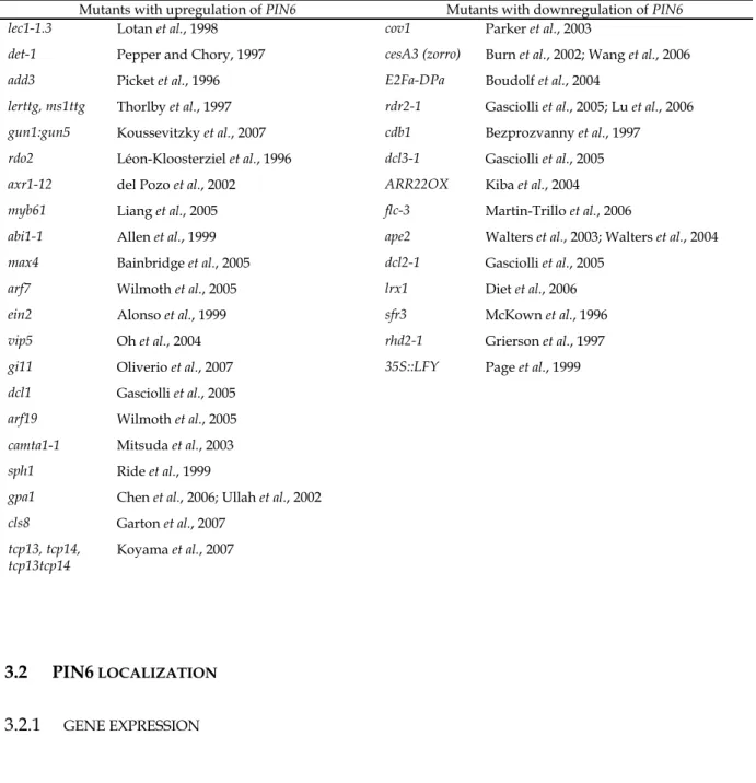
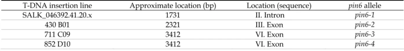
Table 3.1. Partial list of responsive elements present in PIN6 promoter sequence. 51 Table 3.2. List of mutants in which PIN6 expression is up- or downregulated by at least ± 15%. 54Table 3.3. T-DNA insertion lines used for screening pin6 mutants. 62
Table 3.4. Overexpressor and RNAi lines generated for pin6. 65
Table 3.5. Percentage of germinated WT and pin6 seedlings 2 days after vernalisation. 68
Table 3.6. Age of the plant at the transition to reproductive phase. 72
Table 3.7. Numbers of second order stem branches and second order inflorescence branches arising from rosette and cauline nodes in WT and pin6 plants.
A proteína PIN6 é um dos membros menos caracterizados da família de proteínas PIN, associadas ao transporte de auxina em Arabidopsis thaliana. Nesta planta modelo, o transporte polar auxínico tem sido associado a diversos processos fisiológicos. Por exemplo, o movimento acropetal de IAA, do caule para a raíz, tem sido implicado no desenvolvimento de raízes laterais, enquanto que o movimento basipetal de IAA, do ápice radicular para a junção caule-raíz, tem sido associado à resposta à gravidade. As proteínas PIN desempenham um papel crucial no transporte polar auxínico e desta forma medeiam o estabelecimento e manutenção de meristemas, a iniciação e posicionamento de órgãos laterais, a formação do tecido vascular e as diversas formas de tropismo. Neste trabalho procedeu-se ao estudo da função do gene PIN6 recorrendo a diversas técnicas de engenharia genética e de biologia molecular.
O gene PIN6 apresenta níveis de expressão génica relativamente baixos e específicos, sendo detectado apenas em determinadas células e em etapas específicas do desenvolvimento vegetal. O
PIN6 é expresso durante a formação de raízes laterais desde a fase de iniciação da raíz, quando as
células do periciclo, nos vasos condutores, são activadas para entrarem em divisão celular, até fases mais tardias, como a da emergência da raíz lateral. Na fase de indução de raízes laterais o gene PIN6 encontra-se a marcar as células fundadoras da futura raíz lateral no periciclo. Numa fase posterior, de emergência das raízes laterais, PIN6 é expresso nas margens da raíz lateral, na zona de contacto com a raíz principal, formando uma estrutura em forma de anel. O PIN6 está, ainda, presente no meristema apical caulinar, em domínios restritos localizados sob os locais de formação de novos órgãos e nos vasos condutores dos mesmos. Este gene parece estar maioritariamente relaccionado com o desenvolvimento de novos órgãos laterais ao nível dos meristemas.
Os avanços recentes ao nível da Biologia disponibilizaram uma elevada diversidade de estratégias para caracterização funcional de um determinado gene ou família de genes. O recurso a técnicas de genética reversa permite a caracterização funcional de um gene pela determinação dos efeitos causados pela sua ausência. Para a caracterização funcional do PIN6 procedeu-se a uma pesquisa de alelos provenientes de colecções de mutantes com inserções de T-DNA. Foram ainda criadas linhas transgénicas com redução dos níveis de expressão do PIN6. Para o efeito, recorreu-se à estratégia de RNA de interferência (RNAi).
Os fenótipos das diferentes linhas foram analisados em diversas condições de crescimento. As mutações resultaram em taxas de crescimento da raíz primária superiores e num aumento da produção de raízes laterais em relação às plantas de fenótipo selvagem. Tendo em conta esse fenótipo, a proteína PIN6 é, na generalidade, repressora dos processos de crescimento.
O fenótipo dos mutantes pin6 é, ainda, afectado pelo fotoperíodo, uma vez que os fenótipos observados nas plantas crescidas em condições de dia longo foram mais drásticos do que os patentes nas crescidas em presença de luz contínua. Para avaliar uma possível regulação ao nível da
com pigmentos receptores de luz, dos quais se destaca o fitocromo A.
Adicionalmente, os mutantes germinaram mais cedo e fazem a transição meristemática da fase vegetativa para a fase reprodutiva antes do fenótipo selvagem, produzindo menos folhas roseta e menos ramificações secundárias nos caules e nas inflorescências. Estes resultados apontam para uma relação estrita entre o PIN6 e genes de identidade dos meristemas vegetativo e floral.
Os mutantes pin6 são ainda hipergravitrópicos, sugerindo um papel para PIN6 na percepção da gravidade, provavelmente em conjunto com PIN2. A proteína PIN2 foi já caracterizada como envolvida na resposta à gravidade e o seu gene é expresso juntamente com o PIN6 nos tecidos na raíz. Os níveis de expressão destes dois genes são afectados por alguns factores comuns.
Actualmente está disponível uma vasta gama de informação proveniente de experiências de análise de transcritos a larga escala e de pesquisa in vitro de parceiros de interacção, especialmente em Arabidopsis thaliana. Cada vez mais se torna necessário recorrer a estratégias de biologia de sistemas que permitam análises transversais dos resultados obtidos, cruzando dados de fontes diversas para obtenção de informações sobre determinados genes ou processos de interesse. Uma análise in silico dos dados existentes forneceu informação adicional necessária à caracterização do gene PIN6 no que diz respeito à compreensão da sua função e dos mecanismos de regulação que o regem nos processos de desenvolvimento vegetal mencionados, nomeadamente no que diz respeito às respostas a outras hormonas vegetais.
Dessa análise concluiu-se que a expressão de PIN6 é afectada a diversos níveis, nomeadamente factores de transcrição para os quais existem elementos reguladores na sequência do promotor de PIN6 e que estão envolvidos na iniciação de fases do desenvolvimento. A nível da iniciação das germinação e desenvolvimento foliar, o PIN6 parece ser induzido pela diminuição dos níveis de LEC1, um factor de transcrição envolvido na dormência e em fases embrionárias do desenvolvimento foliar. MAX4, membro de uma família de genes envolvida na ramificação do caule, parece ser também um repressor da expressão do PIN6, uma relação já descrita para outros genes
MAX e PIN. FLC e VIP estão envolvidos no controlo temporal da floração e percepção da
vernalização, respectivamente, e interagem um com o outro para promover a iniciação floral estando, possivelmente, envolvidos no controlo da expressão do PIN6. Ao nível da formação de raízes laterais, a proteína PIN6 pode interagir com MDR4, uma proteína envolvida no transporte basipetal auxínico. A sobre-expressão de E2Fa-DPa, um factor de transcrição envolvido na divisão celular e ligado à resposta auxínica, resulta na repressão do PIN6. Adicionalmente, estão presentes no promotor do PIN6 elementos reguladores do ciclo celular, bem como da síntese de componentes da parede celular, sugerindo que a função do PIN6 nas células meristemáticas ocorre principalmente ao nível da divisão celular.
expressão génica do PIN6.
Constata-se a existência de redundância aonível dos membros da família de proteínas PIN: no mutante pin1 o domínio de expressão do PIN6 expande-se para filas adicionais de células companheiras dos vasos condutores, de modo a equiparar a falta de proteína PIN1 nessas células.
Com este trabalho pretendeu-se sugerir hipóteses explicativas dos mecanismos pelos quais PIN6 faz a ligação entre o transporte auxínico e os processos de desenvolvimento em que esta proteína está envolvida, nomeadamente na transição de fase, no estabelecimento e manutenção do meristema apical caulinar, na formação de novos órgãos, folhas e raízes laterais, e na resposta gravirópica. Neste trabalho propõem-se, ainda, estratégias para abordagem dos diferentes pontos que ainda carecem de esclarecimento.
Palavras-chave: transporte auxínico; manutenção do meristem; fotoperíodo; transição de fase; gravitropismo.
and in particular cells at distinct time points. This gene seems to be mainly involved with new lateral organ development. It is expressed in lateral roots, since early stages when pericycle cells are activated for cell division, and in the shoot apical meristem, in restricted domains directly below sites of new organ formation. Screening for T-DNA insertional mutant alleles and generation of knock-down transgenic lines for PIN6 provided the tools to characterize this gene’s function. Phenotypes of those lines were analyzed under different growth conditions and included faster growth rates, longer roots and production of more lateral roots. PIN6 is therefore likely to be a negative regulator of overall growth processes. pin6 phenotype is regulated by photoperiodism, as phenotypes were more drastic under specific photoperiodic conditions. Furthermore, pin6 mutants germinate and make meristem transition from vegetative to reproductive phase earlier than WT, producing less rosette leaves, less secondary branches and inflorescence stems. These results imply a tight regulation between PIN6 and both vegetative and floral meristem identity genes. In addition, pin6 mutants are hypergravitropic, proposing a role for PIN6 in gravity perception, probably in a concerted fashion with PIN2. Auxin upregulates PIN6 expression levels and induces its ectopic expression in additional root tissues. A certain degree of redundancy exists among PIN protein family members. In fact, in the pin1 mutant PIN6 protein localizes to additional cell files, thus compensating for the absence of PIN1. An additional analysis of in silico available data from microarray experiments provided extra information required to better understand PIN6 function and its regulation, namely by other hormones. Explanations regarding the mechanisms by which PIN6 links auxin transport to developmental processes as phase transition, new lateral organ emergence and gravitropism, are proposed.
µl Microliter
2,4-D 2,4-Dichlorophenoxy acetic acid A*** Absorbance at **** nm
aa Amino acids ABA Abscisic acid
ACC 1-Aminocyclopropane-1- carboxylate
AGI Arabidopsis Genome Initiative Amp Ampicillin
AP Alkaline phosphatase APS Ammoniumperoxodisulfate A.tumefaciens Agrobacterium tumefaciens AVG Aminoethoxyvinylglycine BCIP 5-bromo-4-chloro-3'-
Indolyphosphate p-toluidine salt BFA Brefeldin A
Bisacrylamide N,N’-methylenebisacrylamide bp Basepair
BP BP recombination (Gateway®) BSA Bovine serum albumin
cDNA Complementary deoxyribonucleic acid CDS Coding sequence CSPD Disodium 3-(4-methoxyspiro {1,2-dioxetane-3,2’-(5’-chloro) tricyclo[3.3.1.13,7]decan}-4-yl) phenyl phosphate
CTAB Cetyl trimethyl ammonium bromide
DEPC Diethylpyrocarbonate DMF N,N-dimethylformamide DMSO Dimethylsulfoxide DNA Deoxyribonucleic acid dsRNA Double-stranded RNA DTT Dithiothreitol E.coli Escherichia coli
EDTA Ethylendiaminetetraacetate g Gram GA3 Giberellic acid Gm Gentamycin GUS β-Glucuronidase h Hour
HOAc Acetic acid IAA 3-Indole acetic acid IgG Immunoglobulin G IPTG Isopropyl-ß-D-thiogalactopyranoside J Joule Kan Kanamycin Kin Kinetin KOAc Potassiumacetate kb Kilobase kDa KiloDalton l Liter LR LR recombination (Gateway®) m Meter mg Milligram min Minute ml Milliliter mM Millimolar mol Mole
mRNA Messenger ribonucleic acid 1-NAA [alpha]-Naphthalene acetic acid NaOAc Sodium acetate
NaCl Sodium chloride NaOH Sodium hydroxide
NBT Nitro-blue tetrazolium chloride NH4OAc Ammonium acetate
nm Nanometer
NP40 Nonylphenyl-polyethylene glycol NPA N-(1-naphthyl)thalamic acid
Nr. Number o/n Overnight OD Optical density
oligo(dT) Oligodeoxythymidylic acid PAGE Polyacrylamide gel
electrophoresis
PCIB p-Chlorophenoxyisobutyric acid PCR Polymerase chain reaction PEG Polyethylene glycol
PMSF Phenylmethylsulphonyl fluoride PVDF Polyvinylidene difluoride PVPP Polyvinylpolypyrrolidone QC Quiescent centre Rif Rifampicin R.T. Reverse Transcriptase RNA Ribonucleic acid RNase Ribonuclease
rpm Rotation or revolutions per minute
RT Room temperature SDS Sodium dodecylsulfate ssDNA Salmon sperm DNA TEMED
,N,N´,N´-tetramethylethylenediamine TIBA 2,3,5-Triiodbenzoic acid
Tris Tris(hydroxymethyl) aminoethane Triton X-100 Polyoxyethylene-p-isooctylphenol
tRNA transfer RNA Tween 20 Polyoxyethylene (20) Sorbitan
Monolaurate
U Unit of enzyme activity UTR Untranslated region UV Ultraviolet V Volt vol. Volume X-Gluc 5-bromo-4-chloro-3-indoxyl β–D-glucuronidase w Weight WT Wildtype Ω ohm
A Ala Alanine C Cys Cysteine D Asp Aspartic acid E Glu Glutamic acid F Phe Phenylalanine G Gly Glycine H His Histidine I Ile Isoleucine K Lys Lysine L Leu Leucine M Met Methionine N Asn Asparagine P Pro Proline Q Gln Glutamine R Arg Arginine S Ser Serine T Thr Threonine V Val Valine W Trp Tryptophane Y Tyr Tyrosine NUCLEOTIDES A Adenine C Cytosine G Guanine T Thymine U Uracil R A or G Purine Y C or T Pyrimidine
W A or T Weak hydrogen bonding S C or G Strong hydrogen bonding M A or C Amino group at common position K G or T Keto group at common position B C, G or T not A D A, G or T not C H A, C or T not G V A, C or G not T N A, C, G or T Any nucleotide dNTP 2’-deoxynucleotide-5´-triphosphate dTTP 2’-deoxycitidine-5´-triphosphate UTP uracil-5’-triphosphate
1 INTRODUCTION
“The important thing in science is not so much to obtain new facts as to discover new ways of thinking about them.”
Sir William Bragg
1.1
AUXIN IN PLANT DEVELOPMENTPlants are sessile organisms and their non-motility is reflected in the way they grow. Only through a tight developmental control can they adjust to changes in the surrounding environment. Plants grow toward resources such as light, nutrients or water, adjust to soil changes and resist pathogens, among others. It is the aim of developmental biology to understand how growth, cell differentiation, and pattern formation are regulated at the cellular, biochemical, genetic and molecular levels.
Arabidopsis thaliana is a model plant that provides many advantages for molecular research. It is a
small plant, easy to handle, has a short generation time and produces a large number of offspring. At the moment, there are mutations available for theoretically every gene. Its genome is relatively small and since its complete sequencing (Arabidopsis Genome Initiative, 2000), analysis of specific processes and characterization of individual or families of genes/proteins became more accessible (reviewed by Somerville and Meyerowitz, 2001).
Plant hormones (phytohormones) are small organic molecules that affect diverse developmental processes specifically. In contrast to animal hormones, which are produced in specific organs, phytohormones are produced throughout the plant. Virtually every aspect of plant development from embryogenesis to senescence is under hormonal control. Generally, this developmental control is exerted by controlling cell division, expansion, differentiation and cell death. Many developmental processes can be controlled in this way, including formation of the apical-basal and radial pattern, seed germination, determination of plant architecture, flowering, fruit ripening and shedding (reviewed in Bishopp et al., 2006). For instance, auxin is required for the formation of nodule structures upon wounding stress in hop (Santos, 2006), facilitating mycorrhization in chestnut (Barker and Tagu, 2000), among other processes. Cytokinin is required for calla lily regreening process of the spathe (Pais and Chaves das Neves, 1982/83), a natural process involving plant senescence. Plants have a wide array of hormones, including steroids and peptides, as well as the five classical classes of phytohormones: auxins, abscisic acid, cytokinins, ethylene and gibberellins. There is a certain degree of crosstalk and interaction between pathways downstream of these hormonal signals.
Development and organization of plant structures imply that cells are highly sensitive to positional information. The hormones auxin and cytokinin are primary signalling molecules (Barker and Tagu, 2000;
Gattolin et al., 2006; Jasinski et al., 2005; Kiba et al., 2004; Okada et al., 1991; Uggla et al., 1996; Uggla et al., 1998). Polar auxin transport provides the positional cues required for specifying organized plant structures, acting as a morphogen (Barker and Tagu, 2000; Benková et al., 2003; Bennett et al., 2006; Berleth and Sachs, 2001; Bhalerao et al., 2002; Blilou et al., 2005; Friml et al., 2003; Noh et al., 2003; Reinhardt et al., 2003; Weijers
et al., 2005b). The fate of developing tissue can therefore be determined by the sensitivity of growing cells to
auxin and the relative concentrations of other phytohormones. Auxin is readily conjugated to larger molecules that render it inactive. In fact, the majority of indole-3-acetic acid (IAA) in the plant is in the form of inactive conjugates. Auxin conjugation and catabolism can therefore decrease active auxin levels. De novo synthesis and hydrolysis of conjugates counterbalance the developmental regulation of auxin homeostasis by increasing active auxin levels (Ljung et al., 2005). Young aerial tissues and roots, particularly in the RAM, are sites of auxin synthesis. Auxin is synthesized from indole through tryptophan-dependent and tryptophan-independent pathways (reviewed in Woodward and Bartel, 2005). The existence of multiple pathways for IAA biosynthesis and the absence of fully auxin-deficient mutants identified (suggesting that mutations eliminating auxin are lethal) is a reflection of how important this hormone is in plant development (reviewed in Teale et al., 2006).
In higher plants, auxin is involved in embryogenesis, organogenesis, root meristem maintenance, vascular tissue differentiation, hypocotyl and root elongation, apical hook formation, apical dominance, fruit ripening, growth responses to environmental stimuli, among others.
Perturbations involving auxin may result from interference with auxin signalling at four levels (Berleth and Sachs, 2001): auxin synthesis, and of the relations between the age of the tissue or environmental conditions; auxin transport, which can be critical to the localisation of auxin in responding tissues; changes within cells might affect the activity of auxin on receptors; and auxin metabolism might be disrupted in some way.
The developmental patterning processes appear to be flexible and to emerge from complex intercellular crosstalk. It is plausible that auxins act as intercellular messengers in patterning processes in embryos, meristems and vascular development, and that auxin-mediated long-distance signalling could simultaneously integrate morphogenesis throughout the plant (Berleth and Sachs, 2001). Our understanding of the precise mechanisms by which auxin regulates morphogenic processes is, at best, fragmentary. The development of new technologies for classical biochemical approaches, and the widespread use of model plants such as Arabidopsis thaliana for genetic and molecular studies have led to great advances in the biology of plant hormones. Several genes involved in synthesis, conjugation, transport, perception and/or signal transduction of IAA have already been identified (Abel and Theologis, 1996; Benková et al., 2003; Bennett et al., 1996; Bennett et al., 2006; Dharmasiri et al., 2005a; Friml et al., 2002a; Friml et al., 2002b; Gälweiler et al., 1998; Geisler et al., 2005; Gray et al., 1999; Guilfoyle, 1998; Jaillais et al., 2006; Müller et al., 1998; Noh et al., 2001; Okada et al., 1991; Steinmann et al., 1999; Ulmasov et al., 1997a; Ulmasov et al., 1997b), providing us with both the conceptual structure and the experimental tools to investigate plant structure and morphogenesis at the molecular level. The use of these mutants led to the isolation of genes encoding auxin receptors, such as the TIR1 auxin receptor (Dharmasiri et al., 2005a;
Kepinsky and Leyser, 2005; Tan et al., 2007), transporters and proteins involved in subsequent signal transduction processes, thus linking auxin to several morphogenic processes.
Pleiotropic effects of the exogenous application of hormones complicate full understanding of the underlying mechanisms controlling specific hormone activities in the SAM. Crosstalk between different classes of hormones, such as auxin and cytokinin, or cytokinin and gibberellin, is common in plant development. Both auxin and ethylene contribute to several developmental processes. In root development, ethylene-regulated growth is dependent on auxin transport from the root apex via the lateral root cap and on auxin responses occurring in multiple elongation zone tissues (Swarup et al., 2007). The ability of the ethylene precursor 1-aminocyclopropane-1-carboxylic acid (ACC) to inhibit root cell elongation was significantly enhanced in the presence of auxin. By upregulating auxin biosynthesis, ethylene facilitates its ability to inhibit root cell expansion (Swarup et al., 2007). Synergistic effects of auxin and ethylene have been reported for the regulation of hypocotyl elongation (Smalle et al., 1997; Vandenbussche et al., 2003), root hair growth and differentiation (Pitts et al., 1998), apical hook formation (Li et al., 2004), root gravitropism (Buer et al., 2006), and root growth (Pickett et al., 1990; Rahman et al., 2001), suggesting that these two signalling pathways also interact at the molecular level. A component involved in this auxin-ethylene crosstalk is the POLARIS (PLS) peptide (Chilley et al., 2006). Mutation of PLS results in an enhanced ethylene-response phenotype, defective auxin transport and homeostasis, and altered microtubule sensitivity to inhibitors (Chilley et al., 2006). PLS expression is repressed by ethylene and induced by auxin (Chilley et al., 2006). The authors suggest that PLS is a negative regulator of ethylene response, modulating cell division and expansion via downstream effects on microtubule cytoskeleton dynamics and auxin signalling, thereby influencing root growth and lateral root development (Chilley et al., 2006).
1.2
AUXIN MODE OF ACTION: PERCEPTION AND SIGNALLINGAuxin-regulated gene expression triggers most processes controlled by this hormone. Many auxin-induced genes are regulated by the interplay of two classes of transcription factors: auxin-response factors (ARFs) and the Aux/IAA repressors (Hagen and Guilfoyle, 2002). Several Aux/IAA genes are transcribed within minutes of exposure to auxin or protein synthesis inhibitors. They form homo- and heterodimers with one another, as well as with members of the ARF family (reviewed in Ulmasov et al., 1997b; Kim et al., 1997). Aux/IAA repressors are composed of four conserved domains, of which domain II is essential for the instability of these proteins - it contains a degron sequence (GWPPV) which is the target of the SCF ubiquitin ligase complex (Gray et al., 1999; Kepinsky and Leyser, 2004). ARFs bind to auxin-response promoter elements (AuxREs) of auxin-responsive genes (Kim et al., 1997; Ulmasov et al., 1997b). At basal concentrations of auxin, Aux/IAA repressors are relatively stable – through domains III and IV they can
homodimerize and heterodimerize with ARF. ARF-bound Aux/IAA proteins block transcription from auxin-responsive promoters by controlling the amount of free ARF transcription factors (Ulmasov et al., 1997a). Conversely, when auxin concentrations rise above a certain threshold level, Aux/IAA repressors are destabilized (Tiwari et al., 2001; Zenger et al., 2001). An increase in auxin levels promotes the proteasome mediated degradation of Aux/IAAs, which results in an increasing number of active ARF proteins and transcriptional activation of auxin regulons (Tiwari et al., 2003). In this situation, ARF-ARF dimer binding to AuxREs is facilitated and thereby enables the expression of certain auxin response genes. The large number of Aux/IAA and ARF allow for diverse combinatorial interactions between Aux/IAA and ARF, establishing a regulatory code that programmes auxin responses in a spatial-temporal defined window.
Some of the most related ARF and Aux/IAA proteins share similar expression patterns (Weijers et
al., 2005a). In early embryogenesis, MONOPTEROS (MP)/ARF5 and BODENLOS (BDL) /IAA12 physically
interact and are coexpressed (Hamann et al., 2002). SHORT HYPOCOTYL2 (SHY2)/IAA3 and NON-PHOTOTROPIC HYPOCOTYL4 (NPH4)/ARF7 or ARF19 pairs regulate auxin response in the root (Weijers et al., 2005a), while MASSUGU2 (MSG2)/IAA19 and NPH4/ARF7 interact during hypocotyl growth and lateral root development (Tatematsu et al., 2003).
The SCF (SKP-Cullin-F-box) complex is involved in a wide range of signal transduction processes by ubiquitylating target proteins that are selected by F-box proteins. SCF complexes select and covalently modify their target proteins through addition of several ubiquitin peptides, forming a multi-ubiquitin chain that targets them for degradation by the 26S proteasome (reviewed in Teale et al., 2006). The F-box protein TRANSPORT INHIBITOR RESPONSE1 (TIR1) was identified in the past few years as a receptor for IAA (Dharmasiri et al., 2005a; Kepinski and Leyser, 2005). There are over 700 F-box proteins in Arabidopsis (Gagne et al., 2002) and TIR1 belongs to a small subfamily of seven related genes (Dharmasiri et al., 2005b). TIR1 is a component of the SCFTIR1 ubiquitin-protein ligase protein complex (Dharmasiri et al., 2005a;
Kepinski and Leyser, 2005), which also includes the scaffold protein cullin (CUL1), SKP1-like proteins (ASK1/ASK2), and the ring-domain protein RBX1 (Gray et al., 1999; reviewed in Moon et al., 2004). The core cullin, SKP1-like and RBX1 proteins provide the catalytical activity necessary for the transfer of the activated ubiquitin to the target protein, whereas target specificity is conferred by the F-box protein (Gray et
al., 2001).
Loss-of-function tir1 mutants show mild alterations in auxin response and development (Dharmasiri et al., 2005b; Ruegger et al., 1998). A quadruple mutant for TIR1 and its three most closely related genes, AUXIN SIGNALLING F-BOX PROTEINS 1, 2 and 3 (AFB1-AFB3) shows a more severe phenotype and is auxin insensitive. Moreover, AFB proteins interact with Aux/IAAs in an auxin-dependent manner, indicating that auxin binding is collectively mediated by TIR1 and the AFB proteins (Dharmasiri et al., 2005b). The phenotypic variability and presence of some auxin signalling in the quadruple mutant may be explained by residual AFB activity or by the presence of other genes coding for auxin receptors (reviewed in Bishopp et al., 2006).
TIR1 recruits its substrates, Aux/IAA repressors, in an auxin concentration-dependent manner, targetting them for degradation. More recently, its crystal structure was resolved, revealing how auxin
molecules fit into a surface pocket of TIR1, filling a hydrophobic cavity at the protein interface, thus enhancing the interactions between Aux/IAA repressors and TIR1 (Tan et al., 2007). Crystal structures revealed the mushroom shaped complex formed between TIR1 and ASK1, with the leucine-rich-repeat domain of TIR1 forming the cap, and the F-box of TIR1 along with ASK1 forming the stem. On top of the TIR1 leucine-rich-repeat domain lays a “pocket” which functions in both auxin binding and substrate recruitment (Tan et al., 2007). The Aux/IAA peptide binds in close proximity to the auxin-binding site in the upper part of the pocket. The GWPPV motif is packed directly against auxin and covers the auxin binding site. This “traps” auxin in the binding pocket until the Aux/IAA peptide is released. Not only does TIR1 bind IAA, it also binds two synthetic auxins, 1-NAA and 2,4-D, with different affinities, IAA showing the highest (Tan et al., 2007).
It is possible that the SCFTIR1 complexes do not account for all of the responses of Arabidopsis to
auxin, such as plasma membrane-associated proton pump. AUXIN BINDING PROTEIN1 (ABP1) 1 is a candidate for such an extracellular receptor, as changes in ion transport associated with early stages of auxin-induced growth can be inhibited by extracellular treatment with anti-ABP1 antibodies (Leblanc et al., 1999). ABP1 is a soluble, ER-located, dimeric glycoprotein, involved in cell expansion, stomatal closure, plasma-membrane hyperpolarization and cell division (Chen et al., 2001; Rück et al., 1993; Steffens et al., 2001). However, there is no data available on downstream signalling events after the binding of auxin to ABP1 or whether there might be a link to the SCFTIR1 pathway. Considering the almost instantaneous auxin
responses that ABP1 can mediate, it is possible that gene expression is not required to be involved in certain aspects of auxin signalling (reviewed in Teale et al., 2006).
1.3
AUXIN TRANSPORTAuxin transport and distribution are essential for polar development in plants, and their capacity to respond to environmental stimuli. Auxin is synthesized in young, apical tissues, but acts in virtually all parts of the plant.
Two main pathways describe the transport of auxin: a fast, non-directional transport in the phloem and a slower, directional, so-called polar auxin transport in various tissues. The phloem transport occurs in both basipetal (from the apex towards the base) and acropetal (from the base towards the apex) directions, proceeds relatively fast (5-20 cm/h) and correlates well with transport of assimilates. This transport often corresponds to the transport of inactive auxin conjugates. In contrast, polar auxin transport is specific for active free auxins, occurs in a cell-to-cell manner and has a strictly unidirectional character. Polar auxin transport requires energy, is saturable and sensitive to protein synthesis inhibitors, which taken together suggest the existence of specific auxin transport proteins. The Chemiosmotic Hypothesis (Rubery and Sheldrake, 1974; Raven, 1975) explains polar auxin transport: in the relatively acidic environment of the cell
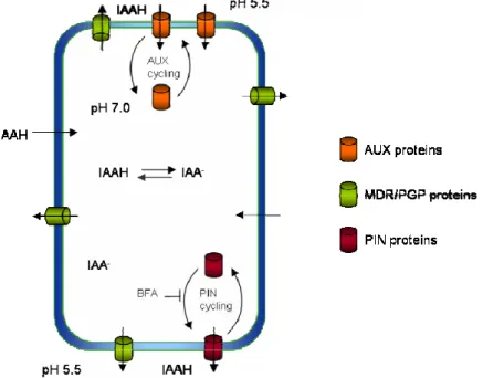
wall (pH around 5.5) about 15% of IAA exists in its protonated form (IAAH). This non-charged, lipophilic molecule passes easily through the plasma membrane by diffusion. In the cytoplasm, where the pH is more basic (around 7) IAAH dissociates and the resulting IAA- anion is imprisoned inside the cell due to its low
membrane permeability (Fig. 1.1.). Therefore, the existence a specific efflux carrier was postulated and the polarity of the flux is explained by the protein’s asymmetric distribution in cells.
Fig. 1.1. Chemiosmotic hypothesis of polar, cell-to-cell, auxin transport. A pH gradient across the plasma membrane leads to the accumulation of IAA in the cell. A higher pH inside the cell promotes auxin molecules (IAAH) dissociation, rendering them unable to pass passively back through the membrane. Therefore, IAA becomes trapped inside the cell. Auxin efflux carriers (PINs, MDR/PGPs) are thus required to transport auxin out of the cell. In addition, auxin influx carriers (AUX) transport auxin anions (IAA ) into the cell. The polar subcellular localization of PIN proteins is important for directional auxin transport, and is accompanied by constitutive endocytic cycling of the PIN proteins. Auxin itself inhibits endocytosis of PINs, increasing their levels at the cell surface. The inhibitor of vesicle trafficking BFA represses the exocytosis of PINs but not of AUX1. The subcellular dynamics of PGP proteins is as yet unclear.
-Polar auxin transport is then required for local accumulation of auxin, necessary for developmental processes such as embryo axis formation (Friml et al., 2003), organ development (Reinhardt et al., 2000), root meristem maintenance (Sabatini et al., 1999) or tropic growth responses (Friml et al., 2002b; Ottenschläger et
al., 2003). Several approaches enable us to visualize such local accumulations of auxin and the gradients
created thereby, which include the auxin responsive promoter DR5 (Ulmasov et al., 1997b), immunolocalization of IAA (Avsian-Kretchmer et al., 2002) or direct auxin measurements in tissue sections (Casimiro et al., 2001; Marchant et al., 2002; Ljung et al., 2005).
1.3.1
INFLUX AND EFFLUX TRANSPORTERSEven though the rates of auxin synthesis and conjugation are important for the overall auxin status of the plant, it is the fine concentration gradients across only a few cells that have powerful effects on plant development, as they provide vectorial information to the tissues (reviewed in Teale et al., 2006).
AUX1 is an auxin cellular influx carrier present in positions consistent with vascular loading at the sources of synthesis (in leaves) and unloading at the sink tissues (in roots; Marchant et al., 2002). Establishment of apical-basal epidermal polarity in Arabidopsis roots is also dependent on AUX1 activity, since aux1 mutant displays apical shifting of root hair initiation consequence of change on trichoblast polarity (Grebe et al., 2002). aux1 shows a strong agravitropic phenotype, suggested to be a result from disruption of basipetal auxin transport in root and/or loss of AUX1 expression in gravi-sensing columella cells (Marchant et al., 1999; Swarup et al., 2001). AUX1 transports auxin directly (Yang et al., 2006) and its asymmetric subcellular localization is dependent on an ER protein AXR4 specifically involved in AUX1 trafficking to the plasma membrane (Fig. 1.1.; Dharmasiri et al., 2006). The other members of the LAX (AUX--like) gene family remain to be characterized.
Polar auxin transport is also dependent on a group of ABC transporters belonging to the MULTIDRUG RESISTANCE (MDR)-like family, also known as the P-GLYCOPROTEINS (PGPs; Sanchez-Fernandez et al., 2001; Martinoia et al., 2002; Noh et al., 2001). Knockout mutations in the
Arabidopsis thaliana MDR1 gene blocks 80% of basipetal transport in seedling hypocotyls and in the
inflorescence stem (Noh et al., 2001). PGP1, the family member most closely related to MDR1, is also involved in basipetal auxin transport in stems (Noh et al., 2001). These MDRs may directly transport IAA, as shown in protoplasts, cell suspension cultures or by expression in mammalian cell lines (Geisler et al., 2005; Bouchard et al., 2006; Petrášek et al., 2006), or aid in polar localization of PIN efflux facilitators (Noh et al., 2003), or both. MDR1’s promoter is active throughout the root (Noh et al., 2001), and MDR1 is one of the highly expressed family members in most root tissues (Birnbaum et al., 2003). MDR4 may also contribute to auxin transport in the root, as it is expressed in the root cap and the epidermal cells of the root apex (Birnbaum et al., 2003; Terasaka et al., 2005). A 30% reduction in root basipetal auxin transport was measured in pgp4-1 mutant, an allele of mdr4 (Terasaka et al., 2005).
1.4 ATPIN FAMILY OF AUXIN EFFLUX CARRIERS
The PIN gene family of Arabidopsis consists of eight transmembrane proteins involved in auxin efflux and whose sequences differ mainly in the central hydrophilic region. Homologous genes have already been found in other plant species (reviewed in Paponov et al., 2005). The PIN family shows a relatively high
similarity in the two groups of membrane-spanning domains, located at the N- and C-termini of the proteins, and a high heterogeneity in the central hydrophilic region (Fig. 1.2.). PIN5 and PIN8 are different in that they lack a central loop domain. The identity between any two members of the family ranges from 32% to 85% (PIN3 and PIN7; Paponov et al., 2005).
The predicted topology of the PIN proteins is similar to several membrane transporters in bacteria and prokaryotes. Indeed, immunolocalization assays located PIN1 to the plasma membrane, in a polar fashion, at the basal side of xylem and cambial cells in the inflorescence axis (Gälweiler et al., 1998). This pattern supports further the hypothesis that PIN1 is regulating auxin transport throughout the plant, as proposed by the chemiosmotic hypothesis (Rubery and Sheldrake, 1974; Raven, 1975). Recently, a set of experiments performed in HeLa cells, a non-plant system, resolved the question of whether PIN proteins play a catalytic role in auxin efflux or solely act as positive regulators of endogenous plant auxin efflux carriers (Petrášek et al., 2006). Indeed, PINs are transporting auxin, given that cells transfected with PIN2 and PIN7 showed strong PIN expression and a substantial increase in the net flux of natural auxin IAA (Petrášek et al., 2006). Whether or not other plant-specific co-factors are required for auxin efflux is not yet known. Furthermore, at least four of the other PINs (PIN1, PIN4, PIN6 and PIN7) are rate-limiting for auxin efflux from plant cells (Petrášek et al., 2006).
Fig. 1.2. Typical PIN protein topology: two membrane-spanning domains composed of five to six transmembrane sequences linked by a central hydrophilic loop.
The five PIN genes characterized so far have been linked to organogenic (PIN1 and PIN4), embryogenic (PIN1, PIN4 and PIN7), gravitropic (PIN2 and PIN3) and phototropic (PIN1 and PIN3) processes (Friml et al., 2002a; Friml et al., 2002b; Müller et al., 1998; Paponov et al., 2005; Sabatini et al., 1999). PIN2 localizes to the upper membrane of the epidermal and cortical cells of root apices, regulating acropetal auxin transport essential for gravitropic responses (Müller et al., 1998). PIN2 protein in roots is predominantly present at the basal side of epidermal cell files oriented towards the elongation zone and further on the apical side in root cortical cells. PIN3 is mainly localized in the lateral walls of the endodermal cells in the shoot and in root columella cells it has a uniform distribution (Friml et al., 2002b). Upon gravistimulation, PIN3 becomes polarized to the lateral side of the columella cells and later to lateral root cap (Friml et al., 2002b). The polar localization of PIN4 in the root quiescent centre directs the auxin flow towards the initial columella cells, the location of main auxin accumulation in the root apex (Friml
et al., 2002a; reviewed in Friml, 2003; Sabatini et al., 1999). PIN7 is apically localized in the basal cell of the
the apical-basal axis by efflux-dependent auxin gradient during embryogenesis, as it is expressed immediately after the zygotic division, being the first marker for polarity (Friml et al., 2003).
Absence of PINs results in aberrant local auxin accumulation patterns and most aspects of the respective mutant phenotypes can be phenocopied by chemically inhibiting polar auxin transport (Friml et
al., 2003; Friml et al., 2002a; Friml et al., 2002b; Luschnig et al., 1998). Loss-of-function pin1 mutants grow a
single naked pin-shaped stem after floral transition, without any floral organs (Okada et al., 1991), a phenotype due to lower rates of auxin transport (Gälweiler et al., 1998). In fact, application of auxin to the
pin1 stem results in promoting lateral growth (Reinhardt et al., 2003).
Members of the PIN protein family are considerably homologous and show significant functional overlap among them, as shown by the increasingly severe phenotypes of the multiple pin mutants (Friml
et al., 2003; Blilou et al., 2005; Vieten et al., 2005). Moreover, the most similar family members can
complement each other in knockout mutants. For example, in the pin1 background, PIN4 expression domain extends into the cells where PIN1 would have been present (Blilou et al., 2005).
To this moment, all PIN proteins analyzed show subcellular asymmetric localization within auxin transport-competent cells, even though some may be found in specific cell types without pronounced polarity. Polarity of PIN localization correlates with the direction of auxin transport and/or with the local accumulation of auxin in adjacent cells, suggesting that PIN polar localization directs the intercellular auxin flow (Benková et al., 2003; Blilou et al., 2005; Friml et al., 2003; Friml et al., 2002a; Friml et al., 2002b; Gälweiler
et al., 1998; Heisler et al., 2005; Müller et al., 1998). Recently, by manipulating PIN polarity and monitoring
auxin transport it has been proposed that PIN localization is sufficient to direct auxin flow in plants (Wiśniewska et al., 2006).
Auxin induces the expression of many PIN proteins in an Aux/IAA-dependent manner (Vieten
et al., 2005). However, correct expression of PIN proteins is dependent on pre-existing cell patterning rather
than auxin concentration (Xu et al., 2006). Moreover, auxin alone is not sufficient for root specification in the absence of PIN-mediated auxin transport (Weijers et al., 2006). It is therefore unlikely that PINs alone determine cell specification or polarity. Taken together, the results suggest that PINs mediate distinct developmental signals as part of a wider developmental programme (Xu et al., 2006).
It is essential to understand the molecular mechanism of targetting auxin transport components to opposite sides of the cell. The relationship between PINs and other auxin transporters is still unclear. It seems that there are at least two different polar targetting machineries, as AUX1 and PIN proteins are controlled by different subsets of vesicle trafficking pathways, showing different sensitivities to various inhibitors (Kleine-Vehn et al., 2006). PIN1 is mislocalized in the pgp1 mdr1 double mutant, suggesting a certain extent of control of the MDR/PGPs over PIN localization (Noh et al., 2003). PGP1 and PGP19 co-localize with PIN1 in the shoot apex and with PIN1 and PIN2 in root tissues. In addition, PGPs and PINs interact in yeast 2-hybrid and co-immunoprecipitation essays, suggesting that PIN-PGP interactions are required to enhance auxin transport activity (Blakeslee et al., 2007). Quite contradictory, it has also been shown that PIN1 function is not dependent on the presence of other factors, which includes MDR/PGPs (Petrášek et al., 2006).
Active auxin transport mediates cellular auxin concentration and is therefore a determinant part in the creation of gradients crucial for coordination of plant development. There are additional factors contributing to the relationship between auxin signalling and transport. Many kinases are regulated by auxin (Beltran-Peña et al., 2002; Kovtun et al., 2000; Mockaitis et al., 2000) and members of the MAP kinase cascade, including MAP KINASE KINASE-7 (MKK7), have been demonstrated to negatively regulate polar auxin transport (Dai et al., 2006). RAC-LIKE (ROP) GTPases may also have a role in auxin action. Auxin activates ROP3 (Tao et al., 2002), causing Aux/IAA proteins to aggregate. These nuclear protein bodies activate the 26S proteasome to mediate degradation of Aux/IAA proteins. Consequently, this could be the connection between auxin perception at the plasma membrane and control of gene regulation in the nucleus (Tao et al., 2002; Tao et al., 2005).
1.4.1 PINs
IN PLANT DEVELOPMENT1.4.1.1 PINs in leaf development and phyllotaxis
During embryogenesis the basic plant body plan is established, including the formation of meristems that will perpetuate cell division and originate new organs in the mature plant. The apical-basal axis is defined, with the shoot apical meristem being located at one end of the axis and the root apical meristem in the opposite. While during embryogenesis all cells undergo division, after germination further growth and development becomes restricted to special areas of the plant that maintain embryonic character - the meristems. Meristem cells divide to create the tissues and organs responsible for the general architecture, shape and size. Primary root and shoot meristems are formed during embryogenesis. Most plants also develop secondary meristems during postembryonic development, such as axillary meristems, floral meristems, intercalary meristems and lateral meristems (e.g. lateral roots). The vegetative shoot apical meristem is usually indeterminate in its development, i.e., shows no predetermined limit to growth. In contrast, floral meristems are determinate: all meristematic activity stops when the last floral organs have been generated.
Meristematic undifferentiated cells that retain the ability for cell division indefinitely are stem cells. Similarly to animal stem cells, when plant stem cells divide, one of the cells retains the identity of the mother stem cell while the other undergoes a specific developmental program leading to its differentiation.
The shoot apical meristem (SAM) is located at the shoot apex and leaves, stems and axillary meristems are produced from its derivative cells. The SAM is composed of three concentric tissue layers: L1 (outermost), L2 and L3 (innermost). It can also be divided into different histological zones. The peripheral zone (PZ) flanks the central zone (CZ). Underneath lies the rib zone (RZ), which gives rise to the internal tissues of the stem. Lateral organs initiate from the PZ, at the meristem flanks. The CZ contains
self-maintaining, slowly dividing cells – the stem cells, which provide initials for both the PZ and the RZ (Bowman and Esched, 2000).
Coordinating the formation and differentiation of pluripotent stem cells in apical meristems seems to depend on transcriptional regulation and intercellular signalling. Several classes of transcription factors have been shown to take part in SAM maintenance. The indeterminate nature of the meristem and the formation of organ boundaries require class I KNOTTED1-like homeobox (KNOX1) genes, expressed in many plant species in specific patterns in the SAM (reviewed in Hake et al., 2004). Members of the NAC group of transcription factors, including NO APICAL MERISTEM (NAM), CUP-SHAPED COTYLEDON1 (CUC1), CUC2 and CUC3, are also essential for meristem establishment and organ boundary formation. Their expression in narrow strips in the SAM will correspond to the future organ–organ and meristem-organ boundaries (Laufs et al., 2004; Mallory et al., 2004).
The patterning of the SAM and its stem cell niche is tightly controlled by a central regulatory mechanism in which the homeodomain protein WUSCHEL (WUS) specifies stem cell identity. In the active shoot meristem, WUS is expressed in a small cell group underneath the presumed position of the stem cells and positively regulates the synthesis of the small secreted peptide CLAVATA3 (CLV3) ligand at the stem cell niche (Laux et al., 1996). CLV3, in turn, acts together with the receptors CLV1/CLV2, found mostly in an underlying domain of the L3 layer, to repress WUS expression, thereby creating a negative feedback loop that restricts the size of the stem cell population (Clark et al., 1997; Fletcher et al., 1999; Mayer et al., 1998; Schoof et al., 2000; Trocochaud et al., 2000). In addition, CLV3 has been shown to be required for the dynamic regulation of meristem size, restricting the domain of its own expression in the CZ by preventing re–specification of PZ cells as CZ cells (Reddy and Meyerowitz, 2005). CLV3 has a long-range effect on cell division rate to restrict SAM size. Increasing CLV3 levels at its native expression domain results in repression of WUS expression. Balance signalling between CLV3/WUS seems to coordinate the cell-fate at the meristem: cells with higher CLV3 expression allow cells to adopt peripheral cell identity while WUS-expressing cells keep meristematic identity (Müller et al., 2006). Additional signalling components may be necessary to stabilize WUS expression at the meristem organizing center.
The formation of new organs during plant development obeys predetermined genetic programmes, as well as possesses an inherent plasticity allowing the plant body to organize and adapt in response to environmental cues. Correct SAM function requires the maintenance of a delicate balance between the production of lateral organs from its flanks and indeterminate growth at its center. Leaf primordia arise from a small subset of cells in the L1 and L2 layers of the PZ that acquires leaf founder cell identity. Cells from the L1 layer originate epidermis, cells from the L2 layer differentiate as photosynthetic mesophyll cells and cells from L3 give rise to vascular elements and bundle sheath (reviewed in Sinha, 1999). These leaf primordia are separated from the main shoot through the establishment of a boundary between the organ-forming cells and the meristem cells. Once leaf primordia initiate, leaves grow rapidly through active cell division and expansion from the
initial primordia cells. Mature organs present three developmental axes of polarity: the proximodistal, the dorsoventral (adaxial-abaxial), and mediolateral axes.
A number of mutants displaying leaf formation defects have been isolated in Arabidopsis thaliana. Plants mutated in either ASYMMETRIC LEAVES1 (AS1) or AS2 form asymmetric, rumpled, lobed leaves with ectopic leaflet-like organs on the petioles (Byrne et al., 2000; Semiarti et al., 2001; Sun et al., 2002). AS1 encodes a MYB domain–containing putative transcription factor (Byrne et al., 2000) and AS2 is a member of the plant-specific LATERAL ORGAN BOUNDARIES (LOB) domain (LBD) family of proteins, encoding a widely expressed protein with a Leu zipper motif (Iwakawa et al., 2002). AS1 and AS2 function in overlapping developmental pathways (Serrano-Cartagena et al., 1999; Ori et al., 2000; Semiarti et al., 2001) and physically interact with one another (Xu et al., 2003). Both genes repress expression of three class I
KNOX genes, BREVIPEDICELLUS (BP), KNAT2, and KNAT6, which promote the activity and maintenance
of the SAM (Byrne et al., 2000; Ori et al., 2000; Semiarti et al., 2001). While AS1 and AS2 in turn repress BP,
KNAT2, and KNAT6 activity in the initiating primordia, class I KNOX gene STM negatively regulates AS1
and AS2 expression in the SAM maintaining a pool of meristematic cells (Byrne et al., 2000, 2002). This results in a reciprocal negative molecular interaction that promotes leaf initiation at the periphery of the meristem.
Phyllotaxis, the regular way in which leaves are arranged around the stem, is therefore a consequence of the precise spatial regulation of growth within the apex, reflected in the number and order in which leaf primordial form. The relative stage of lateral primordium development is described in terms of plastochron (P), whereby the latest emerging primordium is termed P1, the next oldest primordium is P2 and so forth. The region within the meristem from which the next lateral organ primordium will be formed is then termed P0. Phyllotaxis is characterized by the divergences angles between two consecutively generated organs. The most common patterns in nature are spirals with divergence angles of 137.5º. Mathematical modelling suggests that spiral leaf arrangements are superior for light capture or as a solution to a packing problem (reviewed in Kuhlemeier, 2007).
The current model - the Canalization Hypothesis - explaining the regulation of leaf primordium formation and phyllotaxis by auxin transport proposes that auxin is transported by PIN1 in epidermal cells towards the SAM, where it further induces PIN1 expression, which will, in turn, promote auxin accumulation at the site of incipient primordium formation (Reinhardt et al., 2003). Leaf primordia then drain auxin through their central midveins, causing auxin depletion in the surrounding cells. Through a combination of positive feedback (i.e. auxin accumulation) and lateral inhibition (i.e. depletion of auxin from adjacent tissues), auxin accumulates at certain distances from the existing primordia, allowing for the phyllotactic patterning. Consistently with this model, direct auxin application or auxin transport inhibition enlarges primordia or reduces their lateral separation, eventually leading to primordium fusion (Reinhardt
et al., 2000). Additionally, inhibition of polar auxin transport through chemicals or by mutations in PIN1
specifically inhibits organogenesis. A naked meristem that grows normally but forms no lateral organs will arise. The defect can be rescued by the application of a microdroplet of auxin to the peripheral zone of such a pin-shaped meristem (Reinhardt et al., 2000). Auxin accumulates at sites of incipient organ formation