UNIVERSIDADE DE LISBOA
FACULDADE DE MEDICINA DE LISBOA
Social memory in zebrafish: behavioral assessment and the role of
Brain-Derived Neurotrophic Factor (BDNF)
Natália da Costa Madeira
Orientador: Professor Doutor Rui Filipe Nunes Pais de Oliveira Co-Orientador: Professora Doutora Maria José de Oliveira Diógenes
Master Degree in Neurosciences 2015
Todas as afirmações efectua das no presente documento são da
exclusiva responsabilidade do seu autor, não cabendo qualquer
responsabilidade à Faculdade de Medicina de Lisboa pelos
conteúdos nele apresentados.
A impressão desta dissertação foi aprovada pelo Conselho Científico da
Faculdade de Medicina de Lisboa em reunião de 23 de Julho de 2015
AGRADECIMENTOS
A realização deste trabalho não teria sido possível sem a colaboração, compreensão e ajuda de inúmeras pessoas. Após este período de intensa aprendizagem científica e pessoal, quero deixar o meu profundo agradecimento a todos os que me apoiaram ao longo deste percurso académico:
Ao Instituto Gulbenkian de Ciência, por me ter acolhido durante o decurso da tese e pelas aprendizagens multidisciplinares que adquiri;
Ao Professor Doutor Rui Oliveira, pela oportunidade de desenvolver o meu trabalho no seu grupo de investigação e por todo o conhecimento que me transmitiu;
À Professora Doutora Maria José Diógenes, por me ter aceite a co-supervisão deste trabalho;
À Magda Teles, por todo o apoio e dedicação durante este percurso; por todas as técnicas e conhecimento que me transmitiu. Principalmente por toda a amizade que me ofecereu.
À Júlia Pinho, pelo apoio árduo e incansável de uma verdadeira amiga. Sem dúvida que foste um elemento chave para o sucesso deste trabalho.
Aos membros do IBBG (Ana Faustino, Rodrigo Abreu, João Lopes, Ana Rita Nunes, André Monteiro, Ana Sofia Félix, António Roleira e Ana Cruz), pelo apoio demonstrado ao longo destes meses;
À minha mãe, por me ter fascinado desde a infância pelo maravilhoso mundo do conhecimento. Pela maravilhosa forma como me fez ver a natureza, as pessoas os animais e o mundo.
À minha familía, pelo apoio incondicional;
Ao meu namorado, por todo o amor e companheirismo;
À Mariana Marques, pela enorme amizade. Pelo interesse e satisfação que demonstrava quando falávamos do meu projecto e por todas as dicas. És e continuarás a ser uma das grandes marcas no meu percurso académico;
À Carolina Toste, por todo o apoio, amizade e carinho que demonstrou ao longo do mestrado. Pela sua enorme capacidade crítica e pelo seu infinito conhecimento que nos inspira para continuar neste caminho.
À Rita Belo, pela enorme amizade, carinho e dedicação que demonstrou a longo destes anos.
Ao Luís Ceríaco, pela amizade e carinho. Por me teres conduzido desde cedo ao mundo da investigação e me teres proporcionado muito conhecimento.
Ao Professor Doutor Paulo de Oliveira, por me ter ajudado e incentivado a descobrir o mundo das Neurociências e por toda a confiança depositada em mim.
INDEX
LIST OF TABLES ……….………..…….. xiii
LIST OF FIGURES ………..…. xiv
ABBREVIATIONS ………..…... xvii
RESUMO ……….... xxi
ABSTRACT ……….………….. xxv
CHAPTER 1 – INTRODUCTION ……….…………. 1
1. Behavioral flexibility in social context ………. 3
2. Neuronal plasticity ………... 5
2.1. Molecular mechanisms underlying neuronal plasticity and its influence on memory formation ……….…………..… 6
2.2. Brain-derived neurotrophic factor ……….……….. 9
2.2.1. BDNF and memory processing ……….………... 11
3. Social brain ………..……….. 12
3.1 Hippocampus ………. 14
4. Social recognition memory ……….……… 16
4.1. Neurobiological basis of social memory ……….……… 18
4.2. Zebrafish as a model for study social memory ………. 21
CHAPTER 2 – OBJETIVES ……….……….. 25
CHAPTER 3 – METHODS ……….……….. 27
1. Time dependent expression of c-fos and bdnf ………..……… 29
Animals housing ……… 31
Experimental procedure ………..……….. 31
Sampling ………..……….. 32
Western-blotting ……….. 33
Stripping and re-probing membranes ……….……….. 33
RNA isolation, cDNA synthesis and real-time quantitative PCR amplification ……….………. 34
qPCR data processing ………....……….. 35
Statistics analysis ……….……… 35
Animals housing ………….………..……… 36
Animal tagging ………..………. 37
Experimental tests ……….………. 43
Brain tissue sampling ……….……… 44
RNA isolation, cDNA synthesis and real-time quantitative PCR amplification ……….. 45
Statistics analysis ………..……….. 45
CHAPTER 4 – RESULTS ……….……….. 47
Effects of Kainic acid on c-fos and bdnf gene expression in the telencephalon ………..……… 49
Effects of Kainic acid on C-FOS and BDNF protein levels in the telencephalon ………... 52
Object recognition ……….………. 56
Effects of different sensory modalities in social recognition using the social discrimination test ………..……… 57
Test of social recognition using the habituation-dishabituation paradigm ………..……….. 59
Sociability and memory performance ………..………. 61
Effects of social memory on bdnf gene expression in the hippocampus and amygdala ………...……… 62
CHAPTER 5 – DISCUSSION ………..……… 65
CHAPTER 6 – CONCLUSIONS ……….……….……… 73
LIST OF TABLES
TABLE 1|PRIMER SEQUENCES, ANNEALING TEMPERATURE AND EFFICIENCY.ALL PRIMERS WERE COMMERCIALLY SYNTHESIZED (SIGMA-ALDRICH,GERMANY). ... 35
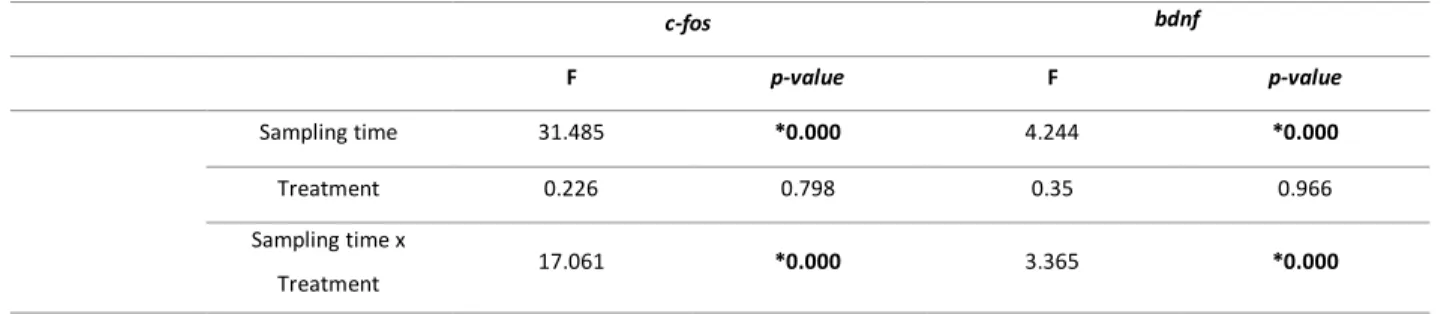
TABLE 2│ANOVA ANALYSES FOR C-FOS AND BDNF GENE EXPRESSION AFTER A TREATMENT WITH KAINIC ACID (KA)
AND SALINE SOLUTION (CT) AT DIFFERENT SAMPLING TIMEPOINTS (0H,0.5H,1H, 2H,4H, 8H, 24H AND
48H}. MAIN EFFECTS AND RESULTS FROM PLANNED COMPARISONS ARE PRESENT IN THIS TABLE. ABBREVIATIONS:B, BASELINE;CT, CONTROL GROUP;KA,KAINIC ACID GROUP.*P<0.005. ... 51
TABLE 3 |ANOVA ANALYSES FOR C-FOS GENE EXPRESSION AFTER A TREATMENT WITH KAINIC ACID (KA) OR SALINE SOLUTION (CT) AT DIFFERENT SAMPLING TIMEPOINTS (0H,0.5H,1H,2H,4H,8H,24H AND 48H). MAIN EFFECTS AND RESULTS FROM PLANNED COMPARISONS ARE PRESENT IN THIS TABLE.ABBREVIATIONS:B,
BASELINE;CT, CONTROL GROUP;KA,KAINIC ACID GROUP.*P<0.005. ... 54
LIST OF FIGURES
FIGURE 1|MODEL OF SYNAPTIC TRANSMISSION AT EXCITATORY SYNAPSES (CITRI AND MALENKA,2008) ... 7
FIGURE 2|MODEL OF BDNF-P75NTR AND BDNF-TRKB INTRACELLULAR SIGNALING PATHWAYS (CUNHA ET AL., 2010) ... 10
FIGURE 3|SCHEMATIC REPRESENTATION OF EXPERIMENTAL PROCEDURE.AFTER ADMINISTRATION OF KA OR SALINE SOLUTION, ANIMALS WERE SACRIFICED AT DIFFERENT TIMEPOINTS. ... 32
FIGURE 4|BRAIN AREAS COLLECTED.A)MID SAGITTAL CUT; RIGHT SIDE FOR QPCR AND LEFT SIDE FOR WESTERN
-BLOTTING;B)IN EACH HALF PART OF THE BRAIN, FIVE MACROAREAS WERE COLLECTED. ... 32
FIGURE 5|TOP VIEW OF EXPERIMENTAL SET-UP.THE GREY DOTTED LINES REPRESENT REMOVABLE PARTITIONS AND THE YELLOW SQUARES REPRESENT THE LOCALIZATION OF DEMONSTRATORS’ FISH. ... 37
FIGURE 6|SCHEMATIC REPRESENTATION OF BEHAVIORAL PROCEDURE ... 38
FIGURE 7 | DIAGRAMS OF THE RECOGNITION MEMORY TASKS. A) OBJECT DISCRIMINATION TEST; B) SOCIAL DISCRIMINATION TEST AND C) SOCIABILITY TEST. ... 42
FIGURE 8 │TEMPORAL CHANGES IN IEG’S EXPRESSION AFTER A TREATMENT WITH KAINIC ACID.ANIMALS WERE INJECTED WITH EITHER SALINE OR KAINIC ACID SOLUTION AND SACRIFICED IMMEDIATELY,30 MIN.,1HR,2HR, 4HR, 8HR, 24HR OR 48HR AFTER INJECTIONS. THE TIME COURSE, AFTER EITHER SALINE OR KA
ADMINISTRATION, FOR INCREASES IN RELATIVE LEVELS OF MRNA IN TELENCEPHALON ARE SHOWN FOR C-FOS
(A) AND BDNF (B).ACUTE INTRAPERITONEAL KAINIC ACID (0.5MG/KG) INCREASES (A) C-FOS EXPRESSION 30
MINUTES AFTER TREATMENT;(B) BDNF EXPRESSION 0 MINUTES AFTER SALINE SOLUTION ADMINISTRATION.THE RESULTS ARE THE MEAN±SEM OF SEVENTEEN DIFFERENT EXPERIMENTS PERFORMED IN INDEPENDENT ANIMALS.*P≤0.05;***P≤0.001. ... 49
FIGURE 9|TEMPORAL CHANGES IN C-FOS PROTEIN LEVELS AFTER A TREATMENT WITH KAINIC ACID.ANIMALS WERE INJECTED WITH EITHER SALINE OR KAINIC ACID SOLUTION AND SACRIFICED IMMEDIATELY,30 MIN.,1HR,2HR, 4HR, 8HR, 24HR OR 48HR AFTER INJECTIONS. THE TIME COURSE, AFTER EITHER SALINE OR KA
INTRAPERITONEAL KAINIC ACID (0.5MG/KG) DOES NOT INDUCES AN INCREASE IN PROTEIN LEVELS.THE RESULTS ARE THE MEAN±SEM OF SEVENTEEN DIFFERENT EXPERIMENTS PERFORMED IN INDEPENDENT ANIMALS. *P≤0.05;**P≤0.01;***P≤0.001 ... 52
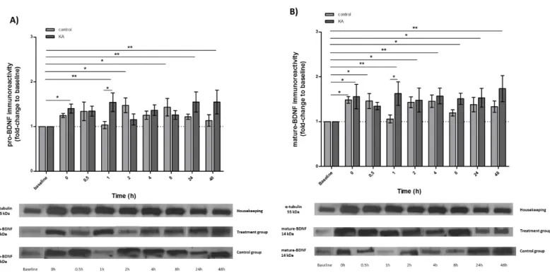
FIGURE 10|TEMPORAL CHANGES IN BDNF PROTEIN LEVELS AFTER TREATMENT WITH KAINIC ACID:A) PRO-BDNF IMMUNOREACTIVITY;B) MATURE-BDNF IMMUNOREACTIVITY.ANIMALS WERE INJECTED WITH EITHER SALINE OR KAINIC ACID SOLUTION AND SACRIFICED IMMEDIATELY,30 MIN., 1HR, 2HR,4HR, 8HR,24HR OR 48HR AFTER INJECTIONS. THE TIME COURSE, AFTER EITHER SALINE OR KA ADMINISTRATION, FOR INCREASES IN PROTEIN LEVELS IN TELENCEPHALON ARE SHOWN FOR BDNF. ACUTE INTRAPERITONEAL KAINIC ACID
(0.5MG/KG) INDUCES AN INCREASE IN PRO-BDNF LEVELS 0HRS, 1HRS, 24HRS AND 48HRS AFTER ADMINISTRATION; FOLLOWING AN INCREASE IN MATURE-BDNF LEVELS AT 0HRS,1HRS,2HRS,4HRS,8HRS,
24HRS AND 48HRS.THE RESULTS ARE THE MEAN±SEM OF SEVENTEEN DIFFERENT EXPERIMENTS PERFORMED IN INDEPENDENT ANIMALS.*P≤0.05;**P≤0.01;***P≤0.001 ... 53
FIGURE 11| OBJECT RECOGNITION. THE TIME SPENT IN EACH CHAMBER WERE USED TO INVESTIGATE OBJECT RECOGNITION.ON DAY 1, ANIMALS WERE FREELY TO EXPLORE TWO NAIVE OBJECTS DIFFERENT IN SHAPE BUT WITH THE SAME VOLUME AND COLOR.24HR LATER, INDIVIDUALS WERE FACED WITH A FAMILIAR AND A NON
-FAMILIAR OBJECT.FOCAL FISH CANNOT DISCRIMINATE BETWEEN A FAMILIAR AND A NON-FAMILIAR OBJECT.A) PREFERENCE SCORE AND B) EXPLORATION TIME (%)*P≤0.05 ... 56
FIGURE 12|SOCIAL MEMORY ASSESSMENT FOR OLFACTORY (A), VISUAL (B) AND INTEGRATION OF BOTH (C)
STIMULI. THE TIME SPENT IN EACH CHAMBER WERE USED TO INVESTIGATE SOCIAL RECOGNITION, BASED ON DIFFERENT TYPES OF SENSORIAL MODALITIES. ON DAY 1, ANIMALS WERE FREELY TO EXPLORE TWO NAIVE CONSPECIFICS.24HR LATER, INDIVIDUALS WERE FACED WITH A FAMILIAR AND A NON-FAMILIAR CONSPECIFICS. A) FOCAL FISH CANNOT DISCRIMINATE FAMILIAR FROM NON-FAMILIAR CONSPECIFICS ONLY BASED ON OLFACTORY INTERACTION; B)FOCAL FISH DISCRIMINATE FAMILIAR FROM NON-FAMILIAR CONSPECIFICS ONLY BASED ON VISUAL INTERACTION;C)FOCAL FISH DISCRIMINATE FAMILIAR FROM NON-FAMILIAR CONSPECIFICS BASED ON VISUAL AND OLFACTORY INTERACTIONS.+P<0.06;*P≤0.05 ... 57
FIGURE 13|STIMULUS EXPLORATION (%) FOR SOCIAL DISCRIMINATION MEMORY USING OLFACTORY (A), VISUAL
(B) AND MULTIMODAL (C) CUES.ON THE FIRST DAY ANIMALS EXPLORE ACTIVELY THE CONSPECIFICS.ON THE SECOND DAY INDIVIDUALS REDUCE SIGNIFICANTLY THE EXPLORATION TIME. ... 58
FIGURE 14|SOCIAL MEMORY ASSESSMENT USING A HABITUATION-DISHABITUATION PARADIGM.THE TIME SPENT IN EACH CHAMBER WERE USED TO INVESTIGATE SOCIAL RECOGNITION.ON DAY 1, ANIMALS WERE FREELY TO EXPLORE ONE NAIVE CONSPECIFIC.24HR LATER, INDIVIDUALS WERE FACED WITH A NOVEL (A) OR A FAMILIAR CONSPECIFIC (B). ... 59
FIGURE 15|CORRELATION BETWEEN SOCIAL MEMORY PERFORMANCE AND SOCIABILITY. ... 600
ABBREVIATIONS
AMPA α-amino-3-hydroxy-5-methyl-4-isoxazolepropionic acid
ANOVA Analysis of variance
AVP Arginine vasopressin
BDNF Brain-derived neurotrophic factor
BSA Bovine serum albumin
CaMKII calcium/calmodulin dependent kinase II
cAMP cyclic adenosine monophosphate
CNS Central nervous system
CREB cAMP response element-binding protein
Dl Lateral zone of dorsal telencephalic area
Dm Medial zone of dorsal telencephalic area
EGR-1 Early growth response protein 1
ELF1-a Elongation factor 1
FFA Fusiform face area
HEPES N-(2-hydroxyethyl)-1-piperazine-N’-(2-ethanesulfonic acid)
IEG Immediate-early gene
KO Knockout
LTD Long-term depression
LTP Long-term potentiation
MAPK Mitogen-activated protein kinases
MRS Mesolimbic reward system
NMDA N-methyl-D-aspartate
OT Oxytocin
PCR Polymerase chain reaction
PKA Protein kinase a
PLCγ Phospholipase C gamma
PVDF Polyvinylidene fluoride
SDS-PAGE Sodium dodecyl sulfate polyacrylamide gel electrophoresis
SBN Social brain network
SEM Standard error of the mean
Resumo
Os comportamentos sociais requerem uma elevada flexibilidade comportamental, nos quais conhecimentos adquiridos a priori são fulcrais para a adaptação a novas situações. No reino animal, as espécies diferem na sua capacidade social: espécies sociais (gregárias) formam grupos sociais coesos e demonstram relações afiliativas entre os vários membros do grupo; contrariamente a espécies associais. Desta forma, espécies gregárias interagem diariamente com outras, onde a capacidade de armazenar e recordar informações se torna claramente importante. Na natureza, a capacidade de recordar informações relativas ao meio ambiente, relembrar a localização de estímulos recompensatórios e a identificação de indivíduos familiares, assume extrema importância do ponto de vista ecológico. O conhecimento que os animais adquirem acerca dos seus conspecíficos e a forma como constroem o conhecimento do mundo social circundante envolve, invariavelmente, a capacidade de categorizar os conspecíficos (por exemplo: idade, género, hierarquia, entre outros). A capacidade de distinguir indivíduos e armazenar essa informação durante longos períodos de tempo é uma vantagem social que facilita interacções posteriores e é essencial para os comportamentos sociais. O reconhecimento social tem como base características sensoriais multimodais que, conjuntamente, permitem o reconhecimento de indivíduos. Em condições laboratoriais, a memória social pode ser estudada recorrendo a dois paradigmas amplamente utilizados em roedores: teste de discriminação social binária e teste de habituação-desabituação. Ambos os paradigmas baseiam-se em alterações comportamentais espontâneas na exploração dos indivíduos, quando re-expostos a indivíduos familiares.
O presente trabalho avalia a performance do peixe-zebra nas duas variantes do teste de memória social e num teste de memória asocial. Desta forma, 48 animais foram submetidos a uma série de seis paradigmas comportamentais. Cada indivíduo realizou os seguintes testes experimentais: 1) reconhecimento de objectos (O); reconhecimento social: 2) químico (C); 3) visual (V); 4) visual e químico (V+C); Teste de habituação-desabituação: 5) exploração de indivíduos novos (N+N); 6) exploração de individuos novos e familiares (N+F). Os animais foram inicialmente identificados (código de cores) para possibilitar a sua análise individual em cada teste. Ao longo das experiências os animais encontraram-se agrupados sem manterem contacto com os indivíduos “estímulo”. Os vídeos recolhidos durante aos experiências foram analisados utilizando um programa de video-tracking (Ethovision®). Para cada indivíduo foi calculado um score de
preferência – relaciona o tempo de investigação de um estímulo com o tempo de investigação de ambos – e a taxa de exploração – relaciona a percentagem de tempo de exploração de ambos os estímulos tendo em conta o tempo total do teste. Os resultados comportamentais revelaram que: i) o peixe-zebra não demonstrou preferência por nenhum dos objectos apresentados 24 horas após o primeiro teste; ii) os indivíduos apresentaram preferência por indivíduos não-familiares quando a interação envolveu estímulos visuais e quiímicos; iii) apresentam preferência por indivíduos familiares quando a interacção tem por base estímulos somente visuais; iv) não demonstram preferência por nenhum dos indivíduos quando dispunham somente de estímulos químicos; v) o paradigma comportamental de habituação-desabituação não demonstrou preferência por animais novos ou familiares, 24h após o primeiro teste. Os resultados exploratórios revelaram que durante o primeiro dia de experiência, os indivíduos apresentaram taxas de exploração acima dos 50%, demonstrando a tendência natural do peixe-zebra em explorar novos estímulos. No entanto, 24 horas após o primeiro teste, os animais reduziram significativamente os níveis de exploração; sugerindo habituação ao teste do segundo dia.
Inúmeros estudos têm sugerido a amigdala como a área cerebral onde a memória social está alocada, sugerindo os neuropeptidos (oxitocina e vasopressina) como reguladores deste tipo de memória. No entanto, até ao momento, nenhum estudo demonstrou o envolvimento do BDNF na memória social. Desde que o brain-derived neurotrophic factor (BDNF) provou estar envolvido em mecanismos de plasticidade sináptica, o seu papel em diversos mecanismos de memória e aprendizagem foi amplamente demonstrado. No processamento da memória, o BDNF pode actuar a vários níveis moleculares: regula canais iónicos como o canal de Na+ e canal de K+; modula
receptores glutamatérgicos (NMDA e AMPA) e afecta a síntese proteica. Desta forma, o presente trabalho pretendeu avaliar o papel do BDNF na memória social. Os paradigmas comportamentais foram inicialmente balanceados, o que significa que cada grupo de animais terminaria o conjunto dos testes em paradigmas comportamentais diferentes. Desta forma, 2 horas após o térmito dos testes comportamentais os animais foram sacrificados. As áreas homólogas ao hipocampo (Dl) e à amigdala (Dm) foram extraídas e posteriormente analisados por qPCR. A análise da expressão genética demonstrou que os diferentes testes comportamentais produziam diferenças nos níveis de expressão do bdnf. Na amigdala, não se verificaram diferenças signicativas nos níveis de expressão do bdnf. No entanto, ao nível do hipocampo verificou-se que os indivíduos que realizaram o teste de memória social com base em estímulos químicos (C) possuia níveis de
expressão de bdnf significativamente menores aos testes de memória social com base em estímulos visuais ou visuais (V) e químicos (V+C ).
Por outro lado, foi possível correlacionar o teste de memória social com a sociabilidade. Desta forma, foi possível estabelecer uma correlação significativa entre os indivíduos mais sociais e os que apresentam maior capacidade de reconhecimento na realização do teste de memória social.
O último objectivo do trabalho pretendia perceber a forma como os genes de expressão imediata (immediate early genes – IEG’s) poderão funcionar como marcadores de resposta neural. Desta forma, foram analisados 128 animais submetidos a um tratamento com ácido kaínico para posterior análise do c-fos e bdnf. Os indivíduos foram injectados intraperitonialmente com 0.5mg/Kg de ácido kainico (grupo tratamento) ou solução salina (grupo controlo) e, posteriormente, sacrificados a diferentes tempos de amostragem (imediatamente após a injecção; 0 minutos; 30 minutos; 1 hora; 2 horas; 4 horas; 8 horas; 24 horas; 48 horas após a injecção). Posteriormente, os cérebros foram recolhidos e dissecaram-se as 5 principais macroareas: telencéfalo; diencéfalo; tecto óptico; cerebelo e tronco cerebral. Em cada individuo, foi realizado um corte sagital entre os dois hemisférios cerebrais; metade do cérebro foi alocado para a análise da expressão génica (qPCR) enquanto o restante foi utilizada para avaliar os níveis proteicos (Western-blot). Os resultados revelaram que, após um insulto externo, ocorre um aumento significativo nos níveis de expressão do c-fos após 30 minutos. Subsequentemente, regista-se uma diminuição abrupta da expressão de c-fos que se mantém ao longo de 48 horas. No entanto, os resultados da análise proteica não revelaram diferenças significativas nos níveis de proteinas do C-FOS ao longo do tempo e entre os tratamentos (controlo vs tratamento). Relativamente ao bdnf, verificou-se um aumento significativo nos níveis expressão 0 minutos após a administração de uma substância salina, que se mantém no mínimo até 24 horas. 48 horas após a administração, ambos os tratamentos (ácido kaínico e solução salina) diminuem significativamente os níveis de expressão do bdnf. A nível proteico, avaliou-se as duas isoformas do BDNF – pro-BDNF e mature-BDNF. Os níveis da isoforma pro-BDNF sofrem um aumento significativo 2 e 8 horas após a administração da solução salina; e 0, 1, 4, 24 e 48 horas após a administração do ácido kaínico. A isoforma mature-BDNF revelou um aumento significativo 0 e 30 minutos após a administração da solução salina; e 0 minutos, 1h, 2h, 4h, 8h, 24h e 48h após a administração do ácido kaínico.
Os resultados obtidos pretendem demonstrar a utilidade do peixe-zebra no estudo da memória social, através da validação de paradigmas comportamentais para esse efeito. Por outro lado, demonstrámos que o hipocampo tem um papel neste tipo de memória que é dependente da origem sensorial dos estímulos utilizados.
Abstract
The ability of animals to gather information about their social and physical environment is essential for their ecological function. Animals are often organize conspecifics into categories (e.g. sex, age, hierarchical status). This social organization is underpinned by social recognition. Individuals use an assortment of different cues to obtain information about their environment and to recognize the individuals that they encounter. The present study evaluated the influence of 3 different sensory cues on social recognition: visual-only; olfactory-only and visual + olfactory. We used two different paradigms to assess social recognition memory – social discrimination paradigm and a habituation-dishabituation paradigm – both adapted from mouse studies. We also explored an asocial task – novel object test. A series of six experiments were performed by each individual. Subsequently, bdnf expression levels were evaluated in hippocampus and amygdala.
The behavioral results show that zebrafish: i) did not demonstrate preference for any of the objects presented 24h after the initial test; ii) preferentially associated with conspecifics that are novel, when using both chemical and visual cues; iii) exhibit preference for familiar conspecifics when only visually cues are accessible; iv) did not show preference between two individuals, when only chemical cues were available; v) failed to demonstrate social recognition memory using the habituation-dishabituation paradigm; vi) are highly inquisitive animals. The genetic expression analysis demonstrates no differences in bdnf expression levels in the amygdala. However, in the hippocampus, low-levels of bdnf were present when animals performed a discrimination paradigm based only on olfactory cues; in contrast with individuals that performed the same behavioral paradigm based on visual and visual + chemical cues. ). Here we propose that the high levels of BDNF observed in the Dl could affect LTP and consequently the production and secretion of OT in the Dm. Our findings present a new possibility for the role of neural connections between the Dl and Dm regions, mediated by BDNF, with significant impact on social memory.
We also evaluated the use of immediate early genes (c-fos and bdnf) as neural response markers in the zebrafish telencephalon. We analyzed animals that were submitted to a kainic acid treatment. To study the temporal response of IEG’s (c-fos and bdnf) to the treatment, an analysis of gene expression (qPCR) and protein levels (Western-blot) was performed. Animals were given intraperitoneal injections (0.5mg/Kg) of saline solution (control group) or kainic acid (treatment group) and sacrificed at different sampling time points (immediately after, 0min., 30min., 1h, 2h,
4h, 8h, 24h and 48h). The results show that c-fos suffered an up-regulation 30 min. after treatment; followed by an abrupt decrease in c-fos expression levels. However, protein levels did not show significant differences in C-FOS protein. Regarding bdnf: an up-regulation was observed 0min. after saline solution administration; 48h after administration of either KA or saline solution, a decrease in bdnf expression levels was observed. At the protein level both pro-BDNF and mature-BDNF levels were analyzed. Pro-BDNF levels increase 2h and 8h after saline solution administration; and 0min., 1h, 4h, 24h and 48h after KA administration. In contrast, mature-BDNF levels increase 0min. and 30min. after saline solution administration; and 0min., 1h, 2h, 4h, 8h, 24h and 48h after KA administration.
The present work demonstrates the usefulness of zebrafish in studying social memory, by the validation of paradigms to that effect. This work also suggests that the hippocampus possesses a role in this type of memory, depending on the origin of the cues employed.
C
HAPTER
1
1.
BEHAVIORAL FLEXIBILITY IN SOCIAL CONTEXTEveryday behaviors require a high degree of flexibility, in which prior knowledge is used to adapt to new situations. Understanding the processes and mechanisms by which animals act, learn, remember and use this information to navigate their daily lives, is currently one of the goals in the field of neurosciences. Thus, studying how the brain can produce complex behaviors and cognitive states, and how it can be influenced by social experience is one of key themes in cognitive neuroscience.
Behavior interfaces an animal characteristics – genetic, physiology and personality traits – and its environment. Animals must act to select suitable habitats; to maintain homeostasis; to avoid predation; to find and select mates; to rear their offspring successfully and to manage their social relationships with conspecifics (Kappeler et al., 2013). However, in unpredictable environments animals require an extraordinary ability in modifying behaviors, also known as behavioral flexibility. Across animals, there is a remarkable diversity in naturally occurring behavioral phenotypes. Adaptive behavioral solutions to recurrent, unpredictable environments and social problems should therefore be favored by selection, resulting in robust, species-specific behavior patterns (Duckworth, 2009; Sih et al., 2010). Such flexibility is thought to be supported in part by memory integration.
Social interactions promote changes in morphology, and/or physiology of interacting individuals. There is a large number of gregarious species in the animal kingdom. Contrary to solitary species – with high levels of territorial defenses that lead to the active exclusion of conspecifics – individuals of social species establish relationships with each other. Social species have the ability to adjust their behavior according to previous social experiences and social contexts (Oliveira, 2009). This behavioral flexibility, caused by changes in social environments, results in an optimization of relationships between organisms, known as “social plasticity”. Social plasticity is a process that can be divided into different phases: 1st) animals collect relevant cues
from the social environment; 2nd) they evaluate the salience and valence of social stimulus
(appraisal mechanism); 3rd) mechanisms of cognitive appraisal result in different forms of neuronal
plasticity: short-term changes (activation of proteins that act as transcription factors (CREB); neuronal activity-dependent transcription factors activate IEG’s that can encode other transcription factors (c-fos and egr-1) or synaptic proteins (Arc and Homer1a); and transcription of
miRNAs that regulate translation of synaptic proteins) or long-term changes (epigenetic modifications of genes involved in social behavior (e.g. bdnf)); 4th) temporal and spatial changes in
gene regulation in the social brain network (SBN); 5th) production of hormones or
neuromodulators that can change the strength of connectivity between the nodes of SBN (Oliveira, 2012).
In the specific case of recognition, the identification of conspecifics is essential for the processing of social information. This implies the precise regulation of specific brain mechanisms associated with the recognition, and interpretation of several aspects of social information.
2.
NEURONAL PLASTICITYThe nervous system has the ability to adapt to the environment and to improve its performance with experience. Most learning processes result in long-lasting behavioral changes, but even simple reflexes can be modified transiently. The fact that behavior is learned raises an interesting question: how is behavior modified if the nervous system is wired so precisely? How can changes in neural control of behavior occur when connections between the signaling units, the neurons, are set during early development? The proposal that has proven farsighted is the (neuro)plasticity hypothesis.
Neural plasticity is defined as the brain’s ability to generate and modify neural circuits as a result of experience. The brain is continuously creating new neuronal pathways and altering existing ones in order to adapt to new experiences, learn new information and create new memories. These changes in neural organization may account for various forms of behavior modification, which include adaptation to a mutable environment, various forms of learning and memory and compensatory adjustments in response to functional losses. These changes are a basic requirement for learning and behavioral adaptation in complex organisms (Cowansage, LeDoux, & Monfils, 2010). This dynamic remodeling depends on the ability of environmental stimulation to influence both gene expression and protein activation.
The most well know form of neuronal plasticity is synaptic plasticity. Synapses often have a remarkable capacity for short-term physiological changes (lasting milliseconds to minutes) that refer to activity-dependent modifications of the strength or efficacy of synaptic transmission (at preexisting synapses). Long-term changes (lasting days) can give rise to further physiological changes that lead to anatomical changes, including pruning of preexisting synapses and even growth of new ones (Citri & Malenka, 2008). In this form of plasticity, either the amount of neurotransmitter released from the presynaptic terminal, or its receptor in the postsynaptic neuron are modulated (Colicos & Syed, 2006). Synaptic plasticity possesses a crucial role in the capacity of the brain to incorporate transient experiences into persistent memory traces.
2.1.
MOLECULAR MECHANISMS UNDERLYING NEURONAL PLASTICITY AND ITS INFLUENCE ON MEMORY FORMATIONThe behavior of neural circuits depends on the pattern of synaptic weights that connect individual neurons and consequently define the circuit (Citri & Malenka, 2008). This idea was developed during years, when in the late 1940s Donald Hebb postulated that associative memories are formed in the brain by a process of synaptic modification that strengthens connections, when presynaptic activity correlates with postsynaptic firing (Hebb, 1949). The experimental support for this hypothesis of physiological changes in synaptic strength emerged in 1973 from Timothy Bliss, Tony Gardner-Medwin and Terje Lomo study’s. They reported that a repetitive high-frequency stimulation of excitatory synapses caused an enhancement of synaptic transmission between the stimulated axons and the dentate areas of the hippocampus that could last for hours or even days (Bliss & Lomo, 1973). This phenomena, known as long-term potentiation (LTP) has been object of intense investigation due its important role in molecular and cellular mechanisms by which memories are formed (Martin et al., 2000; Pastalkova et al., 2006; Whitlock et al., 2006). LTP and long-term depression (LTD) are the most extensively studied physiological models of memory formation. The three well-described characteristics of LTP – cooperativity, associativity and durability – have been identified as solid arguments that support the hypothesis that LTP may be a biological substrate for, at least, some forms of memory (Nicoll, Kauer, & Malenka, 1988)
Furthermore, similar to memory, LTP can be generated rapidly and is strength prolonged by repetition. The majority of experimental work aimed to understand the mechanisms of LTP has been performed on excitatory synapses, using high-frequency bursts (tetani), specifically on the synapses between Schaffer collateral and commissural axons and the apical dendrites of CA1 pyramidal cells (Citri & Malenka, 2008; Malenka & Nicoll, 1999).
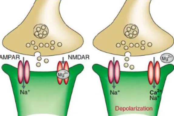
There are two major types of ionotropic glutamate receptors that contribute to postsynaptic response at glutamatergic synapses: AMPA and NMDA receptors; which are often found co-localized on individual dendritic spines. During low-frequency synaptic transmission, glutamate binds to AMPA receptor which has a channel permeable to monovalent cations (Na+ and K+) and
provides the majority of inward current for generating synaptic responses when the cell is close to its resting potential (Figure 1). However, the triggering of LTP requires the activation of NMDA
receptors, a subtype of glutamate receptors, which are voltage-dependent and remain blocked by extracellular Mg2+ (Nowak et al., 1984). Activation of NMDA receptor dissociates Mg2+ from its
binding site allowing the influx of Ca2+ and Na+ to enter the dendritic spine (Lynch et al., 1983;
Malenka et al., 1988) (Figure 1b). This Ca2+ influx through NMDA receptors is responsible for
initiating LTP by activating protein kinases – CaMKII and PKA, and producing cAMP (Citri & Malenka, 2008; Elgersma & Silva, 1999; Lynch et al., 1983). Initially, these kinase proteins phosphorylate receptors and alter the intrinsic proprieties of ligand-gated ion channels; subsequently, they activate local protein synthesis at the synapse and lead to intracellular signaling into the nucleus (via transcription factors), thereby altering gene expression (Alberini et al., 1995; Goelet et al., 1986).
Changes in synaptic strength induced by LTP can be divided into two temporally and mechanistically distinct phases:
Early phase of LTP (E-LTP) – involves modifications of preexisting synapses, as a result of rapid Ca2+influx through NMDA receptor, and subsequent protein phosphorylation
events; induces an increase in synaptic efficacy that lasts for 1-2 hours (Malenka & Nicoll, 1999; Malenka & Bear, 2004; Pang & Lu, 2004).
Late phase of LTP (L-LTP) – requires activation of cAMP-dependent protein kinase (PKA) and the transcription factor CREB (Kandel, 2001) leading to de novo RNA transcription, new protein synthesis and structural changes at synapses; induces an increase in synaptic efficacy lasting over hours or days (Lüscher et al., 2000).
The maintenance of LTP requires pre- and post- synaptic changes that include an increase in neurotransmitter release and a modification of AMPA receptors (Malenka & Nicoll, 1999).
Recently, and elegant study of Nabavi and colleagues showed that fear conditioning – a type of associative memory – can be inactivated by LTD mechanisms and reactivated by LTP, supporting a causal link between synaptic plasticity and memory (Nabavi et al., 2014).
In zebrafish, few studies have been done so far in this field. In 2004, Nam and colleagues showed for the first time that NMDA receptors are synaptically activated and required for induction of LTP in zebrafish telencephalon (Nam, Kim, & Lee, 2004). In zebrafish telencephalon, a repetitive high-frequency stimulation of the connections between Dl and Dm induces a LTP that can be blocked by a NMDA receptor competitive antagonist (APV) (Ng et al., 2012). Further studies have shown the role of NMDA receptors on memory tasks performance, since the uncompetitive antagonist of NMDA receptors MK-801 prevented memory formation (Blank et al., 2009). These findings suggest that molecular and cellular mechanisms underlying learning and memory can be shared by mammals and zebrafish.
2.2.
B
RAIN-
DERIVED NEUROTROPHIC FACTOR(BDNF)
The brain-derived neurotrophic factor (BDNF) belongs to the neurotrophin family of trophic factors. It has multifaceted functions in CNS such as neuronal survival, differentiation, synaptic plasticity and dendritic remodeling (McAllister, Lo, & Katz, 1995; Schinder, Berninger, & Poo, 2000). Among neurotrophins, BDNF and its major receptor TrkB, have the most abundant and widespread expression in the developing and adult brain (Murer, Yan, & Raisman-Vozari, 2001). Secretion of BDNF requires previous expression of BDNF mRNA and the subsequent translation into pre-proBDNF protein.
The BDNF protein is synthesized as a precursor (pre-proBDNF protein) resulting after cleavage in a 32KDa proBDNF protein. ProBDNF is further processed until it is secreted into the extracellular space. This isoform can follow two distinct pathways to leave the intracellular compartment: either the constitutive or the regulated pathway. In the constitutive pathway, proBDNF isoform is secreted as proBDNF and cleaved extracellulary by proteases; whereas in the regulated pathway proBDNF is cleaved intracellulary by enzymes like furin or pro-convertases and originate matureBDNF (14kDa) (Cunha, Brambilla, & Thomas, 2010; Lessmann, Gottmann, & Malcangio, 2003). The biological function of BDNF is mediated by the binding of these secreted homodimeric proteins either to their cognate tropomyosin related kinase (TrkB) receptor or to the common neurotrophin receptor p75NTR (Lessmann et al., 2003). Once released, proBDNF binds
preferentially to pan neurotrophin receptor (p75NTR) and matureBDNF binds preferentially to TrkB
receptors. BDNF is secreted both pre- or post-synpatically in an activity-dependent manner (Pang & Lu, 2004). TrkB and p75NTR play different roles in BDNF function, activating different intracellular
messenger cascades and producing distinct cellular responses. TrkB initiates three major cascades of signaling pathways: PLCγ, PI3K and ERK/MAPK (Cunha et al., 2010), which ultimately lead to the phosphorylation and activation of CREB that mediates transcription of genes essential for survival and differentiation of neurons. p75NTR activation initiates two cascades of signaling pathways: JNK
Figure 2 | Model of BDNF-p75NTR and BDNF-TrkB intracellular signaling pathways (Cunha et al., 2010)
In rat and mice BDNF mRNA is widely distributed throughout the CNS and its presence is correlated with local of protein synthesis. These brain areas include: hippocampus (with highest expression levels), cerebral cortex, thalamus, hypothalamus, olfactory bulb, amygdala, cerebellar granule cell layer and spinal cord (reviewed in: Edelmann, Leßmann, & Brigadski, 2013).
Several studies in teleost fish reported a structural and functional conservation of the amino acid sequence of BDNF throughout evolution. Götz and colleagues cloned BDNF mRNA from platyfish (Xiphorus maculatus) and found that it was 90% identical with mammalian BDNF and had identical biological activity and potency (Götz, Raulf, & Schartl, 1992). Five neurotrophin receptors have been described in zebrafish, two of which have been reported to be isoforms of the TrkB receptor, which share >90% similarity with their mammalian homologous (Martin et al., 1995). An additional BDNF function has been shown by Hashimoto and Heinrich (1997). They reported the involvement of BDNF in fin development of zebrafish, due the presence of BDNF transcripts in the pectoral fin (Hashimoto & Heinrich, 1997). In zebrafish, BDNF and its TrkB receptor are widely distributed along the brain and retina (Germana et al., 2010), playing an essential role during embryonic development (Lum, Huynh, & Heinrich, 2001).
2.2.1. BDNF
AND MEMORY PROCESSINGIt is widely believed that changes in synaptic strength of neuronal connections underlie the formation of memories. The induction of LTP – currently considered to represent the cellular model for memory – is associated with the activation of a large number of signaling cascades, including the ones activated by BDNF. Studies showing the impairment of LTP in hippocampus of heterozygous BDNF knockout animals reported, for the first time, the involvement of BDNF in LTP, which could be rescued by exogenous BDNF (Korte et al., 1995; Patterson et al., 1996). This neurotrophic factor acts through TrkB receptors either pre- or post-synaptically, to modulate LTP (Kovalchuk et al., 2002; Xu et al., 2000). Since BDNF appears to be involved in activity-dependent synaptic plasticity, several studies started to suggest its role in learning and memory mechanisms. In memory processing, BDNF acts at different molecular levels: it regulates ion channels such as Na+ and K+; it modulates glutamatergic receptors (NMDA and AMPA) and it affects protein
synthesis by transcriptional and translational mechanisms (Bekinschtein, Cammarota, & Medina, 2013).
Accumulating evidence shows a correlation between BDNF mRNA expression and behavioral performance in memory tests (Tyler et al., 2002; Yamada & Nabeshima, 2003). Simultaneously, several studies showed that up-regulation of BDNF mRNA expression is increased in the hippocampus after memory tests such as: Morris water maze (Kesslak et al., 1998); radial maze (Mizuno et al., 2000); passive avoidance (Ma et al., 1998); and contextual fear conditioning (Hall, Thomas, & Everitt, 2000). In this context, the hippocampus appears to be involved in the regulation of memory-related BDNF activity. Recently, an increase in BDNF mRNA has been reported in perirhinal cortex 2h after exposure to novel objects (Romero-Granados et al., 2010).
3. T
HE SOCIAL BRAINIn social species, where repeated interactions among the same individuals occur (i.e. social relationships), the success of these relationships depends on specific social skills. One such skill is the ability of individuals to recognize other individuals and to remember past interactions, and adjust their future behavior accordingly (Oliveira, 2013). This ability includes a wide array of cognitive processes such as attention, perception, learning, memory and decision-making. In the past years, several studies have been done to understand how these complex cognitive functions are processed in the social brain (Adolphs, 2010). It has been proposed that the mechanisms responsible for social interactions differ from those involved in non-social interactions (e.g. interactions with physical environment) (Zuberbuler & Byrne, 2006). In mammals, the neuronal circuits that evaluate social stimuli, integrate them and regulate social behavior into adaptive responses have been allocated to a network which integrates the mesolimbic reward system and to social behavior network, that together form the social decision making network (Connell & Hofmann, 2011b). According to this proposal the mesolimbic reward system is responsible for the assessment of the relative value of the social stimuli and the consequences of behaving in dissimilar forms (Connell & Hofmann, 2011a). This circuitry is characterized by massive dopaminergic projections from ventral tegmental area (VTA) to the nucleus accumbens (NAcc), but also includes reciprocal connections between hippocampus (HIP), basolateral amygdala (blAMY), vental pallidum (VP), striatum (Str), lateral septum (LS) and bed nucleus of stria terminalis/medial amygdala (BNST/meAMY) (Connell & Hofmann, 2011b). The social behavior network has been first proposed as the substrate for multiple forms of social behavior in mammals (Newman, 1999). This network compromises six brain nuclei that are reciprocally connected: the lateral septum (LS), preoptic area (POA), anterior hypothalamus (AH), ventromedial hypothalamus (VMH), periaqueductal gray/central grey (PAG/CG) and bed nucleus of stria terminalis/medial amygdala (BNST/meAMY) (Connell and Hofmann, 2011). These areas express sex-steroid hormone receptors and are implicated in a wide range of social behaviors such as parental care, aggression, mating and sexual behaviors, social recognition, affiliation, responses to social stressors and communication (Goodson et al., 2005; Newman, 1999). These multiple forms of social behavior are fundamental and evolutionarily ancient properties of most animal taxa, and as such the brain regions regulating these behaviors are expected to be highly conserved across vertebrates. The
core nodes of Newman’s social behavior network were initially proposed for mammals; however, this framework has been expanded to reptiles, birds and teleosts (Connell and Hofmann, 2011; Goodson et al., 2005). Although the social behavior network and the mesolimbic reward system have been studied as separate circuits, they are anatomically linked by connections between several brain regions and share two nodes: the LS and the BNST. These two circuits complement each other by regulating both the evaluation of the valence and salience of external stimuli and the behavioral output (Connell & Hofmann, 2011b).
3.1.
H
IPPOCAMPUSThe hippocampal formation is one of the most studied neuronal systems in the brain. Its role in memory formation has been studied at almost every level of analysis since the discovery of the patient H.M. with hippocampal damage (Scoville & Milner, 1957). In general, patients with damaged hippocampus show an impairment of new explicit memory acquisition, whereas short-term memory, priming and procedural learning are preserved (reviewed in Bird & Burgess, 2008). The hippocampus appears to have a crucial role in short- and long-term memory (O’Keefe and Nadel, 1978; Anderson et al., 2007).
In nature, the ability to remember environmental information, recall the localization of rewarding stimuli and the identification of familiar conspecifics is clearly adaptive. Several studies have demonstrated the function of hippocampus in different vertebrate taxa (Connell & Hofmann, 2011b; Rodríguez et al., 2002). Although the vertebrates’ forebrain shows an impressive range of morphological variation and specialized adaptations, the close functional similarity of this structure allows the establishment of homologies between mammals and other vertebrates (reviewed in (Connell & Hofmann, 2011a). Developmental studies demonstrate that, contrarily to what happens in mammals, in teleosts there is an eversion of the dorsal part of the neural tube (pallium), resulting in a divergent organization of the mediolateral telencephalon from that observed in mammals (Mueller, Wullimann, & Guo, 2008; Mueller & Wullimann, 2009). In teleosts, the lateral zone of the dorsal telencephalic area (Dl) is currently considered to be the homolog of the mammalian hippocampus (Portavella et al., 2002; Rodríguez et al., 2002; Wullimann & Mueller, 2004). This homology is also supported by behavioral studies that have shown that: Dl ablation leads to impairment in spatial learning acquisition and retention (López et al., 1998); spatial learning acquisition is correlated with an increment of cellular activity in Dl (Vargas et al., 2000); and these effects are similar to those of hippocampal lesions in mammals.
The role of the hippocampus in recognition memory (e.g. object recognition) has been controversial. A study in non-human primates showed a positive correlation between the percentage of damage to the hippocampus and scores on portions of the recognition performance test, suggesting that, the greater the hippocampal damage, the better the recognition (Murray & Mishkin, 1998). In contrast, a convergence of studies using this task show that hippocampal lesions produce recognition memory impairment in monkeys (Beason-Held et al., 1999; Zola et al., 2000),
humans (Reed & Squire, 1997) and rodents (Hampson et al., 1999). Recently, Hitti and Siegelbaum, reported that inactivation of CA2 pyramidal neurons caused a pronounced loss of social memory (Hitti & Siegelbaum, 2014).
4.
SOCIAL RECOGNITION MEMORYWhat animals know about each other, and how they construct and use knowledge of their social world involves at least an ability to recognize different social categories. Animals may categorize, and therefore recognize, individuals according to different social categories – species, group member, kin, age, sex, reproductive status and hierarchical status (Colgan, 1983). These characteristics can be detected through the assessment of cues that do not need to be individual-specific. Social recognition has been defined as “the ability of individuals to categorize conspecifics into different classes (homo- vs heterospecific, same group vs different group, adult vs young, male vs female, kin vs nonkin, dominant vs subordinate, familiar vs unfamiliar) and to recall the learned idiosyncratic identity of a specific individual previously met” (Gheusi et al., 1994). Thus, social memory refers to the ability of animals to change their social behaviors towards a conspecific as a consequence of a previous social encounter with it. To make this possible, a social memory needs to be stored during the initial encounter and retrieved during posterior encounters.
Social memory is a unique form of memory that is critical for reproduction, territorial defense, establishment of dominance hierarchies, pair bonding and allows to understand the structure, organization and evolution of social system (reviewed in (Ferguson, Young, & Insel, 2002; Gheusi et al., 1994). Therefore, the capacity to encode and recall this type of information is required in almost all organisms living in complex social systems. In mammals different species evolved different strategies to encode information: in humans, a specific visual association area (FFA) is responsible for face perception/recognition (Kanwisher, McDermott, & Chun, 1997); in non-human primates, the temporal cortex responds selectively to faces (Perrett, Rolls, & Caan, 1982); in most other mammals, social information is encoded via olfactory (pheromone signaling), auditory or visual signals (reviewed in Ferguson et al., 2002). In birds: long-tailed tits (Aegithalos caudatus) can discriminate between kin and non-kin individuals based on vocalizations (Sharp et al., 2005); in hens discrimination between familiar and unfamiliar conspecifics appears to be visual (Dawkins, 1995; Guhl & Ortman, 1953). In fish kin recognition occurs in Salvelinus alpinus (OlsÉn, Grahn, Lohm, & Langefors, 1998) and nestling recognition in the parental male bluegill (Lepomis macrochirus) (Neff & Sherman, 2003). Also invertebrates (e.g. insects), can distinguish between nestmate and non-nestmate kin based on olfactory cues (Gamboa et al., 1986). As mentioned,
different sensory systems are used in recognition, including olfactory sense, acoustic sense and vision.
The success of social recognition process depends on the integration of various crucial phases: 1) signaling of cues by the stimulus animal (e.g. unique odors, plumage patterns or vocalizations); 2) perception of these cues by other animals; 3) storage of information about familiar individuals cues; and, 4) using this information to discriminate between two individuals.
In laboratory conditions, social memory can be evaluated by changes of spontaneous exploratory behaviors directed towards conspecifics when an individual is re-exposed to a familiar or a novel conspecific. There are two commonly used behavioral tests to study social memory in laboratory animals: the habituation-dishabituation procedure and discrimination procedure. In the former, developed by Thor and Holloway (1982), a juvenile is placed in an adult’s cage for a 5 min encounter. The adult exhibits intense social investigation activity towards the juvenile, and the duration of this investigation reflects the familiarity between the two animals. Thus, a repeated exposure to the same juvenile results in a decrease in investigation time (habituation), which is reversed if a novel conspecific is presented (dishabituation) (Thor & Holloway, 1982). This decrease in social investigation time is taken as evidence of social recognition memory. The discrimination procedure, first described by Mario Engelmann (1995), is based in a binary choice test between a novel and a familiar conspecific (Engelmann, Wotjak, & Landgraf, 1995). Similar to other non-social cognitive tests (object recognition), this paradigm allows the assessment, within the same test, of an animal’s discrimination between two social stimuli. In this test, social recognition is assessed by comparing the difference in the time spent investigating the familiar vs. the unfamiliar conspecific.
Rodents are the most widely study model for this type of memory. Several studies in rodents have demonstrated their remarkable sensitivity to discriminate between familiar and unfamiliar individuals. However, this ability has a limitation in time: it can be a form of short-term memory with a limited duration of 30min to 2hrs (Thor & Holloway, 1982) or it can display robust long-term duration which persists for 24h to 7 days (Kogan, Frankland, & Silva, 2000; Moura, Meirelles, & Xavier, 2010).
4.1.
N
EUROBIOLOGICAL BASIS OF SOCIAL MEMORYSocial memory is an emerging topic of interest in memory research. The neurobiological bases of this type of memory are still poorly understood, with just a few studies addressing this question so far. However, there are several questions that must be addressed: which molecular and cellular mechanisms are involved in its formation? How long can it persist? Which brain areas are involved? What are the physiological differences comparative to others forms of memory?
In the last decade, research in this topic has focused on the role of synaptic plasticity and neuroendocrine responses. Pharmacological studies have shown the role of NMDA receptors, a modulator of synaptic plasticity, in social memory. Hlinák and Krejcí (Hlinák & Krejcí, 2002) showed that administration of NMDA antagonist (MK-801) impairs social recognition in rats when administered immediately after the initial encounter, lasting for more than 30 min. (Gao et al., 2009; van der Staay et al., 2011; Zou et al., 2008). In 2012, Jacobs and Tsien, analyzed how changes in the NMDA receptor composition may change social memory and behavior. They were especially interested in two subunits of NMDA receptor (NR2A and NR2B) that are present in excitatory neurons in the forebrain. They found that the NR2B subunit enhances learning and memory abilities (Jacobs & Tsien, 2012).
Studies conducted in the past decade have yielded several insights about neuroendocrine regulation of social recognition by the neuropeptides oxytocin (OT) and arginine-vasopressin (AVP). OT has been recognized as an important modulator of various aspects of social behavior. OT knockout mice have social memory deficits (Ferguson et al., 2000). In female rats, intra-cerebro-ventricular administration of an OT antagonist impaired social recognition (Engelmann, Ebner, Wotjak, & Landgraf, 1998). Similarly, in male rats administration of low doses of OT after an initial encounter reduced the social investigation upon a second presentation 2hrs later; and this effect could be reversed by administration of an OT receptor antagonist (Benelli et al., 1995). Supporting the involvement of OT in social recognition, an oxytocin receptor knockout mice model showed deficits in social memory, displaying equal levels of investigation of both novel and familiar conspecifics (Lee et al., 2008; Takayanagi et al., 2005). In this context, the most consistent data regarding OT and social memory come from a series of studies focused on the medial amygdala (Choleris et al., 2007; Ferguson et al., 2001). Lukas and colleagues (2013), described the functional involvement of OT in the maintenance and retrieval of social and non-social memory. During
retrieval, in the lateral septum, they found an increase in OT levels that were not present during acquisition an maintenance phases. The posterior blockade of OT activity by an OT receptor antagonist, immediately after acquisition, showed an impairment of social memory (Lukas, Toth, Veenema, & Neumann, 2013). In contrast, non-social memory (object discrimination) was not affected by OT receptor antagonist, indicating that oxytocin is mainly required for memory formation in a social context (Lukas et al., 2013). Recently, Mesic and colleagues, investigated how Gq-protein coupled metabotropic glutamate receptor (mGluR5) and OT receptor affect social memory. Using a KO for these receptors in the lateral septum, they found that the mGluR5 KO did not affect social memory, while the OT receptor KO significantly impaired preference for social novelty. In contrast, non-social memories (object recognition and fear conditioning) were not affect by these genetic manipulations (Mesic et al., 2015). Like OT, acute manipulation of the AVP system also revealed its importance for social recognition memory. In more detail, peripheral administration of AVP enhances recognition responses (Le Moal et al., 1987). Furthermore, the ventricular administration of a selective AVP (V1a) receptor antagonist inhibits recognition (Le Moal et al., 1987). In V1aR KO mice social memory is completely impaired; whereas in V1bR KO mice it is only partially impaired (Wersinger et al., 2002).
Performance on social recognition memory requires the ability to identify and remember information about individuals. In rodents this information can be stored for up to 60 minutes (short-term memory), or maintained for longer periods of time (24h: (Richter, Wolf, & Engelmann, 2005); 7 days: (Kogan et al., 2000)), reflecting long-term memory. A considerable amount of evidence shows that long-term memory (but not short-term memory) depends on de novo protein synthesis. Kogan and colleagues, showed that lesions in the hippocampus disrupt social recognition, and long-term memory was dependent on protein synthesis and cyclic AMP responsive element binding protein function (CREB) (Kogan et al., 2000). In this case, CREB seems to be a gain control device that regulates the expression of genes necessary for memory consolidation (Silva et al., 1998). However, this type of memory requires two stages of protein synthesis: the first stage takes place 1-2h after sampling and is paralleled by an increase in synthesis of c-fos in various brain structures; the second stage takes place between 6-7h after sampling and can be linked with synthesis of proteins that are necessary for enhanced intercellular communication (Richter et al., 2005). However, animals treated with anisomycin (protein synthesis inhibitor) between 9-15 hrs after sampling, showed a block of long-term memory (Wanisch,
Wotjak & Engelmann, 2008). Therefore, these studies show that social memory shared characteristics with other hippocampus-dependent memories.
In rodents, social recognition memory is based mainly on olfactory cues present in the anogenital area (reviewed in: Carr et al., 1976; Ferguson et al., 2002; Gheusi et al., 1994; Popik et al., 1991). It has been demonstrated that sensory inputs from olfactory cues (e.g. urine samples) are sufficiently to promote social recognition.
4.2.
Z
EBRAFISH AS A MODEL FOR STUDY SOCIAL MEMORYIn this thesis, zebrafish (Danio rerio) were used as an animal model. Zebrafish is a small tropical freshwater teleost distributed throughout South and Southeast Asia. In 1930, zebrafish was being used as a classical developmental and embryological model (reviewed in Laale, 1977). In the last decades, Danio rerio have become an important model organism in developmental biology, genetics, neurobiology and biomedicine. Its qualities, such as easy breeding; easy to mimic natural conditions in captivity; short inter-generation and development time; diurnal habits; small size (2-4m standard length) and transparent larvae and eggs, made this teleost fish an excellent model for manipulation studies in laboratory (reviewed in Spence et al., 2008). Additionally, zebrafish display many genetic, neural and endocrine similarities to other vertebrates. Adult zebrafish display a wide repertoire of behaviors and exhibit some similarities in brain function with other vertebrates which lead for their use in translational studies of attention (Braida, Ponzoni, Martucci, & Sala, 2014); memory (Blank et al., 2009; Cognato et al., 2012; Williams, White, & Messer, 2002); learning (Colwill et al., 2005; Xu et al., 2007; Zala & Määttänen, 2013); anxiety (Blaser & Rosemberg, 2012); addiction (Gerlai et al., 2000; Kily et al., 2008) and stress (Barcellos et al., 2007). In the field of memory, several studies clearly demonstrate the mnemonic ability of zebrafish: olfactory conditioning (Braubach et al., 2009); shuttle box learning (Pather & Gerlai, 2009); appetitive choice discrimination (Bilotta et al.,2005); Y-maze memory task (Cognato et al., 2012); aversive reinforcement learning (Aoki et al., 2013).
Zebrafish are highly social, showing preference for the presence of conspecifics. They form aggregates called shoals that offer ecological benefits such as reduction of predation risk, and enhancement of foraging and reproductive success (Krause et al., 2000). Interestingly, interactions early in life shape juvenile shoaling choices. Thus, zebrafish do not associate randomly, rather they show visually mediated preferences for fish of a similar phenotype to the one of their rearing companions (Engeszer, Ryan, & Parichy, 2004a), demonstrating a discrimination process based on recognition. On the other hand, the evolution of social behavior requires mechanisms to avoid investing in conspecifics that are not increasing an individual’s fitness, suggesting the capability of recognizing kin. Thus, zebrafish are able to recognize individuals that share the same genetic relatedness. Gerlach and colleagues (2006) proposed that this process is based on phenotyping matching: zebrafish show preference for unfamiliar kin rather than unfamiliar nonkin and prefer
familiar kin to unfamiliar kin (Gerlach & Lysiak, 2006). This ability is based on a learned olfactory and visual imprinting process that triggers 6 days post-fertilization (Gerlach et al., 2008; Hinz et al., 2013). Research on visual cues suggested that zebrafish prefer to shoal with conspecifics with same size and pattern and that females can distinguish males based on visual cues alone (Hutter, Zala, & Penn, 2011; McCann, Koehn, & Kline, 1971; Rosenthal & Ryan, 2005). These findings suggest that this behaviors are visually based. However, the use of olfactory cues is also used in conspecifics discrimination. As mentioned above, kin recognition is olfactory based (Gerlach & Lysiak, 2006). Thus, zebrafish have the ability to use both visual and chemical cues to base their social preferences. However, it remains unclear under which conditions they use each of these cues to remember specific conspecifics.
All these properties support the use of zebrafish (Danio rerio) as an ideal model for studying social memory mechanisms.
C
HAPTER
2
In the animal kingdom, species differ strongly in their sociality. Social species show group cohesion and social affiliation between their members, whereas asocial species show the opposite. Individuals of gregarious species interact with several others and therefore, the capacity to encode and recall social information is clearly important. Social memory (i.e. discrimination of conspecifics) is a form of memory that is crucial to perform social behaviors. Several studies have reported the occurrence of social memory in different species. These studies suggest that the amygdala is the main brain area where this type of memory is allocated; and demonstrate the essential role of oxytocin and vasopressin for this type of memory. A combination of different sensory modalities can be used in social recognition, including chemical, acoustic and visual. However, relative few studies have examined the contribution of specific sensory modalities to social recognition.
Taking all these points into account, the main goals of this study are: i) to validate the use of immediate early genes as markers of neural activity in the zebrafish telencephalon; ii) evaluate if zebrafish has the ability to recognize conspecifics, and to retain this information and recall it after 24 hours; iii) to evaluate which are the sensory cues that zebrafish uses to discriminate between conspecifics; and iv) to investigate the role of BDNF in social memory, especially in the teleost homologue of the hippocampus. To address these goals two experiments were conducted: experiment 1 addressed aim 1 and the other 3 goals were addressed by experiment 2.