UNIVERSIDADE DE LISBOA
FACULDADE DE CIÊNCIAS
DEPARTAMENTO DE BIOLOGIA ANIMAL
T
HES
PATIOTEMPORALA
CTIVITY OFR
ETINOICA
CIDS
IGNALINGIN THE
A
MPHIOXUSE
MBRYO:
D
EVELOPMENTALF
UNCTIONS ANDE
VOLUTIONARYI
MPLICATIONSJ
OÃOE
MANUELM
ARQUESC
ARVALHOM
ESTRADO EMB
IOLOGIAE
VOLUTIVA E DOD
ESENVOLVIMENTOUNIVERSIDADE DE LISBOA
FACULDADE DE CIÊNCIAS
DEPARTAMENTO DE BIOLOGIA ANIMAL
T
HES
PATIOTEMPORALA
CTIVITY OFR
ETINOICA
CIDS
IGNALINGIN THE
A
MPHIOXUSE
MBRYO:
D
EVELOPMENTALF
UNCTIONS ANDE
VOLUTIONARYI
MPLICATIONSDissertação Orientada pelo Doutor Michael Schubert (IGFL-ENS
Lyon) e Coorientada pelo Doutor Élio Sucena (DBA-FCUL)
J
OÃOE
MANUELM
ARQUESC
ARVALHOM
ESTRADO EMB
IOLOGIAE
VOLUTIVA E DOD
ESENVOLVIMENTOIII
“Ninguém aprende a andar de bicicleta sem cair primeiro!”
“Nobody learns how to ride a bike without falling first!” by JMCC
V
R
ESUMOO ácido retinoico (AR) é um morfogénio derivado da vitamina A, que a par com outras vias de sinalização (e.g. factores de crescimento de fibroblasto (FGF), fatores de crescimento de transformador beta (TGF-β), Sonic hedghog (Shh) e Wnt) está envolvido diretamente no desenvolvimento embrionário sendo, por exemplo, fundamental na especificação do eixo ântero-posterior durante fases precoces da embriogénese. A transdução de sinal da via do AR é feita principalmente através de duas famílias de recetores nucleares, os recetores X de retinoides (RXR) e os recetores de ácido retinoico (RAR). Classicamente, estes recetores atuam sob a forma heterodimérica ligando-se especificamente a regiões promotoras, no ADN. Estas regiões são denominadas elementos de resposta ao ácido retinoico (RARE) e são tipicamente caracterizadas pela dupla repetição da sequência conservada (A/G)G(G/T)TCA, ou na forma mais relaxada (A/G)G(G/T)(G/T) (G/C)A, e encontram-se normalmente separadas por um, dois ou cinco nucleótidos. O heterodímero RXR/RAR encontra-se normalmente ligado ao promotor específico, mas na ausência de ligando este recruta um complexo co-repressor, o qual é responsável pela condensação da cromatina e, consequentemente, pela repressão da expressão génica. Na presença do AR, este liga-se ao complexo RXR/RAR no qual induz uma alteração conformacional o que, por sua vez, leva ao recrutamento de co-activadores permitindo a descondensação da cromatina e a ligação do complexo de pré-iniciação da transcrição à região do promotor.
O AR é endogenamente sintetizado através de um processo que consiste em duas oxidações. A primeira é reversível e origina retinaldeído a partir de vitamina A, também conhecida como retinol. Esta reação é levada a cabo por enzimas da família das desidrogenases de álcoois e por membros da família das desidrogenases de retinol. A segunda oxidação é uma reação irreversível e transforma o retinaldeído em AR, sendo as desidrogenases de retinaldeído as principais enzimas envolvidas nesta reacção. Além disso, a biodisponibilidade de AR é regulada ao nível da degradação. Este processo é principalmente controlado por enzimas da família de citocromos P450
VI subfamília 26 (Cyp26) e transforma AR em metabolitos biologicamente menos ativos.
Até um passado bastante recente, a via AR foi tida como exclusiva na linhagem dos vertebrados. Porém, estudos mais recentes demonstraram que está presente nas diferentes linhagens de cordados (vertebrados, tunicados e cefalocordados). Por outro lado, estudos recentes recorrendo a bioinformática levaram a que a origem evolutiva da via de sinalização do AR fosse alterada uma vez mais, dada a existência de genes ortólogos a componentes básicos desta via em ambulacrários e lofotrocozoários. Assim sendo, a sinalização por AR tem provavelmente a sua origem na base dos bilatérios. Assim, para um escrutínio detalhado da evolução da via de sinalização do AR e dos seus mecanismos reguladores, o cefalocordado anfioxo (B. lanceolatum) foi usado como modelo animal. Este revelou-se o modelo adequado uma vez que possui uma morfologia e um genoma típicos de vertebrados carecendo, no entanto, de caracteres morfológicos e genómicos associados a inovações obtidas por vertebrados ao longo da evolução como as células da crista neural e a duplicações completas do genoma. Apesar de a maior parte dos estudos que abordam a temática do ácido retinoico, em anfioxo, se focar em genes alvo ativados pelo seu único par heterodimérico RXR/RAR, poucas foram as abordagens para revelar como é controlada a biodisponibilidade do AR durante o desenvolvimento embrionário.
Este projecto ambicionou revelar um pouco mais sobre o controlo endógeno de AR, tendo como principal objetivo perceber como é que as três enzimas Cyp26 de anfioxo interagem ao longo do desenvolvimento de forma a controlarem as regiões de influência deste morfogéneo.
Com o objectivo de entender as relações filogenéticas entre os diversos genes Cyp26 em bilatérios, foram realizadas análises in silico contemplando informação genómica, nomeadamente a cadeia de aminoácidos destas enzimas. Para a caracterização do padrão de expressão dos três genes Cyp26 durante o desenvolvimento do anfioxo foi utilizada a técnica de hibridação in situ, a qual marca mARN específico através de sondas de ARN, permitindo inferir a expressão de determinado gene. Adicionalmente, a técnica de PCR quantitativo em tempo real permitiu analisar quantitativamente a expressão dos genes de interesse. O objectivo foi
VII analisar detalhada e informativamente a variação da expressão de Cyp26-1, Cyp26-2 e Cyp26-3 em diferentes pontos do desenvolvimento embrionário, num estádio de diferenciação neural e num estádio larvar. Complementarmente e de forma a estudar a influência de AR, uma análise quantitativa foi também realizada em embriões submetidos a tratamentos farmacológicos com AR e BMS009, um antagonista da sinalização por AR. Em simultâneo, embriões submetidos a tratamentos farmacológicos foram analisados de forma a visualizar os locais de ocorrência de transcrição de Cyp26s recorrendo, uma vez mais, à técnica de hibridação in situ.
Os resultados aqui apresentados revelam, em simultâneo, dados interessantes e limitações que futuramente devem ser contornadas. Do ponto de vista evolutivo, a nossa análise filogenética revela que as sequências de Cyp26 de vertebrados incluídas na análise, devido ao seu arranjo na árvore obtida, recapitulam as duplicações completas do genoma descritas durante a diversificação deste grupo. Adicionalmente, o arranjo filogenético para os genes de anfioxo sugerem que estes terão sido originados através de uma duplicação específica de linhagem ocorrida em cefalocordados. O padrão de expressão dos genes Cyp26 de anfioxo apresenta um arranjo bastante complexo, quer em termos espaciais quer em termos temporais: ambos os Cyp26-1 e Cyp26-3 apresentam uma expressão bastante pouco marcada e que aparenta ser restrita a estruturas mesodermais. Por oposição, Cyp26-2 apresenta uma expressão bastante evidente desde uma fase incial do desenvolvimento (gastrulação) que se mantem até a um estado larvar avançado. Este gene é expresso maioritariamente em duas regiões: posteriormente, onde deverá ser responsável por criar um ambiente permissivo para as células do blastóporo antes da neurulação e numa fase tardia, onde estará envolvido na especificação de estruturas ectodermais da cauda; e anteriormente, onde a sinalização é fundamental para evitar os efeitos deletérios do AR na especificação de estruturas anteriores, derivadas de todas as camadas germinativas e sendo, mais tarde no desenvolvimento, essencial para a degradação do AR em estruturas derivadas da faringe, tal como a boca. Combinando as informações obtidas através de qRT-PCR numa situação controlo e em embriões submetidos a tratamentos de AR e BMS009 com o padrão de expressão obtido com a técnica de hibridação in situ sob as
VIII mesmas condições farmacológicas, é possível sugerir que todos os Cyp26 de anfioxo estão sob o controlo AR, embora nem todos apresentem a mesma sensibilidade. Concomitantemente, Cyp26-2 aparenta ser o principal regulador da padronização dependente da degradação de AR durante o desenvolvimento embrionário e, por sua vez, Cyp26-1 e Cyp26-3 devem efetivamente apresentar uma função quase nula no que toca a este capítulo. No entanto, devem ser as principais enzimas recrutadas no caso de uma situação na qual exista excesso de retinóides. Assim, estes dois genes parecem ter desenvolvido uma função marginal que funciona como um tampão interno para a manutenção da homeostasia de AR.
Em suma, este estudo apresenta as primeiras evidências experimentais do envolvimento de Cyp26s no desenvolvimento embrionário de anfioxo revelando, simultaneamente, novos detalhes sobre a história evolutiva da cascata de sinalização do AR.
Degradação de ácido retinóico, Cefalocordados, Duplicação específica de linhagem, Evolução, Desenvolvimento
IX
A
BSTRACTRetinoic acid (RA) is a potent morphogen, derived from vitamin A, which exhibits crucial functions during development. Transduction of the RA signal is driven by two nuclear receptors, working as heterodimers, the retinoid X receptor (RXR) and the retinoic acid receptor (RAR), that are responsible for activation of the transcription of target genes. Endogenous RA is synthesized by the irreversible oxidation of retinal to RA by retinaldehyde dehydrogenases, and is degraded by Cyp26 enzymes. Initially described as being specific to vertebrates and subsequently to chordates, recent in silico studies locate the evolutionary origin of the RA signaling cascade at the base of the bilaterian animals.
To fully understand the evolution of RA signaling as well as the mechanisms governing its developmental functions, the cephalochordate amphioxus constitutes an ideal model. Amphioxus is characterized by a vertebrate-like morphology and genome, yet lacking key morphological innovations of vertebrates, such as definite a neural crest, and the whole genome duplications characterizing vertebrates. Most studies on RA signaling in amphioxus have focused on the targets activated by the single amphioxus RXR/RAR heterodimer and not much is known about the bioavailability of endogenous RA in the amphioxus embryo.
Here, we address this question from the perspective of RA degradation. We demonstrate that, in amphioxus (Branchiostoma lanceolatum), there are three Cyp26 genes which have originated by lineage-specific duplication. In situ hybridization studies show that only one of the three Cyp26 paralogs is characterized by conspicuous developmental expression indicating that it may single-handedly mediate the majority of Cyp26-dependent developmental patterning functions. Concomitantly, qPCR analyses suggest that the lineage-specific duplication of Cyp26 genes, which has occurred in the cephalochordates, might have contributed to the development of a mechanism that controls and avoids the teratogenic effects associated with excess or lack of endogenous RA.
Retinoic acid degradation, Cephalochordates, Lineage-specific duplication, Evolution, Development
XI
ACKNOWLEDGEMENTS
To Maria Theodosiou, a former post-doc fellow in our lab that had a huge contribution for my head start in this project which, in the end, allowed me to put this entire thesis together.
To Michael Schubert, for the availability to receive me as an intern, for the helpful discussions and for the constant support during this long journey.
To Vincent Laudet for make this opportunity a real option.
To Florent Campo-Paysaa for his great importance to get me started in the lab and to show me “the French way”.
To all the Molecular Zoology of the IGFL, for the friendliness and support since my arrival. Especially to Claire, Basile and Ricardo for collaborating directly with me in the hard duty of running the amphioxus facility.
To Élio Sucena for the supervision and availability to help and solve the arrising problems.
To the international offices, both at the ENS Lyon and at FCUL, for their great effort to make this Erasmus Placement a reality in all its facades.
To the “Erasmus crew” (JG, FB, JL, PA, AS, LG, MP, VF, RB and IG) for being like a young, cute and sometimes not very functional family, especially to Vanessa and Isa for making the difference in the most critical months.
To you my friends (“Inêses”, Djix, Ricardo, Ângela, Carla, Sofia, Cristina, Joana, Miguel, Cátia, Patrícia, Sami, Célia and Ana) for being always there for me, for understanding this urge that impels me to go away and try new things and for contributing to my constant happiness, each one of you, in your particular way. Wherever you all are it is my home, it is my shelter. Thank you all very much! I would like to thank specially to Inês M for being my “administrative slave” and Sofia for being my confessor of never ending nights.
Also, in particular, I would like to register my eternal gratitude to a few people that changed my life: to you Diana for our great discussions about future perspectives and for opening my eyes to this new world full of opportunities. To you Mário, for being an example of kindness and strength
XII showing always a peaceful and joyful side. To you Capela, for being, at your own way, a small genius that marked my university life.
To all the great and brilliant scientific minds that cross my path and contribute directly or indirectly to the prospect of a scientist that I am today (Rita Zilhão, Graça Vieira, José Feijó, Erwan Michard, Carla Pereira, Sólveig Thorsteindóttir, Élio Sucena and Florian Raible) my many thanks.
Finalmente, mas nunca menos importante gostaria de deixar aqui bem clara a minha gratidão para com a minha família. Pelo apoio constante e incondicional, independentemente da dor causada pelas minhas decisões mais ou menos ortodoxas. “Dona” Aida, “Sô” Jorge e Diogo muito obrigado por serem os pilares que são na minha vida, espero ter-vos a meu lado e partilhar convosco sucessos e alegrias, sempre com a humildade que vos caracteriza.
XIII
ABBREVIATIONS LIST
ADH – alcohol dehydrogenase AP – anterior-posterior
ASW – artificial sea water
BMP – bone morphogenetic protein
CRABP – cellular retinoic acid binding protein CRBP – cellular retinoid binding protein CYP – cytochrome P450
DR – direct repeats DV – dorsal-ventral
FGF – fibroblast growth factor hpf – hours post-fertilization ISH – in situ hybridization KO – knockout
LR – left-right
NCC – neural crest cell PSM – presomitic mesoderm
qRT-PCR – quantitative real time polymerase chain reaction RA – retinoic acid
RALDH – retinaldehyde dehydrogenases RAR – retinoic acid receptor
RARE – retinoic acid response element RBP – retinol binding protein
RDH – retinol dehydrogenases RXR – retinoid X receptor Shh – sonic hedgehog
1
TABLE OF CONTENTS
Resumo ... V Abstract ... IX Acknowledgements ... XI Abbreviations list ... XIII
Table of Contents ... 1
I. Introduction... 3
i. General topics in Evolutionary Developmental Biology ... 3
ii. Evo-Devo and Retinoic Acid ... 4
iii. Retinoic Acid Synthesis, Degradation and Trafficking ... 4
iv. RA Signaling Integration, the Paradigm of the RXR/RAR Heterodimer 8 v. RA Functions during Embryonic Development ... 8
vi. The Beast and the Beauty: Amphioxus as a Model System ... 11
vii. RA Signaling in Amphioxus ... 13
viii. Objectives... 14
II. Material and Methods ... 15
i. Phylogenetic Analysis ... 15
ii. Maintenance and Spawning of Amphioxus in the Laboratory ... 16
iii. Pharmacological Treatments (RA and BMS009) ... 16
iv. In Situ Hybridization (ISH) ... 17
v. RNA Extraction and cDNA Synthesis ... 17
vi. Quantitative RT-PCR and Data Analysis ... 17
III. Results ... 19
i. Cyp26 Phylogenetic Analysis ... 19
ii. Cyp26 Expression Profile During Amphioxus Development ... 21
iii. Quantitative RT-PCR Combined with RA and BMS009 Treatments ... 26
iv. Cyp26 Expression Pattern Induced by Pharmacological Treatments . 28 IV. Discussion ... 31
2 ii. Branchiostoma lanceolatum Cyp26 Genes Display Complex
Expression Patterns, in both Time and Space ... 32
iii. Cyp26s Gene Expression is Dependent of RA Signaling ... 34
V. Concluding Remarks ... 37
VI. References ... 38 Annexe ... A-I
3
I. INTRODUCTION
i.
General topics in Evolutionary Developmental Biology
Developmental biology studies focus on a highly complex question: how to create an entire organism from a single cell? Classically and due to the complexity inherent to this question, it has been approached from three different perspectives: differentiation, morphogenesis and growth1.
Differentiation, once an initial fertilized egg gives rise to different germ layers, which by themselves will derive to different specialized cell types1;
morphogenesis since it is, by definition, the biological process responsible for the rearrangement of these cells to form functional structures (organs) that later will be structured to create an entire functional organism1; and
growth is the paradigmal driving force of expansion of structures, which exhibit a very tight regulation, both spatial and temporal1. All together,
these forces drive this initial cell through the long journey of development. A step forward in the understanding of the remarkable process of embryogenesis was the adoption of the emerging tools of molecular biology. These new tools revealed the involvement of a relatively small number of different signaling pathways. The morphogens, receptors and transcription factors included in these cascades are considered the master regulators of development. Family members of the fibroblast growth factor (FGF), wingless/Wnt (Wnt), hedgehog (Hh), transforming growth factor beta (TGF-β) and retinoic acid (RA) signaling pathways are correlated differentially in different cellular backgrounds, yielding specific functions. In general, embryological processes are very tightly controlled, mostly through elaborate autoregulatory loops in combination with refined interactions between these master signaling cascades1.
More recently, development has coupled its questions with a different field, evolutionary biology. This fusion built a bright new area named evolutionary developmental biology (Evo-Devo) that uses developmental biology as a system to study morphological evolution1. The final aim of
Evo-Devo studies is the design of evolutionary hypoteses able to explain evolution of morphologies, based on the comparison of developmental mechanisms between species.
4
ii.
Evo-Devo and Retinoic Acid
Under an Evo-Devo perspective, where modifications of embryonic development can prompt changes in the body plan of animal species (Fig. 1), the in depth study of the RA cascade might contribute with interesting elements.
RA, more specifically its all-trans conformation, is now generally accepted as the main biologically active form of vitamin A. Vitamin A itself is described as the main precursor of a group of fat-soluble morphogens that enclose functions in fundamental biological processes, ranging from early development to immune functions, including organogenesis, tissue homeostasis, proliferation, differentiation, apoptosis and vision2-10.
Together with the uncountable functions of retinoids, recent studies point to the origin of the RA signaling cascade far earlier in evolution than was initially assumed. The original idea of a vertebrate-specific signaling pathway has been proven wrong by work carried out in tunicates and cephalochordates11. Moreover, recent in silico analyses have revealed the
existence of the molecular components of vertebrate RA signaling cascades outside of chordates7,12. From an evolutionary perspective, the most striking
discovery from this body of work is that orthologs of the key players of RA signaling exist in both ambulacrarians and lophotrochozoans, showing that RA signaling might have already been present in the last common ancestor between protostomes and deuterostomes7,12.
iii.
Retinoic Acid Synthesis, Degradation and Trafficking
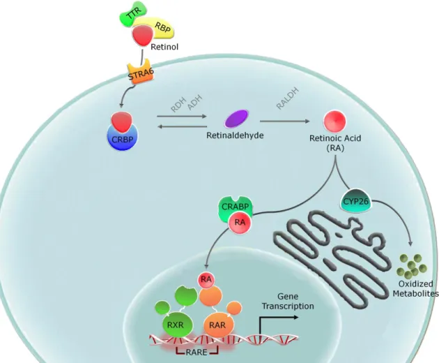
The main source of retinoids in animals is diet dependent, as these compounds cannot be synthesized de novo. In vertebrates, Vitamin A (retinol) binds to retinol binding protein (RBP), a specific transport protein, and this complex is directed to target tissues13 (Fig. 2). Considering that
RBP is the only known specific carrier of retinol, mice lacking RBP have brought unexpected information since neither viability nor morphology are compromised when they are exposed to a vitamin A-sufficient diet14.
Furthermore, a fraction of circulating soluble RBP is associated one-to-one with another serum protein, transthyretin (TTR)15,16. Cellular uptake of
5 Stimulated by Retinoic Acid 6 (STRA6)17 (Fig. 2). This receptor works as
bidirectional retinol transporter, with the internal state of retinoids being responsible for the control of its activity13.
After entering the cell, retinol is transformed to RA by a canonical pathway characterized by a two-step oxidation. The first oxidation represents a reversible reaction that generates retinaldehyde and is carried out by enzymes of two different classes: the cytosolic alcohol dehydrogenases (ADHs) that belong to the medium-chain dehydrogenase/reductase family and microsomal retinol dehydrogenases (RDHs), members of the short-chain dehydrogenase/reductase group18. The
ADH family consists of numerous enzymes with three (ADH1, 3 and 4) highly conserved paralogs in vertebrates19. In extreme conditions of retinoid
Fig. 1 – Simplified schematic phylogenetic tree of the metazoans. Adapted from
6 supply, both deficiency and excess, ADH1 and 4 have been suggested as the main players involved in the first oxidation step, while ADH3 seems to function continously8. Moreover, genetic studies in mice have shown that
loss of function of RDH10 is embryonic lethal20, showing that this enzyme is
essential for RA generation in the developing21 (Fig. 2).
The second reaction that leads, ultimately, to RA production is characterized by the conversion of retinaldehyde during an irreversible oxidation by retinaldehyde dehydrogenase (RALDH) (Fig. 2). There are three main paralog genes characterized, RALDH1, RALDH2 and RALDH3, each with distinct expression patterns6,22. In vertebrates, RALDH2 is present
in very early development and plays a crucial role in early embryogenesis 23-26. In contrast, RALDH1 and RALDH3 are expressed later during
development and they greatly contribute to the patterning of the respiratory and visual systems27,28.
Alternatively, recent data suggest that RA synthesis can also occur through an alternative process by action of cytochrome p450 family of mono-oxygenases2. CYP1B1 has emerged as a very likely candidate, since it
is able to efficiently oxidize retinol into retinaldehyde and subsequently to RA, in vitro, and its expression pattern is consistent with RA synthesis during embryogenesis29.
In addition to RA synthesis, endogenous RA degradation is tightly regulated in space and time during development. This process is mainly driven by proteins of the cytochrome p450 family, chiefly CYP26 enzymes30.
Vertebrates possess three Cyp26 paralogs, Cyp26A1, Cyp26B1 and Cyp26C1, all of which have very well characterized expression patterns31,32.
This family of enzymes promotes the catabolism of RA by production of polar metabolites, including 4-hydroxy RA, 4-oxo RA, 18-hydroxy RA, 5,6-epoxy RA or 5,8-5,6-epoxy RA33. The question of the biological relevance of
these metabolites as signaling molecules is still unclear34 (Fig. 2).
Within cells, RA activity is also modulated by proteins that bind to retinol and RA, the so called cellular retinol binding proteins (CRBPs) and cellular retinoic acid binding proteins (CRABPs)35 (Fig. 2). In vertebrates,
there are two CRBP paralogs, CRBP-I and CRBP-II, that possess very divergent expression patterns35. CRBP-I has high affinity for its substrate
7 key player in the conversion of retinol into retinaldehyde and as regulator of retinol storage. CRBP-II is essentially present in cells of the small intestine, suggesting an involvement in the processing of retinol taken up from food. There are also two CRABP paralogs, CRABP-I and CRABP-II, and these two proteins also exhibit distinct temporal and spatial expression profiles35.
During embryonic development both are widely expressed, however, their expression does not overlap contrasting with the adult stages where CRABP-I expression is almost ubiquitous. CRABP-CRABP-ICRABP-I has been suggested to be responsible for the transport of RA to RAR in the nucleus, fact supported by
Fig. 2 – Overview of retinol metabolism and signaling. Schematic view of the
conversion of retinol into its major active metabolite, retinoic acid, and activation of retinoid-dependent signaling. ADH, alcohol dehydrogenase; CRABP, cellular retinoic acid binding protein; CRBP, cellular retinol binding protein; CYP26, cytochrome P450 family 26; RA, retinoic acid; RALDH, retinaldehyde dehydrogenase; RAR, retinoic acid receptor; RARE, retinoic acid response element; RBP, retinol binding protein; RDH, retinol dehydrogenases; RXR, retinoid X receptor; STRA6, stimulated by retinoic acid gene 6; TTR, transthyretin.
8 the increase of RA signaling when CRABP-II is overexpressed in the frog Xenopus laevis36, as well as in cell lines37. In contrast, CRABP-I seems to
deliver RA to degradation38.
iv.
RA Signaling Integration, the Paradigm of the RXR/RAR
Heterodimer
RA exerts its effects through direct binding to nuclear receptors, classically in association with the heterodimer RXR/RAR (retinoid X receptor/retinoic acid receptor)39. In vertebrates, there are three paralogs
of each receptor (α, β and γ)40. RXR and RAR bind as heterodimers to DNA
in the promoter regions of target genes. After the association of RA and RAR a conformational change is induced in the receptor heterodimers allowing gene-specific transcription41. The RXR/RAR heterodimer interact with
specific DNA regions, the retinoic acid response elements (RARE) (Fig. 2). These RAREs typically comprise two direct repeats (DRs) with the conserved nucleotide sequence (A/G)G(G/T)TCA or of the more relaxed sequence (A/G)G(G/T)(G/T)(G/C)A spaced by one, two or five nucleotides (DR1, DR2 or DR5, respectively)40,42,43. Different RAREs have been identified in
promoters of different RA-target genes involved in a wide variety of biological functions42. According to the canonical model, in absence of the
ligand RXR/RAR can bind DNA associated with a co-repressor complex NCOR/Sin3A/HDAC, responsible for chromatin compaction and therefore target gene repression44. In the presence of ligand, due to a conformational
change within the receptor, there is release of co-repressors and recruitment of co-activator complexes, leading to a decondensation of the chromatin and allowing the assembly of the transcription pre-initiation complex28. Several recent studies have shown that the specific activation
mechanisms of gene transcription programs triggered by RARs are dependent on the target gene promoter45.
v.
RA Functions during Embryonic Development
During vertebrate development, RA exhibits pleiotropic effects, but these outputs are not only RA dependent, they are also the result of
9 interactions with several pathways (FGF, Shh, Wnt and TGF-β). Crosstalk between RA and these pathways can display very different architectures: they can be synergistic or antagonistic, may act directly or through indirect signaling, and can involve one or several regulatory loops46.
Vertebrate embryos treated with exogenous RA lose/reduce a part of the forebrain/midbrain and there is a clear extension of the hindbrain47-50.
Thus, RA function seems fundamental for the establishment of positional information in the central nervous system5. Hox genes are intimately
associated with the establishment of positional information during development of the hindbrain51, with the establishment of the Hox code
itself controlled by RA signaling 52. A very dynamic arrangement of RA
sources, RALDH2, and sinks, Cyp26, regulate the activation or inhibition of specific Hox genes in the hindbrain. FGF information provided by the midbrain and posterior CNS is alsoinvolved in the process53-56. Retinoid
signaling is also important for specification of interneurons and motor neurons across the DV axis during development52. In the spinal cord, RA
interacts with Shh to establish ventral determinants by induction of genes governing the dorsal-ventral (DV) patterning57. In absence of RA, the
ventral neurons are not induced to differentiate from the neuroectoderm of the spinal cord57,58. Neurite outgrowth is also controlled by RA during
development, since a lack of RA signaling is responsible for abnormal axonal projections50,59.
RA signaling plays a fundamental role in AP axis specification, not only in the CNS, but also of the mesoderm. For example, the expression of Cyp26 is suppressed by FGF and Wnt signaling, which are involved in the specification of posterior trunk fate60. Moreover, RA is probably also
involved in a side-specific activation of genes involved in LR specification, such as lefty, pitx and nodal61-63. However, RA action during somitogenesis
is associated with maintenance of somites symmetric formation along the LR axis64,65. Here, RA works as a buffer that is able to balance and prevent
LR asymmetry signaling of occurring in presomitic mesoderm (PSM), which later gives rise to lateral plate mesoderm specification65. Additionally, RA
signaling is an important mediator of cardiac field specification, patterning of the AP axis of the heart tube and heart looping23,66-68. Interestingly, the
10 exogenous supplementation leads to similar defects in mice69, zebrafish70
and chicken71. In this context, RALDH2 is expressed in the PSM and later in
the lateral mesoderm creating a wave of RA that generates cardiac precursors and delimitates the heart field along the AP axis71.
RA also participates in limb development where it is required during forelimb induction, but it is dispensable at later stages when hindlimb budding and patterning are established72. The action of RA seems to be
indirect once it generates a FGF permissive environment that allows development of limb buds72.
RA signaling is also involved in specification and establishment of different endodermal fields. Exposure to exogenous RA prevents the expression of genes normally present in the anteriormost endoderm, while at the level of the pharyngeal arches gene expression is activated and expanded anteriorly73. Interestingly, when RA signaling is disrupted there
are no effects in the first and second pharyngeal arches, but severe defects are observed in structures derived from more posterior pharyngeal arches74.
Some organs derived from the endoderm of the posterior foregut also require RA for proper development, like lungs, stomach, liver and pancreas. Lack of RA signaling causes a disruption of lung bud outgrowth since FGF10, one of the main players in branching morphogenesis (in parallel with BMP4), is absent. In addition, development of stomach and liver requires the control of FGF signaling by RA75. Furthermore, absence of RA signaling is
responsible for the specific inhibition of pancreas development, by disturbing the patterning of its presumptive dorsal bud76,77.
RA interacting with FGF signaling also acts to regulate the epithelial-mesenchymal transition of neural crest cells (NCC)78. RA also has a role in
differentiation of NCC populations during gastrulation events79 and in the
specification of placodes. RA has been shown to have a role in the establishment of in the otic80, optic81, olfactory82, and lateral line placodes83.
Altogether, the above description illustrates that the RA signaling system of vertebrates as well as most of its functions are very well described (Fig. 3). However, relatively little is known about the evolution of this signaling cascade.
11
vi.
The Beast and the Beauty: Amphioxus as a Model System
The phylum Chordata comprises three extant groups: cephalochordates (amphioxus), urochordates (tunicates) and vertebrates. Due to recent data provided by genomic sequencing and molecular phylogeny, the Chordate phylogeny has undergone a considerable change with the proposition that urochordates and not cephalochordates as the sister group of vertebrates84.
Despite this fact, cephalochordates maintain an important position for studies that address Chordate diversification.
One of the main arguments for this leading role concerns the adult body plan of amphioxus. It exhibits striking similarities with the vertebrate body plan, while in tunicates the body plan is more derived and has probably lost some chordate-specific traits84,85. For example, cephalochordates and
vertebrates share a hollow nerve cord dorsal to a notochord, a postanal tail, pharyngeal gill slits and an endostyle (a thyroid gland homologue)85.
However, there are vertebrate-specific characters missing, like paired sensory organs, a cartilaginous or bony skeleton, definitive neural crest cells and a morphologically differentiated brain86. In early embryonic
Fig. 3 – Overview of RA roles during vertebrate embryonic development. Schematic
view of two different stages of vertebrate development, with development of the highlighted being influenced by RA.
12 development, despite some major differences, amphioxus and vertebrates share very specific features. The embryo of amphioxus has thus been proposed to be a realistic approximation of the stem chordate embryo, before the evolution of specific cleavage mechanisms in the tunicates and the evolution of large amounts of yolk in basal vertebrates87.
Recent developments in genomic analysis have indicated that amphioxus possesses an archetypal vertebrate genome lacking whole genome duplications88, contrasting with the massive gene losses and
genome rearrangements associated with urochordate genomes89,90. That
said, the amphioxus genome does exhibit an expansion of several specific gene families that, most likely, do not represent ancestral characters. For example, families of genes encoding opsins, ALDHs and SDRs, together with several homebox genes, have undergone lineage-specific expansions in cephalochordates85,91-93. Apart from this divergence of gene families the
genomes of cephachordates and vertebrates also exhibit an extensive conservation of syntenic regions and gene linkage88.
The subphylum Cephalochordata is divided into three genera: Asymmetron, Epigonychthys and Branchiostoma94. Cephalochordates can
generally be found in the majority of the coastal regions on every continent in relatively shallow marine habitats, burrowing tail-first in the sand and filter-feeding95. The majority of the work on cephalochordates has been
carried out on four Branchiostoma species: the European amphioxus (B. lanceolatum), the Florida amphioxus (B. floridae), the Chinese amphioxus (B. belcheri) and the Japanese amphioxus (B. japonicum). All four amphioxus species present seasonal reproduction periods96,97. After external
fertilization, amphioxus embryos and larvae live in shallow marine water until they undergo metamorphosis, which is characterized by several anatomical remodeling events turning the asymmetric larva into a quasi-symmetric juvenile. After metamorphosis, the amphioxus juveniles alter their lifestyle and become benthic in predominantly sandy habitats98.
Improvements of embryo acquisition and culture have enabled great advances in the development of amphioxus as a usable model to study the evolution of chordates, and particularly the evolution of developmental processes in this phylogenetic branch99. Thus, it is now possible to maintain
13 adult amphioxus in an artificial sea water system and induce on-demand spawning independent from environmental stimuli100.
vii.
RA Signaling in Amphioxus
An approach combining treatments with RA or RA antagonist together with marker gene expression studies yielded a rather detailed description of the roles of RA signaling during amphioxus development. In the amphioxus central nervous system it was shown that RA regulates the expression of key players of AP regional patterning, in particular Hox genes. The domains of Hox1, Hox2, Hox3, Hox4 and Hox6 are expanded anteriorly when RA is added, while RA antagonist shifts the Hox gene expression domains posteriorly101. Likewise, RA strongly upregulates RAR expression, while RA
antagonist has the inverse effect102. Also exogenous RA promotes the loss
of pharyngeal structures whereas RA antagonist treatments yield the opposite outcome103. Morpholino-based functional knockdown experiments
suggest that this role of RA signaling in establishing the posterior limit of the pharynx is directly mediated by Hox1103. Additionally, it has also been
shown that RA and RA antagonist alter the expression domains of Hox genes in the general ectoderm104. Moreover, in the posterior endoderm, RA
signaling controls the expression of two ParaHox genes, Xlox and Cdx, hence mediating AP patterning of the developing amphioxus hindgut105.
Altogether, these data have established that RA, acting via Hox genes, is crucial for AP patterning of the amphioxus early embryo and that this RA-Hox signaling hierarchy plays further, tissue-specific roles, in patterning of the central nervous system, general ectoderm and endoderm106, although
more details on the regulation of amphioxus ParaHox genes by RA must be assessed105.
Albeit the existence of rather detailed concepts about the functions of RA signaling during amphioxus development, with the exception of a study on RA synthesizing enzymes93, virtually nothing is known about the
mechanisms controlling the establishment of RA reactive zones in the amphioxus embryo. There is, thus, a necessity to study the most fundamental components of the RA pathway, including RA binding proteins and enzymes capable of synthesizing and degrading endogenous RA, to
14 assess RA signaling activity in the developing embryo. Detailed data on these key components together with information provided by studies carried out in vertebrates will unravel conserved and/or divergent elements of RA signaling cascade, which in turn will allow the reconstruction of the evolutionary diversification of this morphogen-dependent signaling pathway in chordates.
viii.
Objectives
As previously mentioned, the detailed study of central components of the RA signaling pathway can provide novel insights into the evolutionary diversification of the RA cascade in chordate evolution. In this context, degradation is of vital relevance for RA homeostasis, a process driven by Cyp26 enzymes, which show a very tight regulation30,53. In vertebrates, the
three Cyp26 paralogs present a non-overlaping spatiotempotal expression30,
suggesting that each one of them as a specific function during embryonic development, probably tissue-specific. For example, Cyp26 mutants exhibit severe malformations, such as spina bifida, limb truncations, cerebral dystrophy, caudal regression and respiratory complications after birth30.
Here, we aim to understand how expression of RA degradation enzymes (Cyp26) can be correlated with RA distribution and functions during amphioxus (B. lanceolatum) embryonic development.
To address this question the genes encoding Cyp26 enzymes in B. lanceolatum were cloned and the evolutionary history of Cyp26 genes analyzed by phylogenetic tree reconstruction. Moreover, expression patterns of the amphioxus Cyp26 genes were characterized in detail during development by in situ hybridization (ISH) and expression at key points of development was subjected to quantitative analyses using qRT-PCR. Moreover, the expression patterns were also studied under specific pharmacological conditions, such as RA and BMS009 (a RA antagonist) treatments.
Altogether this work sheds new light on the evolutionary diversification of the Cyp26 family and highlights conserved and divergent aspects of the regulation of endogenous RA levels during development.
15
II. MATERIAL AND METHODS
i.
Phylogenetic Analysis
For the phylogenetic analysis the following list of amino acid sequences has analysed (accession number, and genome web browseres are given with the sequence names): Lottia gigantea Cyp51: 108695; L. gigantea Cyp26: 111029 and 189041 from the genome database (http://genome.jgi-psf.org/Lotgi1/ Lotgi1.home.html). Capitella teleta Cyp51: 173561; C. teleta Cyp26: 150007 and 212322 (http://genome.jgi.doe.gov/Capca1 /Capca1.home.html). Strongylocentrotus purpuratus Cyp51: SPU_025595 (http://urchin. nidcr.nih.gov/) S. purpuratus Cyp26: XP_001194704. Saccoglossus kowalevskii Cyp26s: NP_001161524 and XP_002734884. Branchiostoma floridae Cyp26s: XP_002588109, XP_002588110 and XP_002588112 (http://www.ncbi.nlm.nih.gov/). Gasterosteus aculeatus Cyp26s: ENSGACP00000020277 ENSGACP00000024870 and ENSGACP00000014 658. Tetraodon nigroviridies Cyp26s: ENSTNIP00000007321, ENSTNIP0000 0014173 and ENSTNIP00000021864. Oryzias latipes Cyp26s: ENSORLP0000 0018194, ENSORLP00000004308 and ENSORLP000 00002541 Gallus gallus Cyp26s: ENSGALP00000010871, ENSGALT000000 25943 and ENSGALP00000010878 (http://www.ensembl.org/index.html). Danio rerio Cyp51: AAR89625; D. rerio Cyp26s: NP_571221, NP_997831 and NP_001025122. Xenopus tropicalis Cyp26s: NP_001016147, NP_001072655 and XP_002939137. Monodelphis domestica Cyp26s: XP_001375292 and XP_001375317. Homo sapiens Cyp51: EAL24154; H. sapiens Cyp26s: NP_000774, NP_063938, NP_899230. Mus musculus Cyp26s: NP_031837, NP_780684 and NP_001098671 (http://www.ncbi. nlm.nih. gov/). Ciona intestinalis Cyp26s: ENSCINP00000009998, ENSCINP00000024552. Ciona savignyi Cyp26s: ENSCSAVP00000003718 and ENSCSAVP00000003583.
The sequences were aligned with Clustal as implemented in Seaview107
and refined by eye, the final alignment comprises 661 positions. The molecular phylogeny was reconstructed by Maximum Likelihood (ML) with Phyml108 under the model JTT. The robustness of the node was estimated by
bootstrap (1000 pseudoreplicates) with Phyml. Cyp51 has used as outgroup.
16
ii.
Maintenance and Spawning of Amphioxus in the
Laboratory
Sexually mature animals of the Mediterranean amphioxus (B. lanceolatum) were collected by dredging at Argelès-sur-Mer, France, and retrieved from the sand with a sieve96,97. Following the collection, adult
animals were transported to an artificial sea water facility and then transferred into their respective tanks. All the animals collected were divided accordingly, about 10-15 animals per tank (2,7 L of maximal capacity), both males and females together. Each tank contained a layer of approximately 1cm of washed and sterilized sand collected in Argelès-sur-Mer, France100. Filtered and sterilized (through UV light) artificial sea water
(ASW) with a final salt concentration of 38g/L was pumped every day into each tank, during a controlled time period to renew about 1L of ASW and continuous aeration was supplied100. The facility was equipped with air
temperature control set to 18ºC and the ASW flushed into the tanks had a temperature of approximately 16ºC, allowing a relatively constant water temperature of 17-18ºC inside the tanks. The animals were kept in a spring-like day/night light period (14 hours of light versus 10 hours of absolute darkness) in an inverted illumination cycle100. Animals were fed
with artificial food twice a week100.
Spawning was induced by 36 hours of thermal shock at 23ºC as previously described96,97,100. Males and females with full gonads were
selected (15-20 animals) and put together during the thermal shock in tanks with reduced amounts of sand. The thermal shock was induced one hour after the lights turned on and 36 hours thereafter (1 hour before the lights were turned off) each animal was transferred to an individual clear plastic cup containing about 10 mL of filtered ASW. After spawning, the gametes were collected and fertilization took place in Petri-dishes containing filtered ASW. After fertilization, the embryos were raised in filtered ASW, in the dark, at 19ºC.
iii.
Pharmacological Treatments (RA and BMS009)
Treatments with all-trans RA (dissolved in DMSO) and BMS009, a RA antagonist (dissolved in DMSO), were performed at the late blastula/early
17 gastrula stage, in a small Petri-dish at a final concentration of 10-6M in
filtered ASW. Simultaneously, embryos were treated with DMSO (1:1000) alone in a different dish, as a control102,109. After hatching, at the early
neurula stage, embryos were transferred to untreated filtered ASW and allowed to develop further101,104. During the next two days, developmental
stages (controls, RA-treated and BMS009-treated) were fixed at frequent intervals in 4% paraformaldehyde in MOPS buffer (0.1M Mops, 0.5M NaCl, 2mM MgSO4, 1mM EGTA, pH 7.4) for in situ hybridization110. After fixation
overnight at 4ºC, the embryos were washed with 70% ethanol and stored in 70% ethanol at -20ºC.
iv.
In Situ Hybridization (ISH)
Embryos at different developmental stages were fixed using a similar protocol as described above. For each of the Cyp26 genes of B. lanceolatum, previously cloned in our lab, antisense riboprobes were synthesized accordingly to Holland et al.111 and ISH was performed as
described by Yu and Holland110. ISH was performed in embryos
representative of all stages of development (blastula; early, mid and late gastrula; early, mid, and late neurula; late embryos and different larval stages) and ISH preparations were photographed as whole mounts under DIC (differential interference contrast) settings.
v.
RNA Extraction and cDNA Synthesis
RNA extraction was carried out as described by Yu and Holland112. The
A260/280 ratios and the concentration of all the RNAs prepared were measured using a NanoDrop® (LabTech®) system, and the RNA integrity
was checked by electrophoresis on a 1% agarose gel.
The cDNAs were synthesized with reverse transcription kit (SuperScript®III – Invitrogen®) and the reaction was carried out as
indicated by the manufacturer.
vi.
Quantitative RT-PCR and Data Analysis
The cDNA was used as template for quantitative real-time PCR assay using primers specific for B. lanceolatum glyceraldehyde-3-phosphate
18 dehydrogenase (GAPDH), Elongation Factor 1-α (EF1α), 18S rRNA, β-actin, RAR, Cyp26-1, Cyp26-2 and Cyp26-3 with the QuantiTect SYBR Green PCR reagents (QIAGEN) and the DNA Engine Opticon system (MJ Research) according to the instructions of the manufacturer. The primer sequences used for GAPDH were AGGGTCTCATG ACCACGGTA-3’ and 5’-TTCTTCAGTCGG-CAGGTCAG-3’; for EF1α, 5’-ATGG GCAAGGAAAAGCTTCACA TC-3’ and TCACTTCTTCTTCCCGCCGGCAG-3’; for 18S rRNA, 5’-CATCAGCCATGCCTAAGGTT-3’ and 5’-CTATTCCTTGCTGG CCATGT-3’; for β-actin, 5’-TTACAATGGAAGACGATGTTGC-3’ and 5’-ATCGTTAGCTCCTGACAA GCTC-3’; for RAR, 5’-GTCTGCCATCGGGATAAGAA-3’ and 5’-GCCTCTCTGACCGTGGTTAT; for Cyp26-1, the primers were 5’-CAAGAC GAGGACGAGATCAGTAG-3’ and 5’-CTTCTCGTGGATGTGACGTTTTA-3’; for Cyp26-2, 5’-CAACACCTCACTTTCCTCTTCAC and TCTTCCTCTGAATGTG GTTCATC; and for Cyp26-3, CAAGAGAGATGTCGTTTCAGAGC-3’ and 5’-CTTCCAACCATTGGTCGATACT-3’. The reaction conditions consisted of 95ºC for 15 minutes followed by 40 cycles of 95ºC for 30 seconds, 55ºC for 30 seconds, and 72ºC for 45 seconds. Each reaction was performed in triplicate. After multiple assays, only 18S rRNA was selected as reference for internal standardization of the starting quantity of RNA. This choice was based on our observations in response to the different treatments and is also supported by a recent publication113.
Data were quantified using the 2-ΔΔC
T method. The experimental data
were then pooled, analyzed and presented in relation to the relative quantity of mRNA for RAR in a control situation as mean +/- standard error (SE).
19
III. RESULTS
i.
Cyp26 Phylogenetic Analysis
With the aim to better understand the origin of RA signaling in the diversification of protostomes and deuterostomes as well as to reveal the evolutionary relationships between Cyp26 genes and the three Cyp26s genes identified in amphioxus (both B. floridae114 and B. lanceolatum), we
conducted a phylogenetic analysis including Cyp26 amino acid sequences from different animal groups: mollusks (L. gigantea), annelids (C. teleta), echinoderms (S. purpuratus), hemichordates (S. kowalevskii), vertebrates (G. aculeatus, T. nigroviridies, O. latipes, D. rerio, X. tropicalis, G. gallus, M. domestica, H. sapiens and M. musculus), tunicates (C. intestinalis and C. savignyi) and cephalochordates (B. floridae and B. lanceolatum). Cyp51, the only family of cytochrome P450 that is widely distributed in all the different biological kingdoms115, was used as an outgroup (Fig. 4).
In our analysis, the three amphioxus Cyp26 genes (Cyp26-1, Cyp26-2 and Cyp26-3) are paired together in a strongly supported branch suggesting a common evolutionary origin (Fig. 4). Similarly, all the Cyp26 genes of vertebrates (Cyp26A1, Cyp26B1 and Cyp26C1) show a similar relationship, in a way that all the Cyp26A1s, Cyp26B1s and Cyp26C1s are well organized in very robustly supported groups (Fig. 4). The Cyp26 lineages of cephalochordates and vertebrates are grouped in two different branches, suggesting the evolutionary divergence of the three Cyp26s in amphioxus is independent of those in vertebrates. The duplications of Cyp26 genes in cephalochordates and vertebrates are thus likely independent events.
The invertebrate groups included in the analysis (mollusks, annelids, echinoderms, hemichordates and tunicates) group together in a poorly supported arrangement (Fig. 4). This kind of assemblage can be explained, most likely, as a phenomenon associated with methodological artifacts, called long-branch attraction116.
Taken together, our data reveal in detail the relationship between amphioxus and vertebrates in terms of Cyp26 divergence, however it exposes some weakness resolving the evolutionary origins of this gene family.
21
ii.
Cyp26 Expression Profile During Amphioxus Development
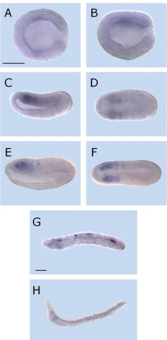
Whole mount in situ hybridization (ISH) was used to assess the expression pattern of the three Cyp26 genes of B. lanceolatum during development. Developmental series were obtained from shortly after fertilization until the 3-day larval stage (Fig. 5, 6 and 7). As a technical comment concerning the expression patterns, representative signaling for Cyp26-1 and Cyp26-3 were very difficult to obtain, in a way that the staining procedure was carried out for several days.
Cyp26-1
ISH results for Cyp26-1 probe exhibit no detectable transcription signal from fertilization to mid gastrulation (data not shown). The Cyp26-1 gene expression is first detected at the late gastrula stage (15 hours post-fertilization or hpf) when the expression is weak in the mesoderm (Fig. 5A, B). Until the early neurula stage, 20 hpf, the signal is expanded in the mesoderm, mainly associated with somite structures (Fig. 5C, D). The Cyp26-1 signal becomes more evident at 24hpf (mid neurula stage) preserving the mesodermal predominance (Fig. 5E, F). At 36hpf, late embryo stage, the signal is rather weak and sparse associated with mesodermal structures (Fig. 5G). At 48hpf, the early larva exhibits a weak mesoderm associated signal (Fig. 5H), which is completely lost in the following stages (data not shown).
Cyp26-2
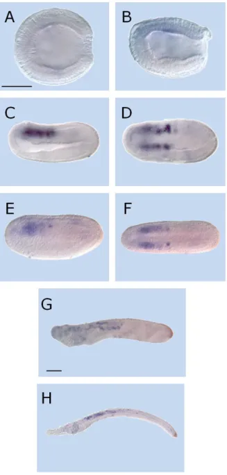
Distinct from Cyp26-1, Cyp26-2 displays a much more complex and conspicuous developmental expression profile. The first appearance of Cyp26-2 expression is at the mid gastrula stage (9 hpf). At this stage, the
Fig. 4 – Cyp26 phylogenetic analysis. Rooted maximum likelihood tree of Cyp26 using
Cyp51 as outgroup. represents branches of low bootstrap support (lower than 70%),
where numbers showed correspond to bootstrap values, and represents branches fully
supported (100%). B.flo, Branchiostoma floridae; B.lan, Branchiostoma lanceolatum; C.int,
Ciona intestinalis; C.sav, Ciona savignyi; C.tel, Capitella teleta; D.rer, Danio rerio; G.acu, Gasterosteus aculeatus; G.gal, Gallus gallus; H.sap, Homo sapiens; L.gig, Lottia gigantea;
M.dom, Monodelphis domestica; M.mus, Mus musculus; O.lat, Oryzias latipes; S.kow,
Saccoglossus kowalevskii; S.pur Strongylocentrotus purpuratus, T.nig, Tetraodon nigroviridies; X.tro, Xenopus tropicalis.
22
Fig. 5 – Expression of Cyp26-1 in amphioxus (Branchiostoma lanceolatum) development. Anterior side to left and, excepting A, D and F, dorsal up. Lateral views in
B, C, E, G, H and A, D, F viewed from dorsal side. Scale bar 50 µm. (A,B) late gastrula – 15hpf; (C,D) early neurula – 20hpf; (E,F) mid neurula – 24hpf; (G) late embryo – 36hpf; (H) early larva – 48hpf.
24
Fig. 6 – Expression of Cyp26-2 in amphioxus (Branchiostoma lanceolatum) development. Anterior side to left and, excepting B, E and G, dorsal up. Lateral views in
A, C, D, F, H, I, J, K and B, E, G viewed from dorsal side. Scale bar 50µm. (A) mid gastrula – 9hpf; (B,C) late gastrula – 15hpf; (D,E) early neurula – 20hpf; (F,G) mid neurula – 24hpf; (H) late embryo – 32hpf; (I) late embryo – 36hpf; (J) early larva – 48hpf; larva – 72hpf.
Fig. 7 – Expression of Cyp26-3 in amphioxus (Branchiostoma lanceolatum) development. Anterior side to left and, excepting A, D and F, dorsal up. Lateral views in
B, C, E, G, H and A, D, F viewed from dorsal side. Scale bars 50µm. (A,B) late gastrula – 15hpf; (C,D) early neurula – 20hpf; (E,F) mid neurula – 24hpf; (G) late embryo – 36hpf; (H) early larva – 48hpf.
25 signal is localized conspicuously around the blastopore (Fig. 6A). At late gastrula, the blastopore signal is still present although less prominently and a new wave of signaling is observed in the region that corresponds to mesoderm (Fig.6B, C). Later, at 20hpf, both expression domains are still present (Fig. 6D, E). The signal associated with the blastopore maintains its posterior identity, but exhibits only weak expression. In contrast, the mesodermal signal is expanded and in this region very strong expression is also detectable in the other tissue layers. At mid neurula stages, the expression in the anterior region is preserved and it displays a strong signal, mostly present in the mesoderm. At this stage the neuroectodermal region also expresses Cyp26-2. Furthermore, a posterior signal appears associated with the forming tail bud and includes the posteriormost ectoderm that already expressed the gene (Fig. 6F, G). Shortly after the process of the neurulation (32hpf), gene expression is restricted to posterior ectodermal structures as well as to the anterior region of the embryo. This latter pattern, thus represents a signal reduction and a more tissue-specific outline when compared to previous developmental stages (Fig. 6H, I). At larval stages (48hpf and 72hpf) the ectodermal region of the tail bud still expresses Cyp26-2. The expression of Cyp26-2 in the anterior region is also maintained with a tendency for tissue-specification, associated here with the pharyngeal ectoderm, mesoderm and endoderm (Fig. 6J, K). Due to the complexity of the general signaling outline, further analyses are required to confirm the tissue specificity during development.
Cyp26-3
The gene expression profile of Cyp26-3 displays a rather similar distribution, both in space and time, as Cyp26-1. From the first cellular divisions until mid gastrulation no expression is detectable (data not shown). Expression commences at the late-gastrula stage with a faint signal in the mesoderm (Fig. 7A, B). Thereafter, at the early neurula stage, the gene expression expands and is localized in the somites (Fig. 7C, D). Amphioxus embryos of 24hpf show an apparent weakening of the signal, albeit keeping the initial somite localization (Fig. 7E, F). In the late embryo, the gene expression of Cyp26-3 exhibits a weak and disperse pattern (Fig.
26 7G) that is almost lost in the early larva (Fig.7H). Finally, no signal is found at later larval stages (data not shown).
iii.
Quantitative RT-PCR Combined with RA and BMS009
Treatments
Quantitative RT-PCR was used to quantify expression of RAR, Cyp26-1, Cyp26-2 and Cyp26-3 as two different stages of amphioxus development: at 20hpf (neurula) and at 48hpf (larva). To obtain an idea of the fluctuations of gene expression levels induced by variations of RA signaling levels, quantitative RT-PCR analysis was coupled with pharmacological treatments with RA and BMS009 (the RA antagonist). The data obtained were normalized to the level of RAR in a control situation, with RAR being a gene shown to be under the direct control of RA in amphioxus.
In a control situation, qRT-PCR results reveal that the effective expression level of Cyp26-1 and Cyp26-3 is much lower that of Cyp26-2 and RAR at 20hpf (Fig. 8A, red bars). Under these conditions, Cyp26-2 expression is generally higher than that of RAR and about 5000 times higher levels than the two other Cyp26 genes (Annex I). When embryos were treated exogenously with RA at 20hpf, RAR expression is increased 2.4 fold (Annex I). Consistently, Cyp26-2 exhibits a similar fold increase of expression. In great contrast, both Cyp26-1 and Cyp26-3 show a huge increase in expression levels: respectively, 350 and 26 fold (Fig. 8A, green bars and Annex I). The addition of BMS009, an RA antagonist, induces a reduction of RAR and Cyp26-3, but on different scales, 2 and 20 fold respectively (Fig. 8A, blue bars and Annex I). Interestingly, under the influence of BMS009, the overall transcription of Cyp26-1 and Cyp26-2 remain almost unaltered when compared to the control situation (Fig. 8A, blue bars and Annex I).
As a later stage of development, 48hpf, small differences in the expression profiles of the Cyp26 genes can be noticed. In a control situation, the different levels of expression between Cyp26-1 and Cyp26-3 (low) and Cyp26-2 and RAR (high) are still evident (Fig. 8B, red bars). That said, the magnitude of the difference is distinct from the situation at 20hpf, with the expression of Cyp26-1 and Cyp26-3 being only about 4 and 8
27
A
B
Fig. 8 – Relative expression of RAR, Cyp26-1, Cyp26-2 and Cyp26-3 in amphioxus (Branchiostoma lanceolatum) embryos. (A) Expression levels at 20hpf
and (B) at 48hpf in a control situation, DMSO 1:1000 (red bars), treatments with 10-6M
RA (green bars) and treatments with 10-6M BMS009, a RA antagonist (blue bars). Data
are expressed as the mean fold change (means +/- SE, n=3) relative to RAR in the control, for detailed check Annex I.
28 times lower than respectively, that of RAR. In addition, expression of Cyp26-1 and Cyp26-3, when compared to Cyp26-2, is only around 2.5 and 5 times lower, respectively (Fig. 8B, red bars and Annexe I). Treatments with 10-6M RA induce drastic changes in the expression profiles of the
genes. The reference gene RAR shows a 3.6 fold increase of expression, while Cyp26-2 only shows an increase of 1.6 fold. When compared to the neurula stage, the increase of expression of Cyp26-1 and Cyp26-3 is less dramatic (101 and 5.2 total fold, respectively), albeit still impressive (Fig. 8B, green bars and Annexe I). In the experiments carried out with the RA antagonist, the general decrease of expression of the Cyp26 genes and of the RAR is confirmed. The expression of RAR is reduced almost 3 times, Cyp26-1 is reduced roughly 6 times and Cyp26-3 shows a 123 fold reduction in its expression levels. Curiously, Cyp26-2 gene expression is almost unaltered after treatment with BMS009 (Fig. 8B, blue bars and Annex I).
Together, these quantitative results illustrate that each of the three amphioxus Cyp26 genes responds differently to alterations of RA signaling levels. Although, the responses of Cyp26-1 and Cyp26-3 are more similar to each other than to the response of Cyp26-2. Ultimately, each of the three genes seems to contribute to the endogenous balance of RA.
iv.
Cyp26 Expression Pattern Induced by Pharmacological
Treatments
To address the spatial arrangement of Cyp26 gene expression changes induced by pharmacological treatments ISH was performed on amphioxus embryos collected at 20hpf and 48hpf (the same stages used in qRT-PCR analysis). Under control conditions (treatment with DMSO 1:1000), the expression profile for Cyp26 genes are unaltered relative to the expression pattern described (Fig. 9B, E, H, K, N, Q). Cyp26-1 expression increases significantly when exogenous RA is applied (as indicated by qRT-PCR) but the signal is not uniformly distributed throughout the embryo with certain regions showing less intense staining, both at 20hpf and 48hpf (Fig. 9A, D). Due to the intensity of the staining, further analyses must be carried out to assess the tissue specificity of the signal. On the other hand, treatments
29
Fig. 9 – Expression of amphioxus (Branchiostoma lanceolatum) Cyp26s in treated embryos. Anterior side to left and dorsal up, all lateral views. (A-F) expression
of Cyp26-1, (G-L) expression of Cyp26-2, (M-R) expression of Cyp26-3. Pharmacological
treatments as follows: 10-6M RA (A, D, G, J, M, P), DMSO control (B, E, H, K, N, Q) and
10-6M BMS009, a RA antagonist (C, F, I, L, O, R) at 20hpf (A-C, G-I, M-O) and 48hpf
30 with RA antagonist lead to a general decrease of the signal. This reduction is less evident at 20hpf than at 48hpf, where almost no Cyp26-1 staining is visible (Fig.9C, F).
The Cyp26-2 signal obtained in embryos treated with RA is also expanded relatively to the control, albeit less extensively than Cyp26-1. At 20hpf, Cyp26-2 expression is strongly increased in the posterior and anterior regions of the embryo. The signal is most conspicuous in the dorsal, anterior and posterior of the embryo (Fig. 9G). At the larval stage the influence of RA has similar effects since in the anterior end and posterior regions of the embryo the signal is broadly detectable (Fig. 9J). BMS009 treatments have a quite evident effect on the Cyp26-2 expression pattern: the posterior signal is completely lost, both at 20hpf and 48hpf, whether the transcription in the anterior region is almost unaffected (Fig. 9I, L).
The Cyp26-3 expression profile is also affected by excess of RA. The expression of this gene is strongly increased at 20hpf being somehow similar to the pattern shown for Cyp26-2 transcription (Fig. 9G vs M). Furthermore, at 48hpf, RA treatments induce a broad Cyp26-3 expression across the embryo (Fig. 9P). As expected, in embryos treated with RA antagonist the Cyp26-3 signal is weaker than in the control at the neurula stage and almost undetectable at larval stages (Fig. 9O, R).