UNIVERSIDADE DE LISBOA FACULDADE DE MEDICINA DENTÁRIA
“THE EFFECT OF AN ADDITIONAL LAYER OF ADHESIVE IN
DENTIN BOND STRENGTH OF A UNIVERSAL ADHESIVE
SYSTEM IN SELF-ETCH MODE”
Madalena de Quinhones Levy Rosa Dias
Dissertação
Mestrado Integrado em Medicina Dentária
UNIVERSIDADE DE LISBOA FACULDADE DE MEDICINA DENTÁRIA
“THE EFFECT OF AN ADDITIONAL LAYER OF ADHESIVE IN
DENTIN BOND STRENGTH OF A UNIVERSAL ADHESIVE
SYSTEM IN SELF-ETCH MODE”
Madalena de Quinhones Levy Rosa Dias
Orientação por:
Professor Doutor Alexandre Cavalheiro
Co - orientação por:
Mestre Ana Luísa Silva
Dissertação
Mestrado Integrado em Medicina Dentária 2014
“Learn from yesterday, live for today, hope for tomorrow. The important
thing is to not stop questioning.”
Albert Einstein
“Live as if you were to die tomorrow. Learn as if you were to live forever”
Mahatma GandhiAGRADECIMENTOS
Ao Professor Doutor Alexandre Cavalheiro, pela sua orientação, apoio, incentivo e total disponibilidade ao longo da realização deste trabalho.
À Dra. Catarina Coito e à Dra. Ana Luísa Silva pela paciência e inestimável ajuda durante o procedimento laboratorial.
À Professora Doutora Manuela Lopes pela incansável disponibilidade, ajuda e simpatia durante a realização do ensaio laboratorial.
À Catarina por ter sido a minha companhia, a minha família, a minha dupla, o meu incentivo e o meu maior apoio durante todo este ano.
À Marialice, Inês, Catarina e Soraia por terem partilhado comigo estes cinco anos, por todos os bons e maus momentos, pelo carinho mas principalmente por saber que vão estar sempre aqui.
À minha família, em especial à minha Titi Joana, por me lembrarem todos os dias o quão rodeada de amor, carinho e paciência eu estou. Principalmente pelo apoio que desde o inicio me deram e me continuam a dar todos os dias.
À minha irmã, por ser o meu maior apoio, a minha melhor amiga e principalmente pelo seu amor insubstituível e pela motivação constante.
À Maria, a quem espero um dia poder ensinar tudo o que me ensinaram a mim. À minha mãe, por me ter proporcionado tudo o que tenho e consegui até hoje, sem ela nada disto seria possível. Pela sua enorme paciência e por sempre me ter ensinado a lutar pelos meus sonhos.
Ao João, por ser o amor da minha vida, por estar do meu lado todos os dias e por fazer de mim uma pessoa melhor.
GENERAL INDEX
TABLES INDEX ... viii
GRAPHICS INDEX ... ix FIGURES INDEX ... x ABREVIATIONS ... xi ABSTRACT ... xiii RESUMO ... xv I- LITERATURE REVIEW...1 1. TOOTH ADHESION...1 a. Adhesion to Dentin...1
2. EVOLUTION OF ADHESIVE SYSTEMS...1
3. CLASSIFICATION OF ADHESIVE SYSTEMS...2
a. Etch-and-rinse/Total-etch...2
b. Etch-and-dry/Self-etch...3
c. Universal Adhesives……….4
4. EFFECT OF A HYDROPHOBIC COAT...6
5. BOND TESTING METHOD...7
II- PURPOSE...8
III- MATHERIALS AND METHODS...9
1. Type of study...9
2. Design of the study...9
3. Teeth selection and preparation...9
4. Distribution and treatment of the crown segments………..12
5. Restorative Procedures………14
6. Specimens preparation for micro-tensile tests……….15
7. Micro-tensile bond strength tests (μTBS)………...16
8. Statistical Analysis………..17
IV- RESULTS………18
V- DISCUSSION……….22
a. Limitations of the study and Future Research………29
VI- CONCLUSION………...30
VII- REFERENCES………31 APPENDIX……….I APPENDIX 1……….II
b. Materials and Components………..II
TABLES INDEX
Table 1 - Number of sticks (N); micro-tensile bond strengths (μTBS) mean values; standard deviation (Std. Deviation) and standard error mean (Std. Error Mean)………18 Table 2 – Test of Normality: Kolmogorov-Smirnov and Shapiro-Wilk tests……...18 Table 3 – Results of Levene’s Test and t-test……….19 Table 4 – T-test for Equality of Means………...20 Table 5 and 6 – Number of sticks in each failure mode: A – adhesive failure; CC– composite cohesive failure; CD – dentin cohesive failure; M – mixed failure…..…...21 Table 7 - Materials used, components, manufacturers and lot numbers ……….II
GRAPHICS INDEX
Graphic 1 and 2 – Tests of Normality for the SBU SE D and SBU+A SE D groups…………...………...19 Graphic 3 – Box-whisker plot of the μTBS for SBU SE D and SBU+A SE D: x axis represents the group and y axis represents the MPa………20 Graphic 4 – Failure mode distribution: A – adhesive failure; CC– composite cohesive failure; CD – dentin cohesive failure; M – mixed failure………21
FIGURES INDEX
Figure 1 - Diamond Wafering Blade - 10,2cmx0,3mm - Series 15HC, Buehler Ltd.,
Lake Bluff, IL, USA………..………9
Figure 2 - IsometTM 1000 Precision Saw, Buehler Ltd. Ltd., Lake Buff, IL, USA….…9 Figure 3 - Tooth fixed to an acrylic holder with sticky wax………..…10
Figure 4 - First cut 1-2 mm below the CEJ………..…..10
Figure 5 - Pulp chamber’s exposure………...…10
Figure 6 - Removal of pulp tissues……….………..…..11
Figure 7 - Filling the pulp chamber with cyanoacrylate glue (737 Black Magic Toughened adhesive, Permabond, Hampshire, UK)………...…11
Figure 8 - Crowns fixed with cyanoacrylate glue to the acrylic holder……….11
Figure 9 - Removing the occlusal enamel and superficial dentin.……….………11
Figure 10 - Mid-coronal dentin surface…..………....…11
Figure 11 - Mechanical grinder (Lunn Major, Struers, Denmark)………...12
Figure 12 - Scotchbond Universal Adhesive.………...13
Figure 13 – Adper Scotchbond Multipurpose Adhesive……….…...14
Figure 14 - Resin composite ENAMEL plus HRi………..14
Figure 15 - Resin composite build-up with 6 mm………..14
Figure 16 and 17 - Teeth after being sectioned in both ‘x’ and ‘y’ directions………..15
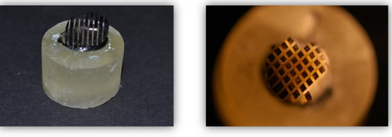
Figure 18 - Sticks………16
Figure 19 - Sticks attached to Geraldeli’s jig with cyanoacrylate glue………..16
ABREVIATIONS
% - per cent
10-MDP - 10-Methacryloyloxydecyl dihydrogen phosphate Bis-GMA – bisphenol-A diglycidyl methacrylate
CEJ - cementoenamel junction cm – Centimeter
Et al. – Et alli
HEMA - 2-hydroxyethyl methacrylate
ISO/TR - International Organization for Standardization/ Technical Report mm – Millimeter
mm/min – Millimeter per minute mm2 - Square millimeter
MPa - MegaPascal
mW/cm2 - milliWatt per square centimeter N – Newton
nm - Nanometer p – Significance value pH – Power of hydrogen
SBU SE D – Scotchbond Universal Adhesive (3M ESPE, St. Paul, MN, USA) applied to dentine in self-etch mode
SBU+A SE D - Scotchbond Universal Adhesive (3M ESPE, St. Paul, MN, USA) applied to dentine in self-etch mode with a hydrophobic resin layer of Adper Scotchbond Multi-purpose Adhesive (3M ESPE, St. Paul, MN, USA) SEM – Scanning Electron Microscopy
TEM – Transmission Electron Microscopy μm – Micrometer
ABSTRACT
A new generation of one-bottle self-etch dental adhesives is currently being used. The literature refers to them as universal or multi-mode adhesives. Universal adhesives are indicated as either self-etching or etch-and-rinse adhesives. Some clinical studies and laboratory evaluations have been performed, in fact, demonstrating that some universal adhesives may perform at the same level of previous adhesives.
Purpose: To evaluate the effect of an additional hydrophobic resin layer (Adper Scotchbond Multipurpose) on the resin-dentin bond strength (μTBS), of the universal adhesive Scotchbond Universal used in a self-etch mode compared to the manufacturer’s instructions.
Materials and Methods: A total of six extracted human teeth (n=6) were randomly distributed between two groups for bond strength testing, according to the different adhesive strategy used: Scotchbond Universal applied as a one-step self-etch adhesive as per manufacturer’s instructions (SBU SE D) and Scotchbond Universal applied as a one-step self-etch adhesive followed by one layer of the hydrophobic resin Adper Scotchbond Multipurpose. After composite restoration, specimens composed of sticks with 1mm2 were stored in distilled water (37ºC/24h) and then tested at 1mm/min using micro-tensile tests (μTBS) to assess dentin bond strength. Failure modes were analyzed under a stereomicroscope. Data were analyzed with a parametric paired-sample t-test since the assumptions of normality were valid.
Results: For dentin, the use of a hydrophobic resin layer (SBU+A SE D) resulted in a statistically significantly higher mean µTBS value (38,59 ± 22,21 MPa) than the SBU SE D group (27,66 ± 13,22 MPa), (p<0,05) with a 95% confidence interval.
Conclusions: Within the limitations of this in vitro study, it can be concluded that the use of a hydrophobic resin layer may be beneficial to the resin-dentin bond strength when applied to dentin with the self-etch mode.
Keywords: universal adhesives; self-etch; hydrophobic resin coat; micro-tensile bond strength.
RESUMO
Introdução: Devido à grande procura de tratamentos restauradores estéticos, tornou-se importante, para a maioria dos estudos em dentisteria adesiva, melhorar a resistência da união de resinas compostas à dentina. Além disso, a fiabilidade da adesão à dentina demonstrou ser um fator importante em restaurações adesivas. Isto deve-se ao facto de, enquanto que a adesão ao esmalte tem sido demonstrada como sendo fiável ao longo do tempo, a adesão à dentina ainda é um grande desafio.
Novos adesivos estão continuamente a ser testados, lançados e comercializados para a adesão de resinas compostas na dentina e no esmalte. A simplificação dos sistemas adesivos pelos fabricantes é uma tendência contínua. Ao reduzir as etapas de aplicação, reduzindo o tempo de aplicação clínica e diminuindo a sensibilidade da técnica, estes novos adesivos dizem corresponder à eficácia da adesão dos adesivos ditos gold-standard de múltiplos passos.
É também do conhecimento geral que, o mecanismo básico de adesão ao esmalte e à dentina é essencialmente um processo de troca, envolvendo a substituição de minerais retirados do tecido duro por monómeros de resina. Estes tornam-se micro-mecanicamente interligados nas microporosidades que foram sendo criadas no substrato dentário após a desmineralização.
Atualmente, os sistemas adesivos são classificados, mais frequentemente, em dois grupos, dependendo da sua interação com a estrutura do dente como etch-and-rinse e self-etch. Cada um deles é ainda subdividido de acordo com o número de passos de aplicação.
Os adesivos self-etch mostraram ser fáceis de usar, com um procedimento de aplicação mais rápido quando comparado com os adesivos etch-and-rinse. Embora possuam algumas vantagens clínicas, o desempenho a longo prazo destes adesivos simplificados é muito inferior em termos da durabilidade da adesão, em especial quando em comparação com os adesivos de três passos etch-and-rinse.
Recentemente, uma nova geração de adesivos de um passo está a ser utilizada. A literatura refere-se a este tipo de adesivos como adesivos universais ou adesivos multi-mode. Estes adesivos universais são por sua vez indicados tanto em modo self-etch como em modo etch-and-rinse. Alguns estudos clínicos e laboratoriais foram já realizados, de facto, demonstrando que estes adesivos podem ter um desempenho ao
mesmo nível dos adesivos já lançados anteriormente. No entanto, como estes adesivos ainda são novos, a literatura publicada sobre o seu desempenho é escassa e carece de uma investigação mais profunda.
Devido à composição hidrofílica dos adesivos self-etch, tem sido proposta em numerosos estudos uma abordagem diferente à das instruções do fabricante. Esta é a de uma aplicação de uma camada adicional de resina hidrofóbica sobre o adesivo em questão. Este revestimento de resina adicional permite a redução da permeabilidade da camada híbrida, através do aumento da espessura e uniformidade da camada adesiva, bem como a redução do fluxo de fluído através da interface adesiva.
No entanto, embora esta abordagem já tenha sido utilizada para melhorar o desempenho de sistemas adesivos self-etch de um passo e, devido à recente introdução de adesivos universais, poucos estudos têm abordado até agora o efeito de uma camada de resina hidrofóbica sobre a resistência da união resina-dentina de adesivos universais.
Objetivo: O objetivo do presente estudo in vitro foi o de avaliar a influência da aplicação de uma camada adicional de resina hidrofóbica (Adper Scotchbond Multi-Purpose, 3M ESPE, St Paul, MN) sobre as forças de adesão à dentina de um adesivo universal no modo self-etch (Scotchbond Universal Adhesive, 3M ESPE, St Paul, MN), em comparação com as instruções do fabricante, medida por testes de microtração. A hipótese nula testada neste estudo foi a de que não existem diferenças nas forças de adesão à dentina entre a aplicação do adesivo universal (Scotchbond Universal) de acordo com as instruções do fabricante em comparação com o mesmo sistema adesivo universal aplicado com uma camada adicional de resina hidrofóbica.
Materiais e métodos: Uma amostra conveniente de seis terceiros molares (n=6) recém-extraídos, íntegros e sem evidência macroscópica de cáries ou restaurações, foi utilizada neste estudo. As amostras foram armazenados numa solução de Cloramina T a 0,5% a uma temperatura de 4ºC e posteriormente colocados em água destilada a 4ºC por um período de não mais de 3 meses, de acordo com as normas ISO/TR 11405. Um segmento de coroa foi obtido expondo a dentina média através de dois cortes paralelos à face oclusal do dente utilizando um disco diamantado a baixa rotação e sob refrigeração com água num micrómetro de tecidos duros. Com o objetivo de formar uma smear layer semelhante à que é obtida em situações clínicas, a superfície dos dentes foi polida com discos de papel abrasivo de carbureto de silício (SiC) de grão 600. Os espécimens foram
então, aleatoriamente distribuídos em dois grupos segundo o tipo de sistema adesivo utilizado. Em um dos grupos (SBU SE D) foi aplicado o adesivo Scotchbond Universal segundo as instruções do fabricante no modo self-etch. No segundo grupo (SBU+A SE D), para além da mesma aplicação do adesivo universal foi adicionada uma camada de resina hidrofóbica do adesivo Adper Scotchbond Multi-purpose. Após polimerização dos adesivos, os segmentos de coroa foram restaurados utilizando a resina composta ENAMEL Plus Hri em três camadas de 2mm, sendo que cada uma foi polimerizada por 20 segundos. Uma polimerização adicional de 10 segundos em cada uma das faces mesial, distal, vestibular e lingual foi executada. Posteriormente são efetuados cortes na direção “x” e “y” de forma a obter palitos com uma área de 1mm2. As falhas pré-teste, como os palitos descolados ou perdidos, foram registados. Após um período de 24h de armazenamento, cada palito foi colado individualmente num GIG de Geraldeli, com cola de cianoacrilato, e testado um a um sob uma força de tração numa máquina de Teste Universal, a uma velocidade de 1 mm/minuto até ocorrer fratura. A secção de cada espécimen fraturado foi medida com uma craveira digital e a área foi determinada em milímetros quadrados (mm2). As forças de adesão (μTBS) foram calculadas a partir da divisão entre a força (N) no momento da fratura e a área de cada palito. Cada fratura foi observada através de um estereomicroscópio com uma ampliação de 10X para se caracterizar o tipo de fratura ocorrida (coesiva, adesiva ou mista).
A análise estatística dos resultados foi realizada através de métodos descritivos e de inferência. Um Teste-t foi realizado após ser verificado a existência de uma distribuição normal das amostras. As falhas pré-teste que ocorreram durante a preparação de amostras foram previamente excluídas e não foram tidas em conta para a análise estatística.
Resultados: Uma amostra com um total de 133 (N=133) palitos foi analisada: cinquenta e oito utilizando o Scotchbond Universal em modo self-etch segundo as instruções do fabricante (SBU SE D) e setenta e cinco utilizando o adesivo Scotchbond Universal com uma camada extra de resina hidrofóbica Adper Scotchbond Multi-Purpose (SBU + A SE D).
Após verificação da existência de uma distribuição normal em cada grupo através dos testes de Kolmogorov-Smirnov e Shapiro-Wilk, foi realizado um teste paramétrico de amostras emparelhadas, o Teste-t. Para além disso, um teste de Levene
foi executado para avaliar a homogeneidade das variâncias e, uma vez que o valor de p foi inferior a 0,05, as variâncias foram assumidas como sendo diferentes.
O grupo SBU + A SE D resultou num valor µTBS médio significativamente maior (38,59 ± 22,21 MPa) do que o grupo SBU SE D (27,66 ± 13,22 MPa), sendo que p se mostrou ser inferior a 0,05 (p <0,05), com um intervalo de confiança de 95%.
Quanto ao tipo de fraturas predominantes nos grupos, em ambos os grupos (SBU SE D e SBU A + SE D) a maioria das falhas foram adesivas.
Conclusão: Os resultados deste estudo permitem rejeitar a hipótese nula. Podemos então concluir que a aplicação de uma camada extra de resina hidrofóbica sobre o adesivo universal Scotchbond Universal, quando aplicada na dentina em modo self-etch, pode levar a um aumento da sua resistência adesiva imediata, em comparação com as instruções do fabricante por si.
Será importante realizar no futuro um estudo de envelhecimento dos espécimes para confirmar se esta abordagem melhora a durabilidade a longo prazo da adesão entre a resina composta e a dentina.
Palavras-chave: adesivos universais; modo self-etch; camada adicional de resina hidrofóbica; testes de microtração.
I – LITERATURE REVIEW
1. TOOTH ADHESION
a. Adhesion to Dentin
Due to a large demand of esthetic restorative procedures, improving the bond strength of resin composites to dentin has become one important subject of most research in adhesive dentistry. Furthermore, the reliability of bonding to dentin has become an important factor in bonded restorations.
While bonding to enamel has been shown to be reliable over time, bonding to dentin is still a great challenge as it is more difficult and less predictable than that achieved by enamel (Van Meerbeek B et al., 2003; Cardoso MV et al., 2011). This may be due to its complex structure, heterogeneous composition, hydrophilicity and the presence of smear-layer, giving rise to large differences in bond strength (Pashley DH, 1992; Van Meerbeek B et al., 2001). Besides that, dentin is also intimately connected with pulpal tissue by numerous fluid-filled tubules which make dentin surface naturally moist and thus intrinsically hydrophilic (Cardoso MV et al., 2008).
Currently, this hydrophilicity represents one of the major challenges for the interaction of modern adhesives with dentin and this led to the different bond strategies currently available.
2. EVOLUTION OF ADHESIVE SYSTEMS
Bonding systems have been improved dramatically throughout the past decades. It is generally accepted that adhesive restorative dentistry was introduced when Buonocore (1955), suggested that acids could modify the enamel surface, allowing it to become more receptive to adhesion. Since then, adhesives are commonly used in restorative dentistry and advancements to improve current products are constantly being studied.
New bonding agents are continually being tested, released and marketed towards restorative dentistry for bonding composite resins to dentin and enamel. Simplification of adhesive systems by manufacturers’ is a continuous trend. By reducing application
steps, shortening clinical application time and decreasing technique sensitivity it is claimed to match the bonding effectiveness of multi-step adhesives (Van Landuyt KL et al., 2006a; Reis A et al., 2008; Marchesi G et al., 2014).
3. CLASSIFICATION OF ADHESIVE SYSTEMS
The basic mechanism of bonding to enamel and dentin is essentially an exchange process involving replacement of minerals removed from the hard tissue by resin monomers, which, upon setting, become micro-mechanically interlocked in the created microporosities of the dental substrate (Nakabayashi N et al., 1982; Van Meerbeek B et al., 2003). This interlock was first describe by NakaBayashi et al. (1982) and is commonly referred as “hybridization” or the formation of a “hybrid layer” .
Adhesive systems are often classified in generations, according to their chronological and historical development. However, this classification can suggest that the latest generations are better than the previous ones, misleading the clinicians. Furthermore, it is not based on any objective criteria. Hence, currently, the adhesive systems are classified, more often, in two groups depending on their interaction with tooth structure and each is subdivided according to the number of clinical application steps (Van Meerbeek B et al., 2001; Van Meerbeek B et al., 2003):
a. Etch-and-rinse/Total-etch
Etch-and-rinse systems treat the tooth tissues with phosphoric acid (35% to 40%) prior to the application of a primer and adhesive, as a separate step, in order to remove the smear-layer completely and partially demineralize the hydroxyapatite (Pashley DH et al., 2011). This technique permits the etching of enamel and dentin simultaneously (Fusayama T, 1980). After rinsing the etchant, the collagen fibrils on dentin are exposed to the surface. Acid etching in dentin surface leads to a demineralization of 5–8µm of the intertubular dentin matrix thus creating nanometersized porosities within the underlying collagen fibrillar matrix (Perdigão J et al., 1996).
The primer is, then, applied in the dentin surface. This primer contains specific monomers with hydrophilic properties, such as 2-Hydroxy ethyl methacrylate (HEMA), dissolved in organic solvents like acetone, ethanol or water. This monomer is
responsible for improving the wettability and promoting the re-expansion of the collagen network (Nakabayashi N et al., 1992).
This step is then followed by the application of a solvent-free adhesive resin, resulting in a three-step application procedure (De Munck J et al., 2005; Pashley DH et al., 2011). Hydrophobic monomers will penetrate into the interfibrilar spaces of the collagen network but also into dentin tubules thus resulting in the formation of a hybrid layer, as we can see in scanning electron microscopy (SEM) and transmission electron microscopy (TEM) examination of the interface layer (Van Meerbeek B et al., 1993).
Unfortunately, the complete replacement of the superficial mineral content, lost due to acid etching, remains practically unattainable resulting in the formation of voids within the polymerized hybrid layer that are water-rich and resin-poor. So called nanoleakage can be identified using water-soluble tracers under TEM. Nanoleakage seems to play a negative role in bonding, especially when the durability of the adhesive interface is concerned (Sano H et al., 1995; Pashley DH et al., 2011).
This approach has been simplified over the years with reduction of some of these steps. Simplified two-step combines the primer and adhesive into one application (Van Meerbeek B et al., 2001; Van Meerbeek B et al., 2003; De Munck J et al., 2005). Regardless of how many steps there is in the adhesive system, acid etch is the first step and is always followed by thorough rinsing.
b. Etch-and-dry/Self-etch
Self-etching adhesive systems have a primer with acidic monomers in their composition that do not require rinsing as they condition and prime enamel and dentin simultaneously, relying on their ability to infiltrate through smear layers and partially dissolve hydroxyapatite to generate a resin-infiltrated zone with minerals incorporated (Tay FR et al., 2000; Van Meerbeek B et al., 2003).
In these systems, manipulation has been further simplified by reducing the number of steps from the initial two solutions, where the acidic primer and the adhesive are dispensed separately as the application of a relatively hydrophobic bonding resin is made on top of the primed surface; to a one-step system, in which all components (etchant, primer, and bonding resin) are incorporated into a single solution in the same bottle, or in separate bottles but mixed before application on the tooth structure (Van Meerbeek B et al., 2011).
This type of adhesives can also be subdivided into three-types according to its pH acidity as: a) “strong” self-etch adhesives as they have a very low pH (<1) and exhibit a bonding mechanism and interfacial ultra-morphology in dentin comparable to that produced by etch-and-rinse adhesives, dissolving the smear layer completely and forming a thick hybrid layer in the intact dentin, despite the fact that the products originated from demineralization are not rinsed away; b) intermediate (pH ≈ 1.5); and c) “mild” self-etch adhesives (pH around 2) as they dissolve the dentin surface only partially incorporating the smear layer as part of the bonded interface, so that a substantial number of hydroxyapatite crystals remain within the hybrid layer (Van Meerbeek B et al., 2001). Specific carboxyl or phosphate groups of functional monomers can then chemically interact with this residual hydroxyapatite (Yoshida Y et al., 2004).
Because of their higher pH, self-etching adhesives do not etch enamel to the same depth as that attained with phosphoric acid. Furthermore, the increase in surface area in intact and ground enamel obtained with self-etch adhesives is lower than that obtained with phosphoric acid, thereby resulting in lower bond strengths and high occurrence of enamel marginal discrepancies (Perdigão J et al., 2005; Peumans M et al., 2010).
Self-etch adhesives turn out to be easy-to-use, with a faster application procedure when compared with multi-step etch-and-rinse adhesives (Marchesi G et al., 2014). This approach eliminates the rinsing phase, reducing clinical application time, but also the technique-sensitivity or the risk of making errors during application, rinsing or drying (Boillagguet S et al., 2001; De Munck J et al., 2005; Peumans M et al., 2005). Despite of some clinical advantages, the long-term performance of simplified one-step adhesives is inferior in terms of bond durability, in particular when compared to the gold-standard three-step etch-and-rinse approach (Peumans M et al., 2005; Van Meerbeek B et al., 2011; De Munck J et al., 2012). In a critical review perform by De Munck et al. (2005), he concluded that any kind of simplification in the clinical application procedure of adhesives results in a loss of bonding effectiveness .
c. Universal Adhesives
Recently, a new type of adhesive has been released in the market clamming to make clinical procedure more user-friendly (Mena-Serrano A et al., 2013; Perdigão J et
al., 2013). These new materials are called “Universal”, purpose” or “Multi-mode” adhesives as they can be applied either with the etch-and-rinse or the self-etch technique (Hanabusa M et al., 2012; Mena-Serrano A et al., 2013; Muñoz MA et al., 2013; Perdigão J et al., 2013; Marchesi G et al., 2014).
This multi-approach enables the practitioner to decide for a specific adhesive protocol combining the advantages of the etch-and-rinse technique on enamel, by using the enamel etching technique, with the simplified self-etch approach on dentin with additional chemical bonding on the remaining hydroxyapatite crystallites (Hanabusa M et al., 2012; Marchesi G et al., 2014).
Manufacturers also claim that one monomer solution can be used for either adhesive strategy without compromising the bonding effectiveness, therefore being able to replace existing simplified adhesives (Muñoz MA et al., 2014).
Scotchbond Universal is one of the current universal adhesives available in the market. He is considered to be a mild self-etch adhesive as its pH is around 3 (Muñoz MA et al., 2013). SE adhesives within this pH range partially demineralize dentin, leaving a substantial amount of hydroxyapatite crystals around the collagen fibrils (Tay FR et al., 2001; Muñoz MA et al., 2013), possibly remaining available for additional chemical interaction. This two-fold micromechanical and chemical adhesion is believed to be advantageous in terms of bonding effectiveness and durability (Cardoso MV et al., 2011).
The composition of Scotchbond Universal may also play an important role on its adhesive performance. This universal adhesive contains a functional monomer methacryloyl-oxydecyl dihydrogen phosphate (MDP) to provide acidity for its self-etching capability and that allows the chemical interaction between the adhesive and the hydroxyapatite (Inoue S et al., 2005; Peumans M et al., 2010). Yoshida et al. (2012) showed that an effective chemical interaction occurs between MDP and hydroxyapatite forming a stable nano-layer at the adhesive inter-face thus increasing its mechanical strength . In addition, stable MDP-Ca salt deposition along with nano-layering may explain the high bond stability involving these adhesives (Yoshida Y et al., 2012).
Scotchbond Universal also contains a polyalkenoic acid copolymer known as Vitrebond Copolymer (3M ESPE) (VCP), which creates an additional bond to the calcium in hydroxyapatite (Lin A et al., 1992; Taschner M et al., 2014). For self-etch adhesives, chemical bonding between polycarboxylic monomers (such as VCP) and hydroxyapatite plays a crucial role in their bonding mechanism as over 50% of the
carboxyl groups in the polyalkenoic acid copolymer are capable of bonding to hydroxyapatite (Lin A et al., 1992; Yoshida Y et al., 2000). Carboxylic groups replace phosphate ions on the substrate and make ionic bonds with calcium.
The bonding capacity of Scotchbond Universal may be a result of the chemical bonding ability of both 10-MDP monomer and VCP copolymer to hydroxyapatite, the protective effect of the calcium-MDP salt and, the formation of a submicron micromechanical interlocking at the dentin surface (Perdigão J et al., 2012; Mena-Serrano A et al., 2013).
These molecules are usually associated with improved adhesive performance (Van Meerbeek B et al., 2011) which may explain the good performance associated to Scotchbond Universal in other studies (Mena-Serrano A et al., 2013; Perdigão J et al., 2013; Muñoz MA et al., 2014).
However, as these universal adhesives are still new, the information in the literature about their performance is scarce and in needing of deeper investigation and further studies (Mena-Serrano A et al., 2013; Muñoz MA et al., 2013; Perdigão J et al., 2013).
4. EFFECT OF A HYDROPHOBIC COAT
One-step self-etch systems are composed of high concentrations of hydrophilic monomers (Van Landuyt KL et al., 2007) and this fact associated with the lack of a hydrophobic resin coat on top of the hybrid layer turned them into a semi-permeable membrane even after polymerization (Tay FR et al., 2002; Tay FR et al., 2004a; Tay FR et al., 2004b). Tay et al. (2002) have suggested that these porous structures act as semi-permeable membranes allowing bidirectional water movement across the adhesive layer. Concern about accelerated degradation of the tooth-resin bonds of these adhesives exists, as this permeability may increase the hydrolytic degradation of the hybrid layer, the disruption of the collagen, suboptimal polymerization, phase separation and the leaking of resin components compromising the adhesion interface over time (Tay FR et al., 2003b; Cadenaro M et al., 2005; Breschi L et al., 2008).
Since universal adhesives have the equivalent water contents as one-step self-etch adhesives, we might think that degradation of the bonding interface might also occur to them (Perdigão J et al., 2014).
Different clinical approaches have been proposed to improve monomer infiltration. Between them are, multiple-layer application (Hashimoto M et al., 2004), enhanced solvent evaporation (Van Landuyt KL et al., 2005) or prolonged curing-time intervals (Cadenaro M et al., 2005). One of the most promising ones is the use of an additional layer of hydrophobic resin coating over the unpolymerized adhesive (King NM et al., 2005). This additional resin coat enables the reduction of the permeability of the hybrid layer by increasing the thickness and uniformity of the adhesive layer, as well as reducing the fluid flow across the adhesive interface (King NM et al., 2005; Van Landuyt KL et al., 2006b).
The beneficial effect of converting one-step self-etch adhesives into two-step self-etch adhesives by applying an additional coat of hydrophobic resin has already been proven by some authors (Reis A et al., 2008; Sartori N et al., 2013). In fact, recently, in an in vitro study (Muñoz MA et al., 2013), it was showed that an universal adhesive applied as a two-step self-etch resulted in higher bond strengths, compared with other simplified universal adhesives.
However, although these approaches were shown to improve the immediate performance of one-step self-etch systems, and due to the recent introduction of universal adhesives, few studies have so far addressed the effect of a hydrophobic resin layer on the resin–dentin bond strength of universal adhesives (Perdigão J et al., 2014).
5. BOND TESTING METHOD
The constant development of dental adhesive materials over the past decades has resulted in the launching of adhesives without reliable clinical validation (Perdigão J et al., 2013). In vitro bond strength tests are important screening tests for new dentin adhesives prior to their introduction to the market (Perdigão J et al., 2006).
Bond strength tests seem to be the most frequently used tests to screen adhesives. The idea behind these testing method is that the stronger the adhesion between tooth and biomaterial, the better it will resist stress imposed by resin polymerization and oral function (De Munck J et al., 2005).
II – PURPOSE
The aim of the present in vitro study was:
• To evaluate the influence of the application of an additional hydrophobic resin layer on the immediate dentin micro-tensile bond strength of a universal adhesive (Scotchbond Universal Adhesive, 3M ESPE, St Paul, MN) compared to the manufacturer’s instructions, as a self-etch.
The null hypothesis tested in this study was that:
1. There is no difference on immediate dentin bond strength between a universal adhesive system (Scotchbond Universal) as per manufacturer instructions compared with the same universal adhesive system applied with an additional hydrophobic resin layer (Adper Scotchbond Multi-Purpose).
III - MATERIALS AND METHODS
1. Type of study
This was an experimental in vitro study with the purpose of evaluating micro-tensile dentin bond strength of a universal adhesive used in self-etch mode: 1) as per manufacturer’s instructions, or 2) with an additional layer of hydrophobic adhesive resin.
2. Design of the study
A convenient sample of six recently extracted third molars, intact and without macroscopic evidence of caries or restorations, was used in this study. Before preparation, the teeth were randomly selected from a group of teeth, firstly stored in a 0,5% chloramine T (Sigma Chemical Co., St Louis, MO, USA) at 4ºC for one week and then, left in distilled water at 4ºC, according to the ISO TR 11405 standard (International Standardization Organization, 2003), no more than three months. All teeth were cleaned under running water using a periodontal scaler before preparation.
3. Teeth selection and preparation
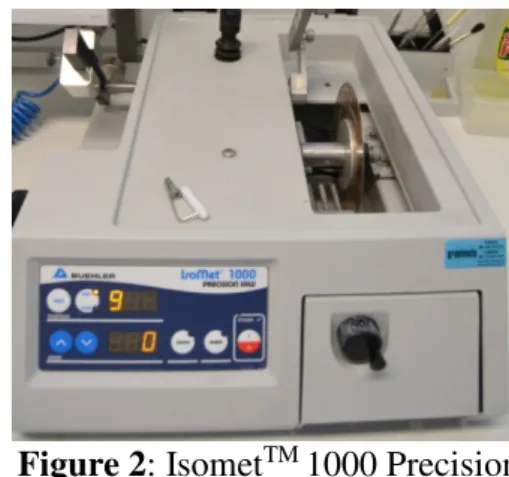
From each tooth, a crown segment was obtained exposing middle dentin by sectioning the crowns with two cuts, a few millimeters apart, parallel to the occlusal surface, with a precision diamond disk at low speed (Diamond Wafering Blade -10,2cm*0,3mm- Series 15HC, Buehler Ltd., Lake Bluff, IL, USA – figure 1) on a hard tissue microtome (IsometTM 1000, Buehler Ltd. Ltd., Lake Buff, IL, EUA – figure 2) under distilled water irrigation, in the following way:
Figure 1: Diamond Wafering Blade. Figure 2: IsometTM 1000 Precision Saw.
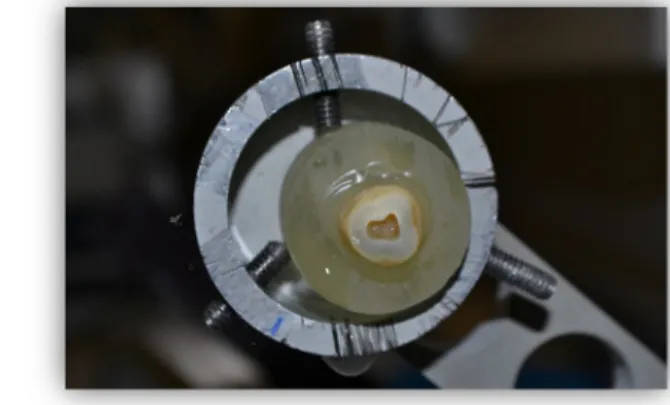
1. The teeth were attached to an acrylic holder with sticky wax, perpendicular to the long axis of the tooth (figure 3);
Figure 3: Tooth fixed to an acrylic holder with sticky wax
2. The first cut was made parallel to the occlusal surface 1-2 mm below the cementoenamel junction (CEJ) to remove the roots (figure 4) and expose the pulp chamber (figure 5);
Figure 4: First cut 1-2 mm below the CEJ. Figure 5: Pulp chamber’s exposure.
3. The pulpal tissues were removed from the pulp chamber with a dentin curette (figure 6) and then filled with cyanoacrylate glue (737 Black Magic Toughened adhesive, Permabond, Hampshire, UK – figure 7);
Figure 6: Removal of pulp tissues. Figure 7: Filling the pulp chamber with cyanoacrylate glue.
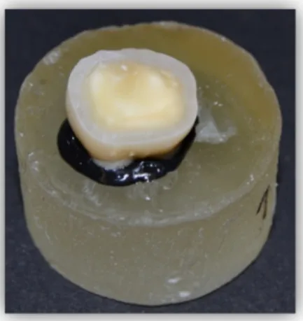
4. The crown segments are then glued with cyanoacrylate glue (737 Black Magic Toughened adhesive, Permabond, Hampshire, UK) to the acrylic holders, by the pulpal side (figure 8);
Figure 8: Crowns fixed with cyanoacrylate glue to the acrylic holder.
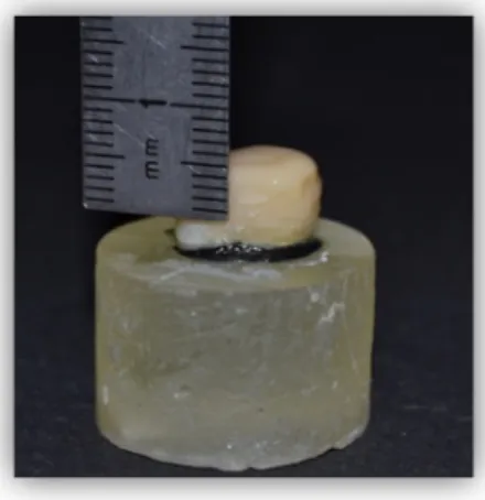
5. Mid-coronal dentin surfaces were obtained by removing the occlusal enamel and superficial dentine of the molar crowns (figure 9 and 10). For that purpose, the second cut, parallel to the first, was made within 1-2mm from the pulp horns, using a diamond disk at low speed, under constant water irrigation;
Figure 9: Removing the occlusal enamel Figure 10: Mid-coronal dentin and superficial dentin. surface.
6. With the purpose of creating a uniform smear layer obtained in similar conditions to those occurring in clinic situations, dentin surface was polished with 600-grit silica-carbide (SiC) sandpaper (Ultra-Prep, Buehler Ltd., Lake Bluff, IL, EUA) on a mechanical grinder (Lunn-Major, Struers, Denmark) during 60 seconds under water irrigation (figure 11) (Pashley DH et al., 1988).
Figure 11: Lunn Major, mechanical grinder.
4. Distribution and treatment of the crown segments
The crown segments were kept in distilled water until the moment of treatment. The six crown segments were randomly assigned to one of the two adhesive groups. The order in which the crown segments were treated was random, to avoid a possible
bias due to any particular sequence of treatment. All the treatment procedures were performed by the same operator in the way that is followed described:
Group 1 – Scotchbond Universal (3M ESPE, St. Paul, MN, USA) (figure 12) as per manufacturer’s instructions – self-etch (etch-and-dry) technique on dentin (SBU SE D):
1. The occlusal surface was rinsed with water being the excess of water removed from the dentin surface using a moist cotton pellet, so that the surface remained shiny and visibly moist.
2. The adhesive was applied, using a disposable microbrush, to the entire dentin surface, scrubbing lightly for 20 seconds.
3. The surface was then gently air-dried until it ceases to show any movement and the solvent was evaporated completely, forming a homogenous and slightly shiny film. Beginning with a soft blow of air from a distance of approximately 10 cm, the air pressure was increased while decreasing distance, finishing at a distance of approximately 1-2 mm from the surface at maximum air pressure.
4. Finally, the surface was polymerized for 10 seconds.
Figure 12: Scotchbond Universal Adhesive.
Group 2 – Scotchbond Universal (3M ESPE, St. Paul, MN, USA) + Adper Scotchbond multipurpose adhesive (3M ESPE, St. Paul, MN, USA) (figure 13) – self-etch (self-etch-and-dry) technique on dentin (SBU+A SE D)*:
* - It is a modification of manufacturer´s instructions. Not recommended by the manufacturer.
1. The occlusal surface was rinsed with water being the excess of water removed from the dentin surface using a moist cotton pellet, so that the surface remained shiny and visibly moist.
2. The adhesive was applied, using a disposable microbrush, to the entire dentin surface, scrubbing lightly for 20 seconds.
3. The surface was then gently air-dried until it ceases to show any movement and the solvent was evaporated completely, forming a homogenous and slightly shiny film. Beginning with a soft blow of air from a distance of approximately 10 cm, the air pressure was increased while decreasing distance, finishing at a distance of approximately 1-2 mm from the surface at maximum air pressure.
4. A layer of Adper Scotchbond Multipurpose adhesive was applied using a disposable microbrush, leaving a uniform, thin and even adhesive layer, removing the excess of adhesive with the same microbrush, as needed. Finally, the surface was polymerized for 10 seconds.
Figure 13: Adper Scotchbond Multipurpose Adhesive.
5. Restorative Procedures
Resin composite build-ups were performed using ENAMEL plus HRi, (Micerium S.p.A. Avegno (GE) Italy), shade UD4 (figure 14), applied in three increments of 2 mm each. Each increment was light cured for 20 seconds, according to the manufacturer's instructions, until reaching a height of 6 mm (figure 15). Additional light polymerization was performed on facial, lingual, mesial and distal surfaces of the composite build-up for 10 seconds each.
Figure 14: Resin composite ENAMEL plus HRi.
All light curing was performed with a light intensity of 600 mW/cm2 using a halogen light-activation unit (ELIPAR S10, 3M ESPE Seefeld, Germany), with the 13 mm light guide held 1-2 mm from the treatment surface. The output of the curing light was periodically verified at >600 mW/cm2 with a radiometer (Curing Radiometer 100, Serial No. 1279, Demetron Research Corporation, Danburry, USA) throughout the procedure.
6. Specimens preparation for the micro-tensile tests
All teeth were painted with different colors with waterproof ink. The exterior surface of the resin composite was also painted, in order to identify, and then, exclude from the study the sticks in which the adhesion was made to enamel.
The teeth were stored in distilled water in an incubator for 24 hours at 37 ° C. Date and time of the restoration are registered.
Posteriorly, the teeth were longitudinally sectioned in both “x” and “y” directions (figure 16 and 17) with a slow-speed diamond disk (Diamond Wafering Blade -10,2cm*0,3mm- Series 15HC, Buehler Ltd., Lake Bluff, IL, USA) under water irrigation, using a microtome (IsometTM, Buehler Ltd. Ltd., Lake Buff, IL, EUA), to obtain sticks with a cross-sectional area of approximately 1 mm2.
Figure 15: Resin composite build-up.
Figure 16 and 17: Teeth after being sectioned in both ‘x’ and ‘y’ directions.
A final cut was made at the base of the root, perpendicular to the long axis of the tooth, to separate the sticks from the acrylic holders (figure 18).
Figure 18: Sticks.
Debonded or lost sticks are registered. Debonded sticks were those separated in the adhesive interface during the cutting procedure. Lost sticks were those which were lost or fractured during test preparation.
The obtained sticks were kept in distilled water for a maximum of 24 hours, until the completion of the micro-tensile tests.
7. Micro-tensile bond strength tests (μTBS)
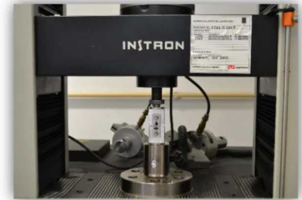
The specimens were individually attached to a stainless-steel grooved Geraldeli´s jig with cyanoacrylate glue (737 Black Magic Toughened adhesive, Permabond, Hampshire, UK) (figure 19) and then submitted one by one to a tension load using a universal testing machine (Instron® 4502 Series, Serial no. H3307, Instron
Corporation, Canton, MA, USA) (figure 20), at a crosshead speed of 1mm/min until
fracture occurred, with the stress to failure expressed in MPa.
Figure 19: Sticks attached to Geraldeli’s jig Figure 20: Instron® 4502, universal with cyanoacrylate glue. testing machine.
A digital caliper (Ficher Darex®, 0-150mm, France) was used to measure the cross section of each bonding interface and calculate the bonding area in mm2. The μTBS (MPa) values were calculated by dividing the load (N) at failure by the area (mm2) of each stick.
The failure modes were analyzed under a stereomicroscope (Nikon, Japan) at 10X magnification to determine the mode of failure. The failure modes were classified as: 1) adhesive (A, failure occurring at the dentin-adhesive interface); 2) cohesive when the failure occurred in dentin (CD) or in composite (CC); and 3) mixed (M, failure with composite and dentin at the interface).
8. Statistical Analysis
The statistical analysis of the results was performed through descriptive and inference methods. A paired-sample t-test was performed since the assumption of normality was valid.
Pretesting failures that occurred during specimen preparation were previously excluded and not taken into account for the statistical analysis.
IV - RESULTS
Micro-tensile bond strength (μTBS) mean values in MPa, respective standard deviations, and the number of sticks tested per group are displayed in Table 1.
A total of 133 (one hundred and thirty three) sticks were analyzed: 58 (fifty eight) using the Scotchbond Universal Adhesive in self-etch mode as per manufacturer’s instructions (SBU SE D, n=58) and 75 (seventy five) using the Scotchbond Universal Adhesive with an extra hydrophobic coat of Adpe Scotchbond Multi-Purpose Adhesive (SBU+A SE D, n=75).
Group N Mean Std. Deviation Std. Error Mean
M
P
a SBU SE D 58 27,6643 13,21793 1,73560
SBU+A SE D 75 38,5869 22,20630 2,56416
Table 1 – Number of sticks (N); Micro-tensile bond strength (μTBS) mean values; Standard deviation (Std. Deviation) and Standard Error Mean (Std. Error Mean).
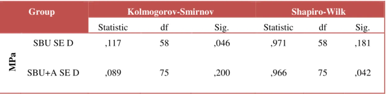
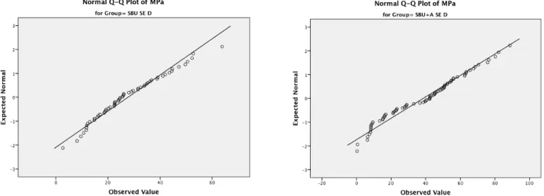
A paired-sample t-test was performed as the assumption of normality (Kolmogorov-Smirnov test and Shapiro-Wilk test - Table 2) in each group was valid (Graphic 1 and 2).
Group Kolmogorov-Smirnov Shapiro-Wilk
Statistic df Sig. Statistic df Sig.
M P a SBU SE D SBU+A SE D ,117 ,089 58 75 ,046 ,200 ,971 ,966 58 75 ,181 ,042
Graphic 1 e 2: Tests of Normality for the SBU SE D and SBU+A SE D group.
Further, the homogeneity of the variances was tested through a Levene’s Test (Table 2). Since the significance value (p) was not superior to 0,05, the variances were assumed as not equal.
Levene’s Test for Equality of
Variances
t-test for Equality of Means
F Sig. t df Sig. (2-tailed) Mean Difference M P a Equal variances assumed 20,110 ,000 -3,317 131 ,001 -10,92264 Equal variances not assumed -3,528 123,645 ,001 -10,92264
Table 3: Results of Levene’s Test and t-test. As p<0,05, it exists a statistically significantly difference between the two groups.
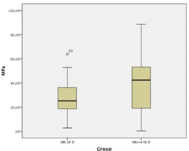
The distribution of μTBS is shown in graphic 3, where the median μTBS is represented by the central line of the box.
Graphic 3 - Box-whisker plot of the μTBS for SBU SE D and SBU+A SE D: x axis represents the group and y axis the MPa.
The SBU+A SE D group (group 2) resulted in a statistically significantly higher mean uTBS value (38,59 ± 22,21 MPa) than the SBU SE D group (group 1) (27,66 ± 13,22 MPa), as showed in the Table 3 (p<0,05) with a 95% confidence interval (Table 4).
t-test for Equality of Means
Std. ErrorDifference
95% Confidence Interval of the Difference Lower Upper M P a Equal variances assumed 3,29258 -17,43615 -4,40914 Equal variances not assumed 3,09633 -17,05131 -4,79398
Table 4 – T-test for Equality of Means.
The type of failures distribution per group is displayed on Table 5 and 6, and graphically displayed on Graphic 4. On both groups (SBU SE D and SBU+A SE D) most failures were adhesive.
Group Failure Mode Number of fractures
SBU SE D A 35 (60,3%)
CC 3 (5,2%)
CD 1 (1,7%)
M 17 (29,3%)
Group Failure Mode Number of fractures
SBU+A SE D A 31 (41,3%)
CC 13 (17,3%)
CD 9 (12%)
M 21 (28%)
Table 5 and 6 - Failure mode: A- adhesive failure; CC- Composite cohesive failure; CD- dentin cohesive failure; M – mixed failure.
Graphic 4: Failure mode distribution: A- adhesive failure; CC- Composite cohesive failure; CD- dentin cohesive failure; M – mixed failure.
0 5 10 15 20 25 30 35 40 A CC CD M SBU SE D SBU+A SE D
V - DISCUSSION
The clinical success of adhesive restorations depends greatly on the proper handling of the adhesive system. However, adhesives have a high degree of technique sensitivity due to a detailed protocol for different systems as well as the possible operator induced variations. Despite that, several studies have already showed that changes to the manufacturer application technique may improve bonding effectiveness (Van Landuyt KL et al., 2006b; Reis A et al., 2008). Thus, it is necessary to test the new universal adhesives placed on the market and how changes in its manipulation can affect its micro-tensile bond strength.
In what may concern the universal adhesive tested in this study, Scotchbond Universal, only a few in vitro and clinical studies have been carried out so far (Mena-Serrano A et al., 2013; Muñoz MA et al., 2013; Perdigão J et al., 2013; Marchesi G et al., 2014; Muñoz MA et al., 2014). However just one of them has yet evaluated the effect of an additional hydrophobic layer on top of the adhesive (Muñoz MA et al., 2014).
This experimental in vitro study evaluated the influence of the application of an additional hydrophobic resin layer (Adhesive of Adper Scotchbond Multipurpose) on the immediate dentin micro-tensile bond strength of a universal adhesive (Scotchbond Universal) used in a self-etch mode (SBU+A SE D – Group 2) compared to the manufacturer’s instructions (SBU SE D – Group 1).
The adhesive chosen to act as a hydrophobic resin layer in this present study was Adper Scotchbond Multipurpose. The purpose of this choice was to use an adhesive system from the same manufacturer thus leading to the same type of chemical reactions. The teeth selected for this study were stored in 0,5% chloramine T at 4ºC for one week and after that, left in distilled water at 4ºC no more than three months, as is required from the ISO TR 11405 standard (International Standardization Organization, 2003). The same procedure was carried out in other studies (Van Landuyt KL et al., 2006b; Van Landuyt KL et al., 2009; Chasqueira AF et al., 2013; Marchesi G et al., 2014; Taschner M et al., 2014)
It is well known that during clinical cavity preparation using rotary instruments, the dentin surface to bond to will be covered by a smear layer. It is also known that, depending on the preparation technique, this smear layer varies significantly in size and structure. It is clear that the main challenge for current self-etch adhesives is to dissolve
the smear layer without demineralizing the tooth surface too profoundly, thereby removing hydroxyapatite at the interface. Preserving hydroxyapatite at the interface not only protects the collagen from external chemical aggression, but the hydroxyapatite will also provide calcium for chemical bonding to the functional monomer (Van Meerbeek B et al., 2011).
As we can see in the meta-analytical review by De Munck et al (2012), the most-used preparation methods identified were preparation by either a carbide or diamond dental bur or by silicon-carbide (SiC) paper. Although in simulation of clinical practice, dentin is best prepared with a bur, most bond-strength studies use silicon-carbide (SiC) paper (Reis A et al., 2008; Muñoz MA et al., 2014; Perdigão J et al., 2014).
Thus, for the purpose of creating a standardized and uniform smear-layer similar to that obtained in clinical situations, exposed dentin surface of each teeth was polished with 600-grit silica-carbide abrasive paper (Buehler, Lunn Major, Struers Denmark) under running water during 60 seconds, on a mechanical grinder (Lunn Major, Struers, Denmark) (Pashley DH et al., 1988). The same procedure was realized in other studies (Pashley DH et al., 1988; Perdigão J et al., 2006; Reis A et al., 2008; Muñoz MA et al., 2013; Muñoz MA et al., 2014; Perdigão J et al., 2014).
All the treatment procedures were performed by the same operator to avoid operator variability as it was done in previous studies (Poitevin A et al., 2008).
Even though the manufacturer´s instructions are well-formulated, they are not very detailed and have some degree of ambiguity. For that reason an effort was made to specify each step as much as possible, so that the protocol would become more consistent and standardized. All of the bonding procedures used for this study are described in the Materials and Methods section.
As we can see in the manufacturer’s instructions displayed in appendix 1, the adhesive is entitled to be rubbed for 20 sec. It is consensual that an active adhesive application could increase the resin–dentine bond strength of SE adhesive systems, as it was already proven in previous literature findings (Pleffken PR et al., 2011). Several factors may account for this effect as the increased penetration of monomers into dentine, the higher solvent evaporation or the improved in polymerization (Muñoz MA et al., 2014).
After the adhesive application, the restorative procedures were performed using ENAMEL plus HRi composite, shade UD4. According to the manufacturer’s
instructions this composite should be polymerized for 20 seconds as it was performed in our study. However, an additional light polymerization was performed on mesial, distal, facial and lingual surfaces, for 10 seconds each, in order to avoid composite cohesive failures. In a previous in vitro study carried out by Proença et al. (2007), a similar process was carried out as they polymerized the resin composite (Tetric Ceram, Ivoclar Vivadent, Schäan, Liechtenstein) for 40 seconds instead of the 20 seconds as is recommended by the manufacturer . As a result they obtained very few specimens with composite cohesive failures as did we in this study since the cohesive failures only represent an insignificant part of the sample (5,2% for SBU SE D and 17,3% for SBU+A SE D). Furthermore, Perdigão et al. (2006) also showed similar results .
In the present study a micro-tensile test was perform to assess the resin-dentin bond strength of the studied adhesives. Although the ultimate test method for the assessment of bonding effectiveness remains a clinical trial, nowadays laboratory bond strength tests are the most frequently used tests to screen adhesives (De Munck J et al., 2005; Peumans M et al., 2005). This may be due to the time taking of clinical trials as well as the number of patients involved, the existence of variables you cannot control and the fact that manufacturers constantly introduce to the market new versions of the same adhesive, before obtaining studies results (Swift EJ et al., 1995; Van Meerbeek B et al., 2003).
In order to measure the bonding effectiveness of adhesives to enamel and dentin, diverse methodologies can today be used. Currently, the shear and micro-tensile bond strength (µTBS) test methods are the most used (De Munck J et al., 2005). Since it was developed by Sano et al. (1994), micro-tensile bond strength tests have been employed in up to 60% of current scientific papers. It was his purpose to measure the bond strength of samples with small bonded surface areas compared to that of macro bond strength tests (about 1mm2 or less). Nowadays, the micro-tensile bond strength test has been accepted as a versatile and reliable in vitro statistic test to quantify the bonding effectiveness and stability of adhesive biomaterials bonded to tooth structures (Poitevin A et al., 2008).
In the present study, teeth were longitudinally sectioned to obtain sticks with a cross-sectional area of approximately 1mm2. According to a previous study carried out by Poitevin et al. (2008), it seems like square specimens of 1mm2 were easier to manipulate, standardize, and reproduce, preserving the desired stress uniformity, which was proven by more regular stress-time graph patterns .
Some advantages can be pointed out for µTBS when compared to macro-bond-strength testing of which are, the possibility of using multiple micro-specimens originating from one tooth, the better stress distribution at the adhesive interface avoiding cohesive failures, the ability to measure adhesion forces in different regions of the same tooth, etc. (Van Meerbeek B et al., 2010). This may be why micro-tensile protocol appears to be able to discriminate adhesives better on their bonding performance than a traditional shear bond-strength approach.
It is also important to note that a bond strength value cannot be considered as a material property. The data recorded depend largely upon experimental factors such as, for example, the type of composite, stress rate, sample size and geometry, and the actual test method (Phrukkanon S et al., 1998; Sudsangiam S et al., 1999).
The prepared specimens in this study were “non-trimmed” as they were in other in vitro studies where the teeth were prepared for micro-tensile tests (Reis A et al., 2008; Muñoz MA et al., 2013; Muñoz MA et al., 2014; Perdigão J et al., 2014; Taschner M et al., 2014).
Specimen preparation for µTBS is often very labor intensive and time consuming. Specimens can be prepared in two different ways: “trimmed” (by free-hand or using a Micro-Specimen Former) (Armstrong S et al., 2010) to an hourglass shape or left “nontrimmed” (stick shape). They both have advantages and disadvantages (Poitevin A et al., 2008).
Trimming is very technique sensitive. It induces additional stress at the interface and involves a more invasive specimen procedure. When this trimming is not carefully performed, interfacial defects may easily be introduced, especially in weaker bonds, thus facilitating pre-testing failures at lower bond strength. The operator’s experience and manual skill will therefore influence the results and the quality of the study (Poitevin A et al., 2008; Armstrong S et al., 2010; Van Meerbeek B et al., 2010).
Non-trimmed micro-specimens are cut out from the restored tooth in the shape of sticks, and directly used in the universal testing machine. They were proven to be easier and faster to prepare (Poitevin A et al., 2008) (Van Meerbeek B et al., 2010). Besides that, the maximum concentration of stress is at the center of the adhesive layer therefore not leading to a preferred point of fracture (Ghassemieh E, 2008).
In the Hanabusa et al. (Hanabusa M et al., 2012) study and others (Van Landuyt KL et al., 2006b; Van Landuyt KL et al., 2009), authors only used nine central sticks from each tooth to reduce substrate regional variability, whereas in our study we used
sticks from the entire interface except those containing enamel. One study carried out by Loguercio et al. (2005), reported lower µTBS for peripheral specimens than for centrally located specimens . Nevertheless, many studies proceeded like the present study and used all the specimens (Reis A et al., 2008; Muñoz MA et al., 2013; Muñoz MA et al., 2014; Perdigão J et al., 2014).
As for the crosshead speed used in the present study for the tension load, it was of 1mm/min. Reis et al. (2004) found no significant difference in the µTBS when non-trimmed, square specimens were tested with a different crosshead speed . However, Poitevin et al. (2008) reported that a more uniform stress-time pattern was observed when 1mm/min was used .
In what may concern the statistical analysis of this in vitro study, pre-testing failures that occurred during specimen preparation were previously excluded and not taken into account. These include debonded and lost sticks. The same approach was done in other studies (Marchesi G et al., 2014; Perdigão J et al., 2014; Taschner M et al., 2014).
Several approaches have been applied to deal with the pre-testing failures. They can be: (a) excluded from further statistical analysis; (b) assigned a bond strength value of 0MPa; or (c) assigned a pre-determined value as for example the lowest µTBS measured within the respective group (Van Meerbeek B et al., 2010). These data transformations may affect the mean µTBS thus subsequently affecting the statistical analysis. For example, the exclusion of the pre-testing failures may overestimate the actual bond strength, however, assigning a value of 0MPa penalizes even more the adhesive analysis as there is a certain bond strength upon the failure (Van Meerbeek B et al., 2010).
Regarding the failure mode analyses, it was done by the same observer under a stereomicroscope at 10x magnification. This could have induced some potential bias concerning the identification of the type of failures as it is desirable to determine the failure mode under a greater magnification as in other studies (Van Landuyt KL et al., 2006b; Muñoz MA et al., 2014; Perdigão J et al., 2014; Taschner M et al., 2014).
In this in vitro study SBU+A SE D showed higher μTBS mean values (38,59 ± 22,21 MPa) than SBU SE D (27,66 ± 13,22 MPa). This resulted in a statistical difference in dentine μTBS between the two groups tested, since p < 0,05. Thus, the null hypothesis must be rejected in this study as the obtained results show that the use of an
additional hydrophobic layer may improve the performance of the universal adhesive system, Scotchbond Universal, when applied as a SE adhesive.
Although the variances were assumed as not equal, the significance value (p) had the same value (p = 0,001) for equal or not equal assumed variances, thus not influencing the results of the study.
For the production of one-step self-etch system, acidic, hydrophilic and hydrophobic functional monomers are dissolved in high concentrations of organic solvents and water, and mixed in a single bottle. This is responsible for the high hydrophilicity of these systems (Tay FR et al., 2003a). As the solvent and functional monomers usually make up almost 50% of the adhesive, the concentration of hydrophobic cross-linking monomers is drastically reduced (Van Landuyt KL et al., 2006b). The presence of such a more hydrophilic layer may thus induce water sorption and water uptake, in turn leading to a subsequent reduction in the mechanical properties of the bonding (Yiu CK et al., 2004).
This is why they are considered permeable membranes to water diffusion from the underlying dentin across the adhesive layer (Tay FR et al., 2002). The retention of water, either from residual water that is incompletely evaporated from the adhesive or from the underlying dentin, as the result of the high osmolarity of the hydrophilic adhesive mixture creates water-filled channels within the adhesive (Tay FR et al., 2002). These findings are not observed in two-step self-etch system, probably due to the placement of a hydrophobic resin coating over the primed surface.
Besides that, Scotchbond Universal also has in its composition a water-soluble methacrylate monomer called HEMA, which is frequently present in the composition of dental adhesives to increase their wettability and hydrophilicity and prevent hydrophilic and hydrophobic phase separation (Nakabayashi N et al., 1992; Van Landuyt KL et al., 2008). However, disadvantages have also been related to the presence of high amounts of HEMA such as, increased water sorption, compromised degree of conversion and hydrolytic degradation adversely influencing the mechanical properties and stability of the adhesive interface (Tay FR et al., 2002; Van Landuyt KL et al., 2008). Theoretically, since Scotchbond Universal and Scotchbond Multipurpose both contain high amounts of HEMA (15-25% and 30-40% respectively), this should result in increased water sorption as seen in a previous study carried out by Takahashi et al.(2011). On the other hand, the lack of HEMA could result in phase separation at the interface, which may be also a limiting factor for improved performance of the material