Clonal evolution of CD8
+
T cell responses against
latent viruses: relationship among phenotype,
localization, and function.
Remmerswaal EB1, Klarenbeek PL2, Alves NL3, Doorenspleet ME2, van Schaik BD4, Esveldt RE2, Idu MM5, van Leeuwen EM3, van der Bom-Baylon N6, van Kampen AH4, Koch SD3, Pircher H7, Bemelman FJ8, Ten Brinke A9, Baas F10, Ten Berge IJ8, van Lier RA11, de Vries N12.
1.Department of Experimental Immunology, Academic Medical Center, Amsterdam, the Netherlands Renal Transplant Unit, Department of Internal Medicine, Academic Medical Center, Amsterdam, the Netherlands e.b.remmerswaal@amc.uva.nl.
2.Department of Experimental Immunology, Academic Medical Center, Amsterdam, the Netherlands Department of Clinical Immunology and Rheumatology, Academic Medical Center, Amsterdam, the Netherlands Department of Genome Analysis, Academic Medical Center, Amsterdam, the Netherlands.
3.Department of Experimental Immunology, Academic Medical Center, Amsterdam, the Netherlands.
4.Department of Clinical Epidemiology, Biostatistics and Bioinformatics, Academic Medical Center, Amsterdam, the Netherlands.
5.Department of Surgery, Academic Medical Center, Amsterdam, the Netherlands.
6.Department of Experimental Immunology, Academic Medical Center, Amsterdam, the Netherlands Renal Transplant Unit, Department of Internal Medicine, Academic Medical Center, Amsterdam, the Netherlands.
7.Institute of Immunology, University of Freiburg, Freiburg, Germany.
8.Renal Transplant Unit, Department of Internal Medicine, Academic Medical Center, Amsterdam, the Netherlands.
9.Sanquin Research at CLB, Amsterdam, the Netherlands.
10.Department of Genome Analysis, Academic Medical Center, Amsterdam, the Netherlands. 11.Department of Experimental Immunology, Academic Medical Center, Amsterdam, the Netherlands Sanquin Research at CLB, Amsterdam, the Netherlands.
12.Department of Clinical Immunology and Rheumatology, Academic Medical Center, Amsterdam, the Netherlands.
Originally published in Journal of Virology. 2015 Jan; 89(1):568-80. Epub 2014 Oct 22.
Doi: 10.1128/JVI.02003-14.
Human cytomegalovirus (hCMV) infection is characterized by a vast expansion of resting effector-type virus-specific T cells in the circulation. In mice, interleukin-7 receptor α (IL-7Rα)-expressing cells contain the precursors for long-lived antigen-experienced CD8+ T cells, but it is unclear if similar mechanisms operate to maintain these pools in humans. Here, we studied whether IL-7Rα-expressing cells obtained from peripheral blood (PB) or lymph nodes (LNs) sustain the circulating effector-type hCMV-specific pool. Using flow cytometry and functional assays, we found that the IL-7Rα+ hCMV-specific T cell population comprises cells that have a memory phenotype and lack effector features. We used next-generation sequencing of the T cell receptor to compare the clonal repertoires of IL-7Rα+ and IL-7Rα− subsets. We observed limited overlap of clones between these subsets during acute infection and after 1 year. When we compared the hCMV-specific repertoire between PB and paired LNs, we found many identical clones but also clones that were exclusively found in either compartment. New clones that were found in PB during antigenic recall were only rarely identical to the unique LN clones. Thus, although PB IL-7Rα-expressing and LN hCMV-specific CD8+ T cells show typical traits of memory-type cells, these
populations do not seem to contain the precursors for the novel hCMV-specific CD8+ T cell pool during latency or upon antigen recall. IL-7Rα+ PB and LN hCMV-specific memory cells form separate virus-specific compartments, and precursors for these novel PB hCMV-specific CD8+ effector-type T cells are possibly located in other secondary lymphoid tissues or are being recruited from the naive CD8+ T cell pool.
IMPORTANCE Insight into the self-renewal properties of long-lived memory CD8+T cells and their location is crucial for the development of both passive and active vaccination strategies. Human CMV infection is characterized by a vast expansion of resting effector-type cells. It is, however, not known how this population is maintained. We here investigated two possible compartments for effector-type cell precursors: circulating acute-phase IL-7Rα-expressing hCMV-specific CD8+ T cells and lymph node (LN)-residing hCMV-specific (central) memory cells. We show that new clones that appear after primary hCMV infection or during hCMV reactivation seldom originate from either compartment. Thus, although identical clones may be maintained by either memory population, the precursors of the novel clones are probably located in other (secondary) lymphoid tissues or are recruited from the naive CD8+ T cell pool.
INTRODUCTION
Adaptive immune responses against transient viral infections typically consist of three phases. First, viral antigens are recognized by naive CD8+ T cells in lymph nodes (LNs), where activated T cells expand vigorously to form effector clones that eliminate virus-infected cells. Second, after clearance of the virus, the majority of the activated CD8+ T cells undergo apoptosis. Third, a proportion of virus-specific T cells survive to provide long-lasting immunological memory (1–3). Although this response is well established for cleared infections, responses against persistent viruses are more complex. The immune surveillance required to control these infections triggers regular activation of virus-specific CD8+ T cells. Persistent infections can therefore challenge the immune system for decades and may be associated with lymphoproliferative disorders and opportunistic infections in immunocompromised patients. Understanding how viral latency is maintained is important in designing strategies that may prevent complications from these infections. Human cytomegalovirus (hCMV) is an attractive virus for the study of persistent infections in humans as the primary infection can be studied longitudinally in recipients of solid organ transplants, such as kidneys. Here, we used this approach to study the clonal and phenotypic relations between peripheral blood (PB) and LN memory- and PB effector-type subsets in primary and latent phases of hCMV infection.
The majority of the latent-phase circulating hCMV-specific CD8+ T cells is CD28−CD27− CD45RA+ granzyme B-positive (granzyme B+) perforin-positive (perforin+) quiescent effector-type cells. These CD8+ T cell populations consist of large clonal expansions that are maintained for many years (4). As such, these cells were thought to be long-lived (5). Recent findings in a murine CMV (mCMV) model, on the other hand, showed that an mCMV-specific effector CD8+ T cell pool was maintained by constant recruitment of CD27-expressing memory T cells and, to a limited extent, naive T cells (6, 7). A “buffered memory” concept was suggested (6), proposing that a memory-like T cell pool, shielded from high antigenic loads by compartmentalization, would be supplementing the effector-type pool at times of rechallenge. Such a concept has not been investigated in hCMV. It has been shown that, besides the prevalent resting effector-type cells, at least two hCMV-specific CD8+ T cell pools with memory-like features can be distinguished. In PB, interleukin-7 receptor α (IL-7Rα) (CD127), a marker that distinguishes memory precursor cells during transient viral infections, is expressed on a minority of the PB hCMV-specific CD8+ T cells (8). Further in LNs, the chemokine receptor CCR7, which can be used to identify central memory T cells, is found on a substantial percentage of the hCMV-specific CD8+ T cells (9). We hypothesized that either one or both of these hCMV-specific CD8+ T cell memory pools might fuel the PB effector-type pool. To address this issue, we combined flow cytometry with next-generation sequencing to investigate phenotype and longitudinal clonal relationships between PB- and LN-derived subsets of hCMV-specific CD8+ T cells.
Subjects.The phenotype and cytokine-producing capacities of IL-7Rα− and IL-7Rα+ hCMV-specific
CD8+ T cells in latency were studied in healthy individuals (n = 7). In order to study the
contribution of acute-phase IL-7Rα-expressing hCMV-specific CD8+ T cells to those present during the latency phase, hCMV-specific CD8+ T cells from two seronegative recipients of an hCMV-seropositive kidney transplant were analyzed longitudinally (Table 1, patients [Pt] 1 and 2). The relationship between paired PB- and LN-derived virus-specific CD8+ T cells was studied in seven hCMV-seropositive renal transplant recipients prior to transplantation (Pt 3 to 9). The contribution of LN-derived hCMV-specific CD8+ T cells upon hCMV reactivation was longitudinally analyzed in four of these hCMV-seropositive renal transplant recipients (Pt 3 to 6); three received an hCMV-seropositive kidney, and one received an hCMV-seronegative kidney. Immunosuppressive drug treatment of these patients is summarized in Table 1. Previously, we did not find an important effect of induction treatment with CD25 monoclonal antibody (MAb) on the immune response against hCMV. These patients were transplanted in the time period from 2000 to 2004, during which no antiviral prophylaxis was administered. Because of clinical symptoms, patients 2 and 4 were treated with valganciclovir until the hCMV PCR was negative at two subsequent time points. None of the patients experienced an acute rejection episode. The study was performed according to the Declaration of Helsinki and approved by the local medical ethics committee. All patients gave written informed consent.
Isolation of mononuclear cells from PB and LN.Heparinized PB samples were obtained from
healthy individuals and from patients before transplantation (pre-TX) and at regular intervals thereafter up to 1 year after transplantation. Peripheral blood mononuclear cells (PBMCs) were isolated using standard density gradient centrifugation. Para-iliac LNs were collected from recipients during living-donor kidney transplantation as described before (9). Directly after extirpation, the gathered LNs were chopped into small pieces. A cell suspension was obtained by grinding the material through a flowthrough chamber. PBMCs and LN mononuclear cells (LNMCs) were subsequently cryopreserved until further analysis.
Virological analysis.Quantitative PCR for hCMV and Epstein-Barr virus (EBV) was performed in
EDTA-treated whole-blood samples, as described previously (10). To determine hCMV serostatus, anti-hCMV IgG was measured in the serum using an AxSYM microparticle enzyme immunoassay (Abbott Laboratories). Measurements were calibrated relative to a standard serum. EBV serostatus was determined by qualitative measurement of specific IgG against the viral capsid antigen (Ag) and against the EBV nuclear Ag (EBNA) using, respectively, an anti-EBV viral capsid Ag IgG enzyme-linked immunosorbent assay (ELISA) and an anti-EBV nuclear Ag IgG ELISA (Biotest). All tests were performed according to the instructions of the manufacturers.
Immunofluorescence staining and flow cytometry.PBMCs were washed in phosphate-buffered
saline containing 0.01% (wt/vol) NaN3 and 0.5% (wt/vol) bovine serum albumin. Two million PBMCs were incubated with allophycocyanin (APC)-labeled tetrameric complexes for hCMV pp65, hCMV IE-1, EBV BZLF-1, EBV BMLF-1, EBV EBNA-1, EBV EBNA-3a, and influenza A virus matrix protein 1 (Table 2), followed by incubation with a combination of the following antibodies: IL-7Rα conjugated with phycoerythrin (PE)-Cy7, IL-7Rα APC-eFluor 780, CD27 APC-eFluor 780, and CX3CR1 PE (eBioscience Inc., San Diego, CA, USA); CXCR3 PE (Caltag, Buckingham, United Kingdom); CCR5-PE and CD45RA Brilliant Violet 650 (BioLegend, San Diego, CA, USA); CD8 Brilliant Violet 421, CD3 V500, and CXCR3 Alexa Fluor 488 (BD Pharmingen, San Diego, CA, USA); CD28 PE, CD45RA PE-Cy7, and CCR7 PE-Cy7 (BD Biosciences, San Jose, CA, USA); and KLRG1 (killer cell lectin-like receptor G1) Alexa Fluor 488 (11). A Foxp3 staining kit (eBioscience) was used for intracellular staining with the following antibodies: granzyme B Alexa Fluor 700 and granzyme A PE (BD Pharmingen); perforin conjugated with peridinin chlorophyll protein (PerCP)-eFluor 710 and eomesodermin PerCP-eFluor 710 (eBioscience); granzyme K PE (Immunotools, Friesoythe, Germany); and T-bet Brilliant Violet 421 (Biolegend). Measurements were done on an LSR Fortessa flow cytometer (BD), and analysis was performed with FlowJo software (FlowJo, Ashland, OR, USA). The gating strategy as well as a representative example of the immunofluorescence staining can be found in Fig. S1 in the supplemental material.
Intracellular cytokine staining.Cytokine release after cognate peptide or phorbol 12-myristate
13-acetate (PMA)-ionomycin stimulation was performed as described by Lamoreaux et al. (12). Freshly isolated PBMCs from hCMV-seropositive healthy individuals were stained with anti-CD3 PE-Cy7 and anti-IL-7Rα APC-Alexa Fluor 750 and sorted into CD3−, IL-7Rα+ CD3+, and
IL-7Rα− CD3+ populations on a FACSAria instrument. Purity of the obtained sorted cells was checked
by flow cytometry and had to be at least 90%. Sorted IL-7Rα+ CD3+ and IL-7Rα− CD3+cells were
rested overnight in suspension flasks (Greiner) in RPMI medium supplemented with 10% fetal calf serum (FCS), penicillin, and streptomycin. Next, IL-7Rα+ CD3+ cells and IL-7Rα− CD3+ cells were
supplemented with the CD3−sorted cells which were used as antigen-presenting cells (APCs) (1
CD3− cell to 2.5 IL-7Rα+ CD3+ or IL-7Rα− CD3+ cells). Subsequently, the cells were stimulated with
PMA-ionomycin or with the viral peptides in culture medium supplemented with anti-CD28 (15E8; 2 μg/ml), anti-CD29 (TS 2/16; 1 μg/ml), brefeldin A (Invitrogen; 10 μg/ml), and GolgiStop (BD Biosciences) in a final volume of 200 μl for 4 h (PMA at 10 ng/ml; ionomycin at 1 μg/ml) or 6 h (1 μg of peptide) in untreated, round-bottom, 96-well plates (Corning). Thereafter, cells were incubated with the appropriate tetramers, followed by incubation with CD8 PE-Alexa Fluor 610 (Invitrogen). The cells were then washed, fixed, and permeabilized (Cytofix/Cytoperm reagent; BD Biosciences) and subsequently incubated with the following intracellular MAbs: tumor necrosis factor alpha (TNF-α) conjugated with fluorescein isothiocyanate (FITC), IL-2 PE, and gamma interferon (IFN-γ) PerCP-Cy5.5 (BD Biosciences). Acquisition was done on a FACSCanto flow cytometer (BD), and analysis was done with FlowJo software. A representative example of a sorting and of the immunofluorescence staining after stimulation can be found in Fig. S2 in the supplemental material.
Isolation of IL-7Rα+ and IL-7Rα− hCMV-specific CD8+ T cells.For isolation of IL-7Rα+ and
IL-7Rα− hCMV-specific CD8+ T cells, at least 1 × 10⁷ PBMCs were first stained with the appropriate
tetramer, followed by anti-CD8 PerCP-Cy5.5 and anti-IL-7Rα PE (Beckman Coulter, Indianapolis, USA). PBMCs were then first sorted into hCMV-specific CD8+ T cells on a FACSAria instrument (BD) and subsequently subjected to a second sorting to obtain high-purity IL-7Rα− and IL-7Rα+ hCMV-specific CD8+ T cells. For isolation of hCMV-specific CD8+ T cells from LNs, paired PB samples, and subsequent longitudinal PB samples, at least 1 × 10⁷ PBMCs were first stained with the appropriate tetramer, followed by incubation with anti-CD3 V500 and anti-CD8 Brilliant Violet 421. CD3+ CD8+ tetramer-positive cells were subsequently sorted, followed by a second sorting if the purity did not exceed 95% after the first sorting (see Fig. S3 in the supplemental material for an example of sorting). The numbers of sorted cells are given in Table 3. Purity of the obtained sorted hCMV-specific cells was checked by flow cytometry and was at least 95%.
RNA isolation and cDNA synthesis.RNA was isolated from PB IL-7Rα− and IL-7Rα+ cells and from total LN and PB hCMV-specific sorted cells by a NucleoSpin RNA XS kit (Macherey-Nagel, Düren, Germany) and subsequently subjected to template switch-anchored reverse transcription-PCR (RT-PCR) by using a SMARTer Pico cDNA PCR synthesis kit and an Advantage 2 PCR kit (both from Clontech, Mountain View, CA, USA) according to the manufacturer's instructions.
Linear amplification, next-generation sequencing, and bioinformatics.Ten microliters of
SMARTer Pico-treated DNA was amplified using a primer set to cover all functional T cell receptor (TCR) Vβ gene segments (13). Here, all Vβ gene segments are denoted according to the HUGO nomenclature (14). The linear amplification, next-generation sequencing protocol (4, 15), and the bioinformatics pipeline (16) have been described previously. Clones were identified by their unique TCR Vβ sequence. The degree of expansion of each clone was expressed as a percentage of all obtained TCR sequences in a sample. To exclude clonal signal from sorting impurities, only clones that exceeded 1% of total were included. Clones that represented less than 1% were included only when the same clone exceeded 1% in the other subsets analyzed.
Statistical analysis.A two-tailed paired t test was used for analysis of differences between groups.
A P value of <0.05 was considered statistically significant. RESULTS
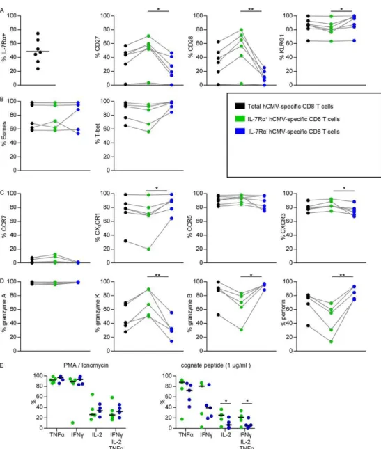
Circulating IL-7Rα+ hCMV-specific CD8+ T cells in healthy individuals have phenotypical traits of memory cells.Murine IL-7Rα+ virus-specific CD8+ T cells have been shown to be antigen-independent, long-lived memory cells (17). We wanted to test whether PB hCMV-specific IL-7Rα+ CD8+ T cells had a true memory phenotype by first measuring their surface expression of several differentiation-associated molecules (Fig. 1A). We found a substantially higher percentage of cells expressing the costimulatory receptors CD27 and CD28 in the IL-7Rα+ hCMV-specific CD8+ T cells. PD1 expression was not different between the IL-7Rα+ and IL-7Rα− subsets (data not shown). Furthermore, neither population appeared to be activated since the expression of
HLA-DR, CD38, and Ki-67 was uniformly low (data not shown). We also tested killer cell lectin-like receptor G1 (KLRG1) expression as the lack of this receptor has been shown to be another feature of memory precursor cells generated during lymphocytic choriomeningitis virus (LCMV) infection in mice (18, 19). Both IL-7Rα− and IL-7Rα+ subsets expressed KLRG1, and although in some individuals the percentages were equal, slightly lower percentages of KLRG1-expressing cells were found in the majority of the IL-7Rα+ hCMV-specific CD8+ T cell populations.
As memory fate and cytotoxic functions have been shown to be linked to the transcription factors eomesodermin and T-bet (18, 20, 21), we analyzed the subsets for expression of these proteins (Fig. 1B). Both transcription factors were expressed on the majority of cells within both subsets, and no significant differences were found.
Finally, we evaluated the expression of homing receptors CCR7 (homing to LN), CCR5, CXCR3 (homing to a wide array of infected tissues) (22–28), and CX3CR1 (homing to stressed endothelium) (22, 29–31). CCR7 was not expressed on either population of PB hCMV-specific CD8+ T cells (Fig. 1C). CCR5 and CXCR3 were found on the majority of cells in both subsets, but the IL-7Rα+ subset contained slightly more CXCR3-expressing cells. Although the differences between the hCMV-specific IL-7Rα subsets varied considerably, in general more CX3CR1-expressing cells were found in the IL-7Rα− subset.
In summary, the PB IL-7Rα+ hCMV-specific CD8+ T cells contained substantially more cells that expressed the costimulatory molecules CD27 and CD28, a typical feature of “classical” memory cells.
Many circulating IL-7Rα+ hCMV-specific CD8+ T cells lack typical effector-type properties.A
feature of hCMV-specific T cells is the abundant expression of components of the cytolytic granule exocytosis machinery (32, 33). We found that within the PB hCMV-specific CD8+ T cell population, the vast majority of the IL-7Rα− cells contained both granzyme B and perforin (Fig. 1D), whereas only half of the IL-7Rα+ hCMV-specific CD8+ T cells contained these proteins. The latter subset, however, did harbor significantly more granzyme K-containing cells. Granzyme A was equally present in both subsets.
We have previously shown that IL-7Rα-expressing hCMV-specific CD8+ T cells have superior proliferation capacities (8). We next wanted to evaluate the ability of both the IL-7Rα+ and IL-7Rα− subsets to produce cytokines after in vitrorestimulation by either PMA-ionomycin or their cognate peptide (Fig. 1E). No difference was detected in IFN-γ and TNF-α synthesis, and although no difference in the production of IL-2 could be detected after PMA-ionomycin stimulation, a significantly higher number of IL-7Rα+ hCMV-specific CD8+ T cells were able to produce IL-2 after stimulation with cognate peptide. Consequently, more polyfunctional hCMV-specific CD8+ T cells (producing IFN-γ, TNF-α, and IL-2 simultaneously) were found in the IL-7Rα+ pool than in the IL-7Rα− pool (Fig. 1E).
In conclusion, with respect to both phenotypic and functional attributes, circulating hCMV-specific CD8+ T cells that express IL-7Rα were found to contain cells with classical memory traits. Novel latent IL-7Rα− effector-type hCMV-specific CD8+ T cell clones seem not to be derived from the circulating acute-phase IL-7Rα+ hCMV-specific CD8+ T cell pool.Since the above
analyses showed that the IL-7Rα+ hCMV-specific CD8+T cell subset contained cells with all typical properties of memory cells, we set out to analyze if they contribute to the establishment and/or maintenance of the IL-7Rα− hCMV-specific effector-type pool. To this end, we studied two patients who were hCMV seronegative prior to kidney transplantation. Each of them received a kidney from an hCMV-seropositive donor, resulting in a primary hCMV infection (Fig. 2 and Table 1). From peripheral blood, IL-7Rα+ and IL-7Rα− subsets of hCMV pp65 tetramer-binding CD8+ T cells were sorted to high purity, and their clonal compositions were determined by next-generation sequencing. Clones were identified by their unique TCRβ sequences.
In agreement with our earlier observations (8), the frequencies of IL-7Rα+ were very low at the early time point, but just after the peak of viral replication enough cells could be isolated to reliably compare the clonal compositions of the IL-7Rα+and IL-7Rα− hCMV-specific cell subsets (Fig. 2A and C). In both patients multiple hCMV pp65-specific clones were identified that carried
diverse Vβ and Jβ genes (see Tables S1 and S2 in the supplemental material). In patient 1, the IL-7Rα−subset at the peak of the primary infection consisted of a single clone that had a frequency of only 6.2% in the IL-7Rα+ population (Fig. 2B), whereas the majority of the clones in the IL-7Rα+ subset were unique. In patient 2 (Fig. 2D) there were three clones that were identical
between the IL-7Rα+ and IL-7Rα− hCMV-specific CD8+ T cells; however, both subsets also contained many nonoverlapping clones. These results showed that during the acute phase, IL-7Rα expression characterized overlapping but not identical hCMV-specific CD8+ T cell pools.
We reasoned that if, after the primary effector phase, the IL-7Rα+ clones did indeed feed the latent-phase hCMV-specific CD8+ T cell population, then the IL-7Rα+ and IL-7Rα− hCMV-specific CD8+ T cell pools at latency would be enriched for clones found in the acute-phase IL-7Rα-expressing CD8+ T cells. We therefore studied the IL-7Rα+ and IL-7Rα− pools 1 year after primary infection (Fig. 2B and D). In patient 1, only 53% and 26% of the sequences of the IL-7Rα+ and IL-7Rα−subsets, respectively, at the 1-year time point were attributable to clones found in the IL-7Rα+ subset at the acute phase. In patient 2, these values were 7% and 73%, respectively; however,
the 73% overlap in the IL-7Rα+ subset was caused by three clones that were also found in the
IL-7Rα− subset from the acute phase. Therefore, it could not be established whether these clones were
derived from the circulating IL-7Rα− or the IL-7Rα+ acute-phase hCMV-specific CD8+ T cell pool.
We cannot exclude the possibility that latent IL-7Rα− hCMV-specific CD8+ T cell clones that are
identical in both the acute-phase IL-7Rα+ and IL-7Rα− pools are maintained from either acute-phase pool. However, the other clones found in the latent IL-7Rα− hCMV-specific CD8+ T cell pool do not seem to be derived from the acute-phase IL-7Rα+ pool.
Lymph nodes contain unique virus-specific CD8+ T cell clones.LN hCMV-specific CD8+ T cells have a (central) memory phenotype (9). As we could not detect the precursor pool of effector-type hCMV-specific CD8+ T cells in PB, we next investigated the possibility that those precursors reside in the lymphoid compartment (34). To this end, we sorted and analyzed total hCMV IE-1- and hCMV pp65-specific CD8+ T cells obtained from paired LNs and PB from seven hCMV-seropositive renal transplant recipients before transplantation (Table 3).
A large variance in the clonal breadth of both LN and PB hCMV-specific CD8+ T cells was observed, and both mono- and oligoclonal responses were found within one individual (Pt 5, for the peptides whose first three residues, as listed in Table 2, are YSE and QIK) or against one epitope (Pt 3, YSE; Pt 5, YSE) (Fig. 3A and B).
When the relationship between the hCMV-specific CD8+ T cell clones of the paired samples was analyzed, again a large variability was observed. In some cases a monoclonal response was found that was identical in both compartments (Pt 6, QIK; Pt 7, VLE), while in other cases highly expanded and unique clones were found in the PB (Pt 3, YSE; Pt 5, QIK) and surprisingly also in the LNs (Pt 3, YSE; Pt 4, NLV; Pt 5, QIK).
These patterns were not unique for hCMV-specific responses as the same variation in breadth and overlap was found for EBV- and influenza virus-specific CD8+ T cells (Fig. 3C to E).
Thus, long after primary infection, LNs often contained unique virus-specific clones.
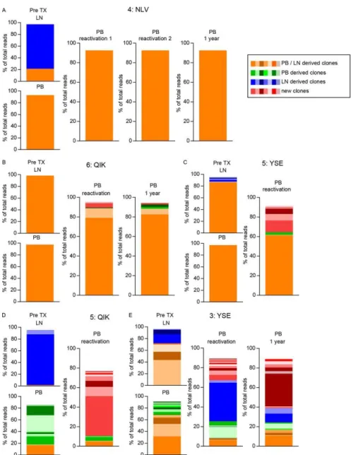
Upon viral reactivation, the majority of the unique LN hCMV-specific CD8+ T cell clones are not recruited to the circulating pool.We next studied the clonal relationships between
hCMV-specific CD8+ T cells from pretransplantation LN samples and those from PB samples taken at the moment of viral replication in four hCMV-seropositive patients who experienced a reactivation (or superinfection) after kidney transplantation (patients 3 to 6) (Fig. 4 and 5 and Table 1). In these patients, five hCMV epitope-specific CD8+ T cell pools could be studied longitudinally. Although the pretransplant overlapping clones (in orange in Fig. 5) most likely have a common ancestor, the fact that they are identical does not allow for the analysis of recruitment of LN clones toward the PB upon viral reactivation. Therefore, we focused on the discrepant clones between LNs and PB as well as any new clone detected during or after reactivation. In only one of five hCMV-specific CD8+ T cell pools studied, two of the four unique clones detected in the LN compartment before transplantation were found at the time of viral replication (Fig. 5E, blue YSE clones of Pt 3). Interestingly, we observed a dichotomy between the responses: if the initial hCMV-specific CD8+ T cell pool was monoclonal, it largely remained so during the antigenic rechallenge (Pt 4, NLV; Pt 6, QIK; Pt 5, YSE) (Fig. 5), while responses that were initially broad led to even broader responses during reactivation, accompanied by the appearance of many new clones (Pt 5, QIK; Pt 3, YSE) (Fig.
5, red clones). Both types of responses can occur within one patient (Pt 5, YSE and QIK), and they
also do not seem to depend on the epitope analyzed (Pt 3, YSE; Pt 5, YSE).
Although the substantial overlap of clones between the pretransplantation PB and LNs might allow the conclusion that these clones during hCMV reactivation are recruited from LNs, we cannot exclude the possibility that they are maintained by proliferation of the PB effector pool or even
may be supplemented from a different lymphoid tissue. We can conclude, however, that novel clones that appear in the PB upon hCMV reactivation are seldom derived from the LNs.
Thus, PB IL-7Rα-expressing hCMV-specific CD8+ T cells contain cells with classical memory-like
features. Identical clones between PB IL-7Rα-expressing and IL-7Rα− hCMV-specific CD8+ T cells
prevent us from drawing definite conclusions on the maintenance of these clones. However, the presence of unique clones in the acute-phase IL-7Rα− and IL-7Rα+ hCMV-specific CD8+ T cells and
the appearance of new clones found in the latent-phase IL-7Rα− hCMV-specific CD8+ T cells suggest
that these clones are not derived from the acute-phase IL-7Rα+ hCMV-specific pool. In a similar
fashion, identical clones found between PB- and LN-derived hCMV-specific clones before transplantation can be substantial, and this finding prevents us from drawing a conclusion on the maintenance of these clones in PB during reactivation. However, the presence of new PB clones during reactivation suggests that these clones are only rarely recruited from the LNs.
DISCUSSION
Although IL-7Rα+ hCMV-specific CD8+ T cells display several typical memory cell features, we did
not find strong evidence that there is recruitment from the acute-phase IL-7Rα+ to the latency
phase IL-7Rα− hCMV-specific CD8+ T cell pool. There are several putative explanations for the
difference between our findings on the human virus-specific T cell pools and data obtained in mice. First, the initial experiments in mice were performed during and following acute viral infections (e.g., LCMV) and not during persistent infection, such as with hCMV. Second, the IL-7Rα+ cells
during and after acute viral infection in mice bear some features that hCMV-specific CD8+ memory T cells do not have. For example, it has been suggested that only IL-7Rα+ cells lacking KLRG1 expression are proper memory precursor cells (18, 19, 35). Although we showed that the IL-7Rα+ subset contained cells lacking KLRG1 expression, the percentage of KLRG1-negative IL-7Rα+ hCMV-specific CD8+ T cells was too low to obtain the required amount of cells to perform TCRβ repertoire analysis. Third, the IL-7Rα+ populations that were used for the murine studies (17) were isolated from the spleen and could thus be quite different from the circulating IL-7Rα+ cells that we analyzed. Indeed, several studies, including those from our own group, have shown that phenotype and function of (virus-specific) CD8+ T cells can differ greatly depending on the tissue studied (9, 28, 36–39).
It should be noted that the diversity of the total hCMV-specific CD8+ T cell pool remained comparable to that found during the acute response but that both the composition and hierarchy of the clones in the IL-7Rα− pools had changed. The latter observation appears to be in contrast with our earlier findings that showed stable clonal composition from the acute response until 5 years of follow-up. First, for this study we used different patients, and we already noted a large variability in immune responses between individuals (4). Second, the largest fluctuation in clones over time in the earlier study was seen in the hCMV pp65-specific CD8+ T cell pool. And perhaps more importantly, in the current study we used samples that were fairly close to the peak of viral replication, which could have resulted in monitoring of immune repertoires in an early stage of clonal competition.
We also found that hCMV-specific CD8+ T cell clones that emerged in PB at the time of reactivation and could not be detected in the PB prior to reactivation were seldom recruited from the LN hCMV-specific CD8+ T cell pool.
This analysis, however, has a number of potential caveats. First, based on the fact that hCMV is a systemic infection, we assume that by sampling a number of para-iliac LNs we sample the full LN compartment in terms of clonal representation. Indeed, at least part of these cells expresses CCR7 (9), and they therefore have the capacity to circulate from LN to LN in search of their cognate antigen. Further, in the context of secondary lymphoid organs, it is worthwhile to analyze if spleen and bone marrow hCMV-specific CD8+ T cells may serve as sources of circulating effector-type cells (28, 36, 40).
Second, three out of four patients received a graft from an hCMV-seropositive donor, making it impossible to conclude whether the T cell response was triggered by superinfection or by reactivation of the endogenous virus. Although the epitopes studied are immunodominant and have not yet been reported to vary between different strains of hCMV, it is feasible that the route of reactivation versus superinfection by the transplanted kidney plays a role, too. Reactivation of
the latent endogenous virus occurred for certain in only one patient, and it is in this patient that we detected unique LN clones in the PB upon reactivation.
Third, we cannot distinguish between LN-derived and PB-derived Il-7Rα− and IL-7Rα+ hCMV-specific CD8+ T cell clones where it concerns overlapping clones. Thus, we are unable to exclude the possibility that overlapping clones are indeed maintained by recruitment from the LNs or from the IL-7Rα-expressing hCMV-specific CD8+ T cell pool. Furthermore, earlier studies have shown that effector-type human CD8+ T cells are long lived (5), and it cannot be excluded that the effector-type pool is, at least partially, maintained by homeostatic proliferation. In this respect, it is of interest that Tesselaar et al. have shown that, different from mice, for the maintenance of the human naive pool, homeostatic proliferation is far more important than influx of new cells from the thymus (41). Moreover, not all clones will respond equally when exposed to their cognate peptides. It is very possible that unique virus-specific CD8+ T cell clones that were found in LNs are clones responding relatively poorly to hCMV reactivation and are just out-competed by the most fit cells. It might be these cells that, because of a lack of strong stimulation, actually resemble memory phenotype cells. It is therefore plausible that unique LN hCMV-specific clones are unique because they do not get involved.
Finally, one can never analyze all virus-specific CD8+ T cell clones in a certain compartment due to limitations in available materials and the techniques used. Although at least 1 × 10⁷ mononuclear cells were used for each analysis and although all clones, including those that comprised less than 1% of the total reads, were considered in the analysis of overlapping clones, it is possible that extremely rare virus-specific clones were missed in the analysis.
Whether a cell will become a memory cell or an effector cell has been shown to depend on many factors, including the amount of antigen, cytokine environment, costimulation, and TCR affinity (2, 42–45). However, it has been shown that one clone can give rise to both acute-phase short-lived effector cells and memory precursor cells (46) and that this balance is altered by the kind of infection, route of infection, tissue-specific events, and other factors (46–48). Price et al. (44) showed that during latency, EBV- and hCMV-specific CD8+ T cells with low-affinity TCRs have a memory-type phenotype, whereas the high-affinity TCR-expressing cells have an effector-type phenotype. However, Griffiths et al. recently reported that, especially in the elderly, the effector-type cells actually have a lower affinity for the major histocompatibility complex (MHC)-peptide complex (49). Our preliminary analyses yielded no apparent evidence for different affinities between the total IL-7Rα+ and IL-7Rα− hCMV-specific CD8+ T cell pools since tetramer binding was indistinguishable between both subsets (see Fig. S4 in the supplemental material). However, it cannot formally be excluded that unique IL-7Rα+clones as well as unique LN clones indeed have different affinities than unique IL-7Rα− PB clones.
Thus, we show that the circulating IL-7Rα+ hCMV-specific CD8+ T cell pool contains memory phenotype cells that lack typical effector features. We cannot exclude the idea that clones that overlap between compartments do harbor the precursors for the circulating effector-type hCMV-specific CD8+ T cell pool. Still, novel PB hCMV-specific CD8+ T cell clones that appeared in PB after primary infection or during reactivation seem to be only rarely recruited from either the LNs or the IL-7Rα+ hCMV-specific CD8+ T cell pool. By characterizing the memory compartment, we aim to provide further tools necessary for optimal vaccine development.
ACKNOWLEDGMENTS
We thank B. Hooibrink for sorting hCMV-specific cells and K. A. M. I. van der Pant and N. van der Weerd for providing clinical data.
We declare that we have no commercial or financial conflicts of interest. REFERENCES AND NOTES
1. Gamadia LE, ten Berge IJ, Picker LJ, van Lier RA. 2002. Skewed maturation of virus-specific CTLs? Nat Immunol 3:203. 10.1038/ni0302-203.
2. Kaech SM, Wherry EJ, Ahmed R. 2002. Effector and memory T-cell differentiation: implications for vaccine development. Nat Rev Immunol 2:251–262. 10.1038/nri778.
3. Zimmerman C, Brduscha-Riem K, Blaser C, Zinkernagel RM, Pircher H. 1996. Visualization, characterization, and turnover of CD8+ memory T cells in virus-infected hosts. J Exp Med 183:1367–1375. 10.1084/jem.183.4.1367.
4. Klarenbeek PL, Remmerswaal EB, ten Berge IJ, Doorenspleet ME, van Schaik BD, Esveldt RE, Koch SD, ten Brinke A, van Kampen AH, Bemelman FJ, Tak PP, Baas F, de Vries N, van Lier RA. 2012. Deep sequencing of antiviral T-cell responses to HCMV and EBV in humans reveals a stable
repertoire that is maintained for many years. PLoS Pathog 8:e1002889.
10.1371/journal.ppat.1002889.
5. Beverley PC. 2004. Kinetics and clonality of immunological memory in humans. Semin Immunol 16:315–321. 10.1016/j.smim.2004.08.012.
6. Snyder CM. 2011. Buffered memory: a hypothesis for the maintenance of functional, virus-specific CD8+ T cells during cytomegalovirus infection. Immunol Res 51:195–204. 10.1007/s12026-011-8251-9.
7. Snyder CM, Cho KS, Bonnett EL, van Dommelen S, Shellam GR, Hill AB. 2008. Memory inflation during chronic viral infection is maintained by continuous production of short-lived, functional T cells. Immunity 29:650–659. 10.1016/j.immuni.2008.07.017.
8. van Leeuwen EM, de Bree GJ, Remmerswaal EB, Yong SL, Tesselaar K, ten Berge IJ, van Lier RA. 2005. IL-7 receptor alpha chain expression distinguishes functional subsets of virus-specific human CD8+ T cells. Blood 106:2091–2098. 10.1182/blood-2005-02-0449.
9. Remmerswaal EB, Havenith SH, Idu MM, van Leeuwen EM, van Donselaar KA, ten Brinke A, Bom-Baylon N, Bemelman FJ, van Lier RA, ten Berge IJ. 2012. Human virus-specific effector-type T cells accumulate in blood but not in lymph nodes. Blood 119:1702–1712. 10.1182/blood-2011-09-381574.
10. Boom R, Sol C, Weel J, Gerrits Y, de Boer M, Wertheim-van Dillen P. 1999. A highly sensitive assay for detection and quantitation of human cytomegalovirus DNA in serum and plasma by PCR and electrochemiluminescence. J Clin Microbiol 37:1489–1497.
11. Voehringer D, Koschella M, Pircher H. 2002. Lack of proliferative capacity of human effector and memory T cells expressing killer cell lectinlike receptor G1 (KLRG1). Blood 100:3698–3702. 10.1182/blood-2002-02-0657.
12. Lamoreaux L, Roederer M, Koup R. 2006. Intracellular cytokine optimization and standard operating procedure. Nat Protoc 1:1507–1516. 10.1038/nprot.2006.268.
13. van Dongen JJ, Langerak AW, Bruggemann M, Evans PA, Hummel M, Lavender FL, Delabesse E, Davi F, Schuuring E, Garcia-Sanz R, van Krieken JH, Droese J, Gonzalez D, Bastard C, White HE, Spaargaren M, Gonzalez M, Parreira A, Smith JL, Morgan GJ, Kneba M, Macintyre EA. 2003. Design and standardization of PCR primers and protocols for detection of clonal immunoglobulin and T-cell receptor gene recombinations in suspect lymphoproliferations: report of the BIOMED-2 Concerted Action BMH4-CT98-3936. Leukemia 17:2257–2317. 10.1038/sj.leu.2403202. 14. Folch G, Lefranc MP. 2000. The human T cell receptor beta variable (TRBV) genes. Exp Clin Immunogenet 17:42–54. 10.1159/000019123.
15. Klarenbeek PL, de Hair MJ, Doorenspleet ME, van Schaik BD, Esveldt RE, van de Sande MG, Cantaert T, Gerlag DM, Baeten D, van Kampen AH, Baas F, Tak PP, de Vries N. 2012. Inflamed target tissue provides a specific niche for highly expanded T-cell clones in early human autoimmune disease. Ann Rheum Dis 71:1088–1093. 10.1136/annrheumdis-2011-200612.
16. Klarenbeek PL, Tak PP, van Schaik BD, Zwinderman AH, Jakobs ME, Zhang Z, van Kampen AH, van Lier RA, Baas F, de Vries N. 2010. Human T-cell memory consists mainly of unexpanded clones. Immunol Lett 133:42–48. 10.1016/j.imlet.2010.06.011.
17. Kaech SM, Tan JT, Wherry EJ, Konieczny BT, Surh CD, Ahmed R. 2003. Selective expression of the interleukin 7 receptor identifies effector CD8 T cells that give rise to long-lived memory cells. Nat Immunol 4:1191–1198. 10.1038/ni1009.
18. Joshi NS, Cui W, Chandele A, Lee HK, Urso DR, Hagman J, Gapin L, Kaech SM. 2007. Inflammation directs memory precursor and short-lived effector CD8+ T cell fates via the graded expression of T-bet transcription factor. Immunity 27:281–295. 10.1016/j.immuni.2007.07.010.
19. Sarkar S, Kalia V, Haining WN, Konieczny BT, Subramaniam S, Ahmed R. 2008. Functional and genomic profiling of effector CD8 T cell subsets with distinct memory fates. J Exp Med 205:625– 640. 10.1084/jem.20071641.
20. Banerjee A, Gordon SM, Intlekofer AM, Paley MA, Mooney EC, Lindsten T, Wherry EJ, Reiner SL. 2010. Cutting edge: the transcription factor eomesodermin enables CD8+ T cells to compete for the memory cell niche. J Immunol 185:4988–4992. 10.4049/jimmunol.1002042.
21. Intlekofer AM, Takemoto N, Wherry EJ, Longworth SA, Northrup JT, Palanivel VR, Mullen AC, Gasink CR, Kaech SM, Miller JD, Gapin L, Ryan K, Russ AP, Lindsten T, Orange JS, Goldrath AW, Ahmed R, Reiner SL. 2005. Effector and memory CD8+ T cell fate coupled by T-bet and eomesodermin. Nat Immunol 6:1236–1244. 10.1038/ni1268.
22. Bolovan-Fritts CA, Trout RN, Spector SA. 2007. High T-cell response to human cytomegalovirus induces chemokine-mediated endothelial cell damage. Blood 110:1857–1863. 10.1182/blood-2007-03-078881.
23. Galkina E, Thatte J, Dabak V, Williams MB, Ley K, Braciale TJ. 2005. Preferential migration of effector CD8+ T cells into the interstitium of the normal lung. J Clin Invest 115:3473–3483. 10.1172/JCI24482.
24. Kohlmeier JE, Cookenham T, Miller SC, Roberts AD, Christensen JP, Thomsen AR, Woodland DL. 2009. CXCR3 directs antigen-specific effector CD4+ T cell migration to the lung during parainfluenza virus infection. J Immunol 183:4378–4384. 10.4049/jimmunol.0902022.
25. Kohlmeier JE, Miller SC, Smith J, Lu B, Gerard C, Cookenham T, Roberts AD, Woodland DL. 2008. The chemokine receptor CCR5 plays a key role in the early memory CD8+ T cell response to respiratory virus infections. Immunity 29:101–113. 10.1016/j.immuni.2008.05.011.
26. Kunkel EJ, Boisvert J, Murphy K, Vierra MA, Genovese MC, Wardlaw AJ, Greenberg HB, Hodge MR, Wu L, Butcher EC, Campbell JJ. 2002. Expression of the chemokine receptors CCR4, CCR5, and CXCR3 by human tissue-infiltrating lymphocytes. Am J Pathol 160:347–355. 10.1016/S0002-9440(10)64378-7.
27. Liu L, Fuhlbrigge RC, Karibian K, Tian T, Kupper TS. 2006. Dynamic programming of CD8+ T
cell trafficking after live viral immunization. Immunity 25:511–520.
10.1016/j.immuni.2006.06.019.
28. Palendira U, Chinn R, Raza W, Piper K, Pratt G, Machado L, Bell A, Khan N, Hislop AD, Steyn R, Rickinson AB, Buckley CD, Moss P. 2008. Selective accumulation of virus-specific CD8+ T cells with unique homing phenotype within the human bone marrow. Blood 112:3293–3302. 10.1182/blood-2008-02-138040. Abstract/FREE Full Text
29. Chi C, Sun Q, Wang S, Zhang Z, Li X, Cardona CJ, Jin Y, Xing Z. 2013. Robust antiviral responses to enterovirus 71 infection in human intestinal epithelial cells. Virus Res 176:53–60. 10.1016/j.virusres.2013.05.002.
30. Sauty A, Dziejman M, Taha RA, Iarossi AS, Neote K, Garcia-Zepeda EA, Hamid Q, Luster AD. 1999. The T cell-specific CXC chemokines IP-10, Mig, and I-TAC are expressed by activated human bronchial epithelial cells. J Immunol 162:3549–3558.
31. van de Berg PJ, Yong SL, Remmerswaal EB, van Lier RA, ten Berge IJ. 2012. Cytomegalovirus-induced effector T cells cause endothelial cell damage. Clin Vaccine Immunol 19:772–779. 10.1128/CVI.00011-12.
32. van Aalderen MC, Remmerswaal EB, ten Berge IJ, van Lier RA. 2014. Blood and beyond: properties of circulating and tissue-resident human virus-specific αβ CD8 T cells. Eur J Immunol 44:934–944. 10.1002/eji.201344269.
33. van Lier RA, ten Berge IJ, Gamadia LE. 2003. Human CD8+ T-cell differentiation in response to viruses. Nat Rev Immunol 3:931–939. 10.1038/nri1254.
34. Torti N, Walton SM, Brocker T, Rulicke T, Oxenius A. 2011. Non-hematopoietic cells in lymph nodes drive memory CD8 T cell inflation during murine cytomegalovirus infection. PLoS Pathog 7:e1002313. 10.1371/journal.ppat.1002313.
35. Rubinstein MP, Lind NA, Purton JF, Filippou P, Best JA, McGhee PA, Surh CD, Goldrath AW. 2008. IL-7 and IL-15 differentially regulate CD8+ T-cell subsets during contraction of the immune response. Blood 112:3704–3712. 10.1182/blood-2008-06-160945.
36. Letsch A, Knoedler M, Na IK, Kern F, Asemissen AM, Keilholz U, Loesch M, Thiel E, Volk HD, Scheibenbogen C. 2007. CMV-specific central memory T cells reside in bone marrow. Eur J Immunol 37:3063–3068. 10.1002/eji.200636930.
37. Piet B, de Bree GJ, Smids-Dierdorp BS, van der Loos CM, Remmerswaal EB, von der Thusen JH, van Haarst JM, Eerenberg JP, ten Brinke A, van der Bij W, Timens W, van Lier RA, Jonkers RE. 2011.
CD8+ T cells with an intraepithelial phenotype upregulate cytotoxic function upon influenza infection in human lung. J Clin Invest 121:2254–2263. 10.1172/JCI44675.
38. Smolders J, Remmerswaal EB, Schuurman KG, Melief J, van Eden CG, van Lier RA, Huitinga I, Hamann J. 2013. Characteristics of differentiated CD8+ and CD4+ T cells present in the human brain. Acta Neuropathol 126:525–535. 10.1007/s00401-013-1155-0.
39. van Aalderen MC, Remmerswaal EB, Heutinck KM, ten Brinke A, Pircher H, van Lier RA, ten Berge IJ. 2013. Phenotypic and functional characterization of circulating polyomavirus BK VP1-specific CD8+ T cells in healthy adults. J Virol 87:10263–10272. 10.1128/JVI.01540-13.
40. Langeveld M, Gamadia LE, ten Berge IJ. 2006. T-lymphocyte subset distribution in human spleen. Eur J Clin Invest 36:250–256. 10.1111/j.1365-2362.2006.01626.x.
41. den Braber I, Mugwagwa T, Vrisekoop N, Westera L, Mogling R, de Boer AB, Willems N, Schrijver EH, Spierenburg G, Gaiser K, Mul E, Otto SA, Ruiter AF, Ackermans MT, Miedema F, Borghans JA, de Boer RJ, Tesselaar K. 2012. Maintenance of peripheral naive T cells is sustained by thymus output in mice but not humans. Immunity 36:288–297. 10.1016/j.immuni.2012.02.006. 42. Badovinac VP, Messingham KA, Jabbari A, Haring JS, Harty JT. 2005. Accelerated CD8+ T-cell memory and prime-boost response after dendritic-cell vaccination. Nat Med 11:748–756. 10.1038/nm1257.
43. Badovinac VP, Porter BB, Harty JT. 2004. CD8+ T cell contraction is controlled by early inflammation. Nat Immunol 5:809–817. 10.1038/ni1098.
44. Price DA, Brenchley JM, Ruff LE, Betts MR, Hill BJ, Roederer M, Koup RA, Migueles SA, Gostick E, Wooldridge L, Sewell AK, Connors M, Douek DC. 2005. Avidity for antigen shapes clonal dominance in CD8+ T cell populations specific for persistent DNA viruses. J Exp Med 202:1349– 1361. 10.1084/jem.20051357. Abstract/FREE Full Text
45. Sarkar S, Teichgraber V, Kalia V, Polley A, Masopust D, Harrington LE, Ahmed R, Wherry EJ. 2007. Strength of stimulus and clonal competition impact the rate of memory CD8 T cell differentiation. J Immunol 179:6704–6714. 10.4049/jimmunol.179.10.6704. Abstract/FREE Full Text
46. Plumlee CR, Sheridan BS, Cicek BB, Lefrancois L. 2013. Environmental cues dictate the fate of
individual CD8+ T cells responding to infection. Immunity 39:347–356.
10.1016/j.immuni.2013.07.014.
47. Buchholz VR, Flossdorf M, Hensel I, Kretschmer L, Weissbrich B, Graf P, Verschoor A, Schiemann M, Hofer T, Busch DH. 2013. Disparate individual fates compose robust CD8+ T cell immunity. Science 340:630–635. 10.1126/science.1235454. Abstract/FREE Full Text
48. Gerlach C, van Heijst JW, Swart E, Sie D, Armstrong N, Kerkhoven RM, Zehn D, Bevan MJ, Schepers K, Schumacher TN. 2010. One naive T cell, multiple fates in CD8+ T cell differentiation. J Exp Med 207:1235–1246. 10.1084/jem.20091175. Abstract/FREE Full Text
49. Griffiths SJ, Riddell NE, Masters J, Libri V, Henson SM, Wertheimer A, Wallace D, Sims S, Rivino L, Larbi A, Kemeny DM, Nikolich-Zugich J, Kern F, Klenerman P, Emery VC, Akbar AN. 2013. Age-associated increase of low-avidity cytomegalovirus-specific CD8+ T cells that re-express CD45RA. J Immunol 190:5363–5372. 10.4049/jimmunol.1203267.
Figure 1 - Flow cytometric analysis of total, IL-7Rα-expressing, and IL-7Rα− hCMV-specific CD8+ T cells. (A to
D) Data represent the percentages of cells expressing the indicated molecules. (E) Percentage of cytokine-producing hCMV-specific CD8+ T cells after PMA-ionomycin stimulation for 4 h (left) or cognate peptide for 6
h (1 μg/ml) (right). Statistical analysis was performed with a paired Student t test: *, P ≤ 0.05; **, P ≤ 0.01. Eomes, eomesodermin.
Figure 2 - Longitudinal analysis of IL-7Rα-expressing and IL-7Rα− hCMV-specific CD8+ T cells following primary hCMV infection. (A and C) Absolute numbers of IL-7Rα-expressing and IL-7Rα− hCMV pp65-TPR-specific CD8+ T cells in two patients (indicated as 1:TPR and 2:TPR) experiencing a primary hCMV infection (arrows indicate time of TCRβ repertoire analysis). (B and D) TCRβ repertoire analysis of IL-7Rα-expressing and IL-7Rα− hCMV pp65-TPR-specific CD8+ T cells just after the peak viral load and at 1 year after transplantation of the same two patients. Clonal populations are differentiated by tonal group and color, as indicated on the figure. Different colors within a tonal group represent different clones. Identical tones within one patient represent identical clones. No overlapping clones between patients were found. (Sequences can be found in Tables S1 and S2 in the supplemental material.)
Figure 3 - TCRβ repertoire analysis of paired PB and LN samples. Different colors within a tone represent different clones. Identical tones within one epitope/patient represent identical clones. No overlapping clones between different epitopes or patients were found. (A) Analysis of hCMV pp65-specific CD8+ T cells. Patients
and epitopes are identified by abbreviations in the form 3:YSE, indicating patient 3 and a YSE-specific response. (B) Analysis of hCMV IE-specific CD8+ T cells. Patients and epitopes are identified as described for panel A. (C)
Analysis of lytic EBV-specific CD8+ T cells: patient 3 BZLF-1-EPL-specific (3:EPL) and patient 4
BMLF-1-GLC-specific (4:GLC). (D) Analysis of latent EBV-BMLF-1-GLC-specific CD8+ T cells: patient 3 EBNA-3a-EPL-specific (3:EPL) and
patient 8 EBNA-1-HPV-specific (8:HPV). (E) Analysis of influenza A virus-specific CD8+ T cells: patient 9
GIL-specific (9:GIL) and patient 4 GIL-GIL-specific (4:GIL). (Sequences can be found in Tables S3 to S14 in the supplemental material.)
Figure 4 - Flow cytometric longitudinal analysis of PB hCMV-specific CD8+ T cells. Left panels show the absolute number (Abs nr) of hCMV-specific CD8+ T cells (left y axis) and hCMV load (right y axis). Right panels show hCMV load (right y axis) and percentages of the indicated cell populations (left y axis) within the total hCMV-specific CD8+ T cell population analyzed. The following T cell subsets were analyzed: CMV pp65-YSE-specific CD8+ T cells in patient 3 (A), hCMV-pp65-NLV-specific CD8+ T cells in patient 4 (B), hCMV pp65-YSE-specific CD8+ T cells in patient 5 (C), hCMV-IE-QIK-specific CD8+ T cells in patient 5(D), and hCMV-IE-QIK-specific CD8+ T cells in patient 6 (E).
Figure 5 - Longitudinal TCRβ repertoire analysis of five patients experiencing hCMV reactivation after transplantation. Pretransplantation (pre-TX) clonal populations are differentiated by tonal group and color, as indicated on the figure. New clones are those that could be detected only at the peak of CD8 expansion after hCMV reactivation and 1 year after. Different colors within tones represent different clones. Identical tones within one epitope/patient represent identical clones. No overlapping clones between different epitopes or patients were found. The following T cell subsets were analyzed: hCMV pp65-NLV-specific CD8+ T cells of patient 4 (4:NLV) (A), hCMV IE-QIK-specific CD8+ T cells of patient 6 (6:QIK) (B), hCMV pp65-YSE-specific CD8+ T cells of patient 5 (5:YSE) (C), hCMV IE-QIK-specific CD8+ T cells of patient 5 (5:QIK) (D), and hCMV pp65-YSE-specific CD8+ T cells of patient 3 (3:YSE) (E). (Sequences can be found in Tables S3 to S7 in the supplemental material.)