i
The effects of aerobic interval training on heart rate
recovery after cardiac resynchronization therapy
Dissertation presented for Master of Science degree in Exercise and Health
Tutor: Professora Doutora Maria Helena Santa-Clara Pombo Rodrigues
Jury
Chairperson: Professor Doutor
Fernando Manuel da Cruz Duarte Pereira
Memeber of the Commitee: Professor Doutor Pedro Xavier Melo Fernando
Castanheira
Eduardo Filipe Bento Mesquitela André
2016
iii
“Porque o homem é sempre mais
iv
Um caminho solitário, reflexivo em essência e que apenas se tornou possível por não estar só. Como tal, agradeço a todos aqueles que direta ou indiretamente me acompanharam durante este processo, e me auxiliaram nesta caminhada:
À Professora Doutora Helena Santa-Clara, a quem reforço a gratidão por me ter facultado a sua orientação e o acesso aos dados que permitiram a realização deste projeto. E ainda, o seu pragmatismo, orientação sólida e bastante sábia aquando das minhas incertezas cientificas.
Ao Professor Doutor Xavier Melo pelo seu papel fulcral na fase de conclusão da tese, particularmente aquando da análise estatistica. À minha família que sempre me apoiou e reforçou a importância do conhecimento, particularmente no âmbito académico. Aos meus pais, Ana Bento e Vitor Mesquitela, que foram sempre firmes e convictos no que concerne ao meu percurso escolar, particularmente quando eu não expressei essa vontade. Aos meus irmãos, Daniel, Sérgio e Maria João, a sua paciência ao longo dos anos, e ainda a importância que foi ver os seus exemplos enquanto ideais de referência.
À minha sobrinha Catarina, o exemplo-mor do que é a dedicação a uma causa maior, marcando em cada passo, com resiliência, a sua identidade. Aos meus sobrinhos Afonso e Maria que olham para os fenómenos e veem… sem condicionamentos, sem constrangimentos e veem. Esta simplicidade ajudou-me tanto no âmbito acadêmico como pessoal, onde a interpretação dos fenómenos, em certos momentos, parece recaír na sua forma mais pura, sem contaminação do pensamento reflexivo.
Ao Professor Doutor Luis Margem, a sua distinção académica e arte no “ganfanso”, duas amigas de tertúlias e tónicos em momentos de introspeção ímpares.
Ao Professor Doutor Jorge Lima, por quem tenho uma estima proporcional ao seu quociente de inteligência, agradeço a sua amizade, e a sua incondicional abenegação ao próximo. E ainda, pela partilha honesta e humilde do seu conhecimento académico e não académico. Por fim, agradeço a todos os alunos com quem tive e tenho o privilégio de trabalhar, a sua compreensão nos momentos em que estive menos disponível e simultaneamente mais ausente.
v
Background: Heart failure is characterized by an autonomic nervous system dysfunction
which leads to sympathetic overactivation and parasympathetic imbalance, culminating in central and peripheral dysfunction. In advanced HF, cardiac resynchronization therapy (CRT) and exercise training seem to improve these conditions and result in improved functional and clinical parameters. A growing body of evidence supports the benefits of aerobic interval training (AIT) in other several HF populations, but less is known about its influence on autonomic function. Here we assessed the effects of AIT on the heart rate recovery (HRR), an indicator of parasympathetic activity. All participants had HF with a reduced ejection fraction, and six days before the intervention, underwent cardiac surgery. Our objective was to compare if the additive effect of AIT to CRT could indeed result in improved vagal reactivation, measured by the difference between the peak heart rate and the HRR at one minute (HRR1diff). Methods: Twenty-nine stable patients (aged 68.96 ± 9.92; VEF< 27 ; and a V O2Peak= 15 mL.kg-1.min-1) who were receiving optimal medical treatment, were randomized either to the control group, or the AIT group. The AIT group exercised twice a week, and began each session with a 10 minute warm-up (50-60% of the peak heart rate), followed by four intervals of 2-minutes (90-95% of the peak heart rate) and a 2-minute recovery (60-70% of the peak heart rate). After the first month, the 2-minute intervals were changed to 4-minute intervals and 3-minutes recovery. After cardiopulmonary exercise testing (CPET) to maximal volitional exertion, using the modified Bruce protocol, patients were seated and the HRR was immediately assessed. Results: After the six months of intervention our main effects were significant for V O2Peak (p= .010) and CPET duration (p= .025). Thus, after testing for simple main effects, only the AIT group depicted significant changes in the post-intervention for: V O2Peak (p= .013), CPET duration (p= .020), heart rate reserve (p= .035), peak pulse pressure (p= .036), and the HRR1diff (p= .025).
Conclusions: After six months of intervention, the simple main effects suggest that AIT
could improve vagal reactivation, assessed through HRR1diff, in patients that underwent CRT and were engaged in optimal medical treatment. Our findings also suggest that differences between groups in exercise capacity could be due to peripheral factors. Keywords: Heart failure; Heart rate recovery; Vagal reactivation; Aerobic interval training; Cardiac resynchronization therapy.
vi
Contexto: A insuficiência cardíaca (IC) é caraterizada por uma disfunção do sistema
nervoso autónomo (SNA) que conduz a uma hiperativação simpática e desiquilíbrio parassimpático, culminando em disfunções centrais e periféricas. Nos casos mais avançados de IC, a terapêutica de ressincronização cardíaca (TRC) e o exercício parecem melhorar estas condições e, outros parâmetros clínicos e funcionais. O emergir de evidência robusta valoriza o treino intervalado aeróbio (TIA) em várias populações com IC, sabendo-se pouco acerca da sua influência sobre o SNA. Nesta análisabendo-se, avaliámos os efeitos do TIA sobre a frequência cardíaca de recuperação (FCR), um indicador de ativação parasimpática. Todos os participantes possuíam uma fração de ejeção diminuída para ventrículo esquerdo, e colocaram o implante cardíaco seis dias antes do início da intervenção. O nosso objetivo foi o de avaliar se o TIA adicionado à TRC poderia melhorar a reativação vagal, medida pela diferença entre a frequência cardíaca pico e a FCR no primeiro minuto (FCR1dif). Métodos: Vinte e nove participantes idade 68 96 9 92; FEVE< 27 ; e o V O2Pico= 15 mL.kg-1.min-1) que estavam a receber tratamento médico otimizado (TMO), foram randomizados diferencialmente para os grupos de TIA e de controlo. O grupo de TIA realizou duas sessões de treino semanais, iniciando as mesmas com 10 minutos de aquecimento (50 a 60% da FC pico), seguido de quatro intervalos de 2 minutos (90 a 95% da FC pico) e 2 minutos de recuperação ativa (60 a 70% da FC pico). Depois de concluído o segundo mês, os intervalos de 2 minutos foram substituídos por intervalos de 4 minutos, enquanto os intervalos de recuperação por outros de 3 minutos. Recorrendo à prova de
stress cardiopulmonar (PSCP), a qual foi efetuada até a capacidade volitiva máxima usando
o protocolo de Bruce modificado, a FCR foi avaliada imediatamente a seguir ao mesmo.
Resultados: A seguir aos seis meses de interven o, os efeitos principais foram
significativos para V O2Pico (p= .010) e a duração da PSCP (p= .025). Contudo, depois de se testarem os simple main effects, apenas o grupo de TIA apresentou altera es significativas no per odo pós-interven o para: V O2Pico (p= .013), duração da PSCP (p= .020), frequência cardíaca de reserva (p= .035), pressão de pulso pico (p= .036), e FCR1dif (p= .025).
Conclusões: Depois de seis meses de intervenção, os simple main effects sugerem-nos
que o TIA pode melhorar a reativação vagal, medida pela FCR1dif a seguir ao exercício em pacientes que se encontram em TRC e TMO. Os resultados sugerem-nos ainda que as diferenças encontradas na capacidade funcional devem-se a fatores periféricos. Palavras
chave: Insuficiência cardíaca; Frequência cardíaca de recuperação; Reativação vagal;
vii
Table 1. American Heart Association/ American College of Cardiology Guidelines – stages of heart failure……… ... 8 Table 2. Comparison of the American College of Cardiology Foundation/ American Heart Association stages of heart failure and New York Heart Association functional classifications… ……… ... 9 Table 3. Inclusion criteria in randomized controlled studies……… … 15 Table 4. Endpoints, and main findings of randomized clinical trials evaluating cardiac resynchronization therapy in sinus rhythm……… ……… 16 Table 5. Baseline characteristics of the patients……… ……… 35 Table 6 Selected variables for Control and Aerobic Interval Training……… ……… 38
Figures List
Figure 1. Flow chart of the inclusion/exclusion criteria. ……… …… … 28 Figure 2.The effects of aerobic interval training in the control group and the exercise group …… ……… …… 42
viii
Acknowledgements……… iv
Abstract……… v
Resumo……… ……… … vi
Tables List……… ……… … vii
Figures List……… ……… … vii
Table of Contents……… ……… ……… ……… viii
Abbreviations……… ……… ... x Preamble… ……… ……… ………… . xi CHAPTER I Introduction…… … ……… …………... 3 CHAPTER II Literature review … ……… ………… .. 7 2 1 Heart failure……… ……… ………... 7
EPIDEMIO OGY AND C INICA CRITERIA……… ……… ... 7
TYPES OF HEART FAI URE………… … ……… ……… ... 10
2.2 Cardiac resynchronization therapy…… ……… …… 12
DYSSYNCHRONY AND RESYNCHRONIZATION… ……… .. 13
TRIALS – WHAT DID WE EARN FROM THEM? ……… … 14
2 3 Autonomic function and resynchronization……… . 17
AUTONOMIC NERVOUS SYSTEM MODU ATION ……… ………… . 17
EXERCISE AND AUTONOMIC FUNCTION……… ……… ... 18
2 4 Exercise and resynchronization……… … 19
PHYSIO OGICA ADAPTATIONS TO EXERCISE PROTOCO S…… ... 20
IN THE URGE OF A NEW EXERCISE METHODO OGY ……… 22
CHAPTER III Methodology……… ……… …… …… 27
3 1 Introduction……… ……… …… ……… ……… … … 27
3 2 Variables in the study……… ……… ……… ……… ……… 27
3.3 Hypothesis……… ……….. 27
3 4 Study design……… ……… ……… 27
3.5 Equipment and protocols of assessment……… ……… ………… 29
ix Results……… ……… ……… 35 4 1 Exercise capacity……… ……… …… …… ……… 36 4 2 Hemodynamics……… ……… ……… ………… …… 36 4 3 Chronotropic capacity… …… ……… ……… 36 4 4 Autonomic function……… ………… ………… …… 37 CHAPTER V Discussion…...……… ……… ……… …… 41 Conclusion……..……… ……… ……… 46 CHAPTER VI References……… ………...……… 49
x
ACE-I Angiotensin-converting enzyme inhibitors AHA American Heart Association
AHF Acute Heart Failure AIT Aerobic interval training ANS Autonomic nervous system AVD Atrioventricular delay Ca2+ Calcium
CHF Chronic HF
CRT Cardiac resynchronization therapy ESC European Society of Cardiology
ExT Exercise training HF Heart failure
HFpEF Heart failure with preserved ejection fraction HFrEF Heart failure with reserved ejection fraction
HR Heart rate
HRR Heart rate recovery HRR1 HRR at 1-minute HRR3 HRR at 3-minute HRR6 HRR at 6-minute
HRR1diff Difference between the peak heart rate and the HRR at 1-minute HRR3diff Difference between the peak heart rate and the HRR at 3-minute
HRR6dif Difference between the peak heart rate and the HRR 6-minute LBBB Left bundle branch block
LV Left ventricle
LVEDV Left ventricle end-diastolic volume LVEF Left ventricle ejection fraction LVESV Left ventricle end-systolic volume
NYHA New York Heart Association QoL Quality of life
RCT Randomized clinical trials RER Respiratory exchange ratio TRIMP Training impulse method V O2Peak Peak oxygen uptake
V O2Max Maximal oxygen uptake
xi
An analysis was carried out based on the variables collected from a financially supported investigation project, by the Portuguese Foundation of Science and Technology i e , PTDC/DES/120249/2010), and was conducted with the authors’ approval. Should the reader feel the need for more detailed information regarding the project or other points of interest, information is available upon request.
With the successful integration of cardiac resynchronization therapy in clinical practice, it is important to explore new paths of unknown knowledge. In this context, the interaction between the autonomic nervous system, exercise and cardiac resynchronization is of extreme importance, as it can improve the patient´s condition.
In this thesis, the aim was: To determine the effects of a long-term exercise training program following cardiac resynchronization therapy, while controlling for autonomic function.
The document is organized by chapters, and the reader can follow our rationale throughout each one of them. Chapter I is the introduction, were we describe the state of the art of the main topics of our theme, and engage through a brief consideration of our problem. At the end we identify our main hypothesis. Chapter II, the literature review, will be dedicated to some of the mechanisms of HF pathophysiology, and how they can benefit from cardiac pacing, exercise or both. The autonomic modulation that seems to occur through pacing will be explored, and detailed attention will be given to the exercise mechanisms responsible for specific central and peripheral adaptations, particularly those related with exercise intensity. The following chapters will then be dedicated to the methodology (Chapter III), the results (Chapter IV), the discussion and main conclusions (Chapter V), and finally, all the references used to elaborate the complete thesis (Chapter VI).
5
3
Introduction
The economic burden of heart failure with reserved ejection fraction (HFrEF) to the western healthcare system has grown and heart transplant inevitability becomes an unsustainable option for every patient (Clarke et al., 2014; Jessup et al., 2011), furthermore, with the increasing ageing population and the growing life expectancy (Beard & Bloom, 2015). Although advances in the treatment and management of heart failure (HF) syndrome can be seen, poor prognosis mirrors an underlying complex pathophysiology (McMurray et al., 2012; Yancy et al., 2013). Indeed, several etiologies, both cardiovascular and non-cardiovascular, have the final common pathway of abnormal cardiac function which results in maladaptive cardiac remodelling. Nevertheless, new methods have been design for accurate diagnosis, prognosis, and risk stratification in clinical practice and experimental field (Brignole et al., 2013; Ponikowski et al., 2016). Considering these epidemiological issues, cardiac resynchronization therapy (CRT) as emerged in a burst of knowledge, technology, and avant-garde approach from distinct areas such as engineering, informatics and medicine (Barold, 2011; Nelson, 1993; Park, Kushwaha, & McGregor, 2012; Zoll, 1973).
The HF syndrome is a continuum and vicious cycle characterized by the hallmarks exercise intolerance and fatigue (Kupper, Bonhof, Westerhuis, Widdershoven, & Denollet, 2015; West, Hernandez, O'Connor, Starling, & Califf, 2010). Concomitantly, form and function become altered, while central and peripheral mechanisms will reflected themselves on several physiological dysfunctions, such as cardiac dyssynchrony (Bank et al., 2015) or muscle myopathies (Piepoli & Crisafulli, 2014). Interestingly, resynchronization seems to improve both of these conditions, as well as challenge the vicious and auto-proliferative cycle of HF. Similarly, exercise training (ExT) has exhibit improvements in clinical and functional parameters in a wide range of HF patients, including those engaged on CRT with optimized therapeutics (Conraads et al., 2007; Haykowsky et al., 2007).
According to Florea & Cohn (2014) the HF syndrome progression is a reflex of the autonomic nervous system (ANS) imbalance. The syndrome seems to be mediated by neurohumoral activation, which has a clinical manifestation of sympathetic overactivation and parasympathetic withdrawal (Levy, 1971; Lymperopoulos, Rengo, & Koch, 2013). In recent years, the heart rate recovery (HRR) has been used as a non-invasive and easy way to assess this interdependent relationship. As suggested by Myers et al. (2007), the HRR has a prognostic value and represents an important clinical index of functional capacity and hemodynamics in HF. There is few information regarding autonomic function and CRT, particularly when assessed through the HRR. Could CRT improve autonomic modulation and improve the HRR? If adrenergic and cholinergic receptors are intimately related and a coordinate sympathetic and vagal control is essential for hemodynamics homeostasis (Levy,
4
1984), as well as for neurohumoral modulation (Arena et al., 2010), could this relationship, altered by HF, be improved by exercise, particularly in the form of aerobic interval training (AIT), and alongside with CRT? Additionally, there are several muscarinic receptors subtypes (Wang et al., 2001) that can actually modulate, and be modulated, in their responsiveness sensibility and specificity to particular amines (e.g., norepinephrine). More, Okutucu et al. (2011) conducted a randomized controlled trial with CRT patients, and suggested that the HRR at 1-minute (HRR1) could reflect vagal reactivation while the HRR at 3-minute (HRR3) could be mediated, predominantly, by sympathetic withdrawal. In this particular, several trials conducted with HF populations and adopting AIT protocol depicted improvement in functional parameters (Guiraud et al., 2013; Johnsen, Hoydal, Rosbjorgen, Stolen, & Wisloff, 2013), vagal tone (Guiraud et al., 2013), and QoL as well (Nilsson, Westheim, & Risberg, 2008). However, the effects of AIT in CRT patients assessed through the HRR are not clearly elucidated yet. Moreover, to our knowledge there is only one trial that addressed CRT and the HRR (Okutucu et al., 2011). Furthermore, to our knowledge, the effect of the AIT on improving vagal reactivation in CRT-patients has never been studied. Therefore, the present analysis will focus on finding evidence that clarifies some of the uncertainty regarding these issues.
1
7
Literature Review
2.1 Heart failure
EPIDEMIOLOGY AND CLINICAL CRITERIA
The incidence and prevalence of heart failure (HF) in modern society is a growing and serious epidemiological issue (Bleumink et al., 2004; Brouwers et al., 2013; Curtis et al., 2008; Guha & McDonagh, 2013; Levy et al., 2002). HF is estimated to affect more than 23 million people worldwide and be accounted for more than 1 million hospitalizations each year (Roger, 2013), and although its prognostic value is dependent upon several variables, 50% of the patients die within 5 years of the initial diagnosis (Bui, Hornich & Fonarow, 2011). Additionally, the prevalence of HF with preserved ejection fraction (HFpEF) is over 50% within symptomatic HF (Edelmann et al., 2011; Frohling, et al., 2011), having superseded other forms of HF such as HF reduced ejection fraction (HFrEF) (Guha & McDonagh, 2013; Kitzman et al., 2014). Furthermore, HFpEF morbidity, mortality, and functional decline are escalating and almost near HFrEF levels (Edelmann et al., 2011; Kitzman, Brubaker, Morgan, Stewart, & Little, 2010). In response to these facts, the HF syndrome implies more knowledge and epidemiological evidence to respond efficiently to the previous progressive and harmful scenario. Nevertheless, some authors (Guha & McDonagh, 2013) prefer to focus on the unclear definition of HF and inclusion criteria in trials, which may question some gushing fears and trends.
HF is a complex clinical syndrome with signs and symptoms that result in congestion, and/or tissues hypoperfusion (McMurray et al., 2012; Saxon & Marco, 2001). As a multifactorial syndrome it has different etiologies that impair both structure and function, with a direct impact over central (Zile et al., 2013) and peripheral (Amann et al., 2014) homeostasis. It seems difficult to formulate a specific definition for HF considering how complex this syndrome is. However, we can observe its natural history, possible etiology and pathophysiology, and proceed to HF stratification by classification (i.e., New York heart association functional class) (Dolgin, 1994) or HF stages as depicted in Table 1 (Hunt et al., 2009).
According to expert consensus from both the American College of Cardiology Foundation (ACCF)/American Heart Association (AHA), and also the European Society of Cardiology (ESC), the New York Heart Association (NYHA) functional class is more than standard policy and refers to an important step both in clinical (i.e., preventive, prognostic, management of HF) and research field (i.e., trials randomization) (McMurray et al., 2012; Yancy et al., 2013). However, some confusion may arise from asymptomatic cardiac
8
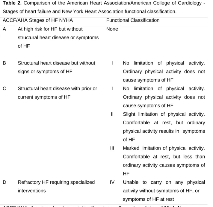
dysfunction, although pathology is present (e.g., systolic dysfunction, diastolic dysfunction). Whereas NYHA functional class refers to HF syndrome symptoms and exercise capacity, which may change rapidly, the stages of HF (Table 1) focus on the development and progression of the underlying disease and objective criteria described elsewhere (Dolgin, 1994; Hunt et al., 2009). Interestingly, these two characteristics are complementary to one another and their intrinsic relation is illustrated in Table 2.
Table 1. American Heart Association/American College of Cardiology guidelines – stages of
heart failure.
Stage Description
A Patients at high risk of developing HF because of conditions strongly associated with the development of HF. Such patients have no identified structural or functional abnormalities of the pericardium, myocardium, or cardiac valves and have never shown signs or symptoms of HF.
B Patients who have developed structural heart disease that is strongly associated with the development of HF but who have never shown signs or symptoms of HF.
C Patients who have current or prior symptoms of HF associated with underlying structural heart disease.
D Patients with advanced structural heart disease and marked symptoms of HF at rest despite maximal medical therapy and who require specialized interventions.
HF-Heart failure.
Adapted from Hunt et al. (2009)
According to the ACCF/AHA (Yancy, 2013) and corroborated by others (McMurray et al , 2012), HF is defined as “a complex clinical syndrome that can result from any structural
or functional cardiac disorder that impairs the ability of the ventricle to fill or eject blood”.
However, to an understanding of HF syndrome, a distinction should be made between acute heart failure (AHF) and chronic HF (CHF). AHF describes patients with tissues hypoperfusion and reduced functional capacity or pulmonary edema that limits active daily living, while CHF describes stable patients with a cardiac dysfunction who may experience decompensation episodes (Pani et al., 2015).
Patients with CHF have received more attention in trials and experimental studies than acute HF patients. In part, this could be due to the difficulty in defining and classifying AHF, although according to ESC guidelines (McMurray et al., 2012) a straightforward interpretation is proposed, namely as: “a rapid onset of, or change in, symptoms and signs of
9
Table 2. Comparison of the American Heart Association/American College of Cardiology -
Stages of heart failure and New York Heart Association functional classification. ACCF/AHA Stages of HF NYHA Functional Classification
A At high risk for HF but without
structural heart disease or symptoms of HF
None
B Structural heart disease but without signs or symptoms of HF
I No limitation of physical activity. Ordinary physical activity does not cause symptoms of HF
C Structural heart disease with prior or current symptoms of HF
I No limitation of physical activity. Ordinary physical activity does not cause symptoms of HF
II Slight limitation of physical activity. Comfortable at rest, but ordinary physical activity results in symptoms of HF
III Marked limitation of physical activity. Comfortable at rest, but less than ordinary activity causes symptoms of HF
D Refractory HF requiring specialized interventions
IV Unable to carry on any physical activity without symptoms of HF, or symptoms of HF at rest
ACCF/AHA=American heart association/American college of cardiology; NYHA=New York heart association; HF=Heart failure.
Adapted from (Yancy, 2013)
Regrettably, cardiac dysfunction is progressive and leans toward the progression of CHF, with episodic manifestations through AHF (Gaasch & Zile, 2011). Ponikowski et al. (2016), refers that miscellaneous etiologies can be observed in different parts of the world- differing from cardiovascular and non-cardiovascular- that cooperate to HF. Regardless of the aetiology, myocardial injury seems to be the beginning process, followed by cardiac dysfunction and finally HF syndrome (Levy, Larson, Vasan, Kannel, & Ho, 1996; Mosterd & Hoes, 2007; Vasan, Larson, Leip, Kannel, & Levy, 2001). Although improvements in the treatment and the diagnosis of clinical or even preclinical HF conditions have been gradually effective, poor outcomes continue to be challenging for clinicians (Ponikowski et al., 2016).
10
HF is a multi-systemic syndrome, and most of HFrEF patients present multiple comorbidities and geriatric syndromes (e.g., Frailty, Sarcopenia, Cachexia), alongside with a highly depressed ventricular function (Cruz-Jentoft et al., 2010; Goldwater & Pinney, 2015; Jermyn & Patel, 2014; Jha et al., 2015; Murad & Kitzman, 2012). These patients can exhibit peak oxygen uptake V O2Peak)≤ 14 m kg-1.min-1, which is associated with poor prognosis (Arena, Myers, Abella, Pinkstaff, et al., 2010), increased mortality (Mancini et al., 1991), and can be a landmark for a differential therapeutic approach such as cardiac resynchronization therapy (CRT). CRT can have a direct impact over central function, but remarkably it also seems to be accountable for indirect improvements on peripheral mechanisms such as skeletal myopathies (Piepoli & Crisafulli, 2014) or even several chronic diseases (Booth, Roberts, & Laye, 2012; Kujala, 2006; Pedersen, 2011). Both of this changes induced by CRT can express themselves as exercise improvement (Jaussaud, Blanc, Bordachar, Roudaut, & Douard, 2011) and decrease baseline sympathetic activation (Hamdan et al., 2002; Middlekauff, 2005).
Interestingly, we have been following a line of thought were we interpreted HF as being solely a unique entity and complex syndrome, and our approach, merely theoretically, must be seen in a whole wide spectrum, as a continuum (Levy et al., 1996; McMurray et al., 2012; Vasan et al., 2001). Actually, in CHF, less physical activity and muscle disuse can be seen. The decreased functional capacity is a result of several factors, and not only cardiomyopathies themselves (Dalal, Doherty, & Taylor, 2015). This concomitant and auto-proliferative effect is meaningful over the natural history of HF syndrome, and a burden that must be taken into account when approaching cardiac rehabilitation.
TYPES OF HEART FAILURE
If we delve into cellular mechanisms or sub-cellular levels, proinflammatory biomarkers also seem to have a role in pathogenesis of HF (Dixon, Griggs, Bersten, & Pasquale, 2011; Fink et al., 2012). Cytokines, chemokines and cell adhesion molecules result in cardiac maladaptive remodelling, which is more than simple heart dysfunction, and involves complex signalling pathways (Briasoulis, Androulakis, Christophides, & Tousoulis, 2016). Evidence supports that CHF exhibit activation of neurohormones (e.g., atrial natriuretic peptide, brain natriuretic peptide, and norepinephrine) and proinflammatory cytokines (e.g., IL-6, TNF-α, I 1-β) Cheng et al , 2013; Ferrari, 2002; Guggilam et al., 2011; Kinugawa et al., 2003). These proinflammatory agents regulate the immune response that will alter cellular metabolism, induce stress and result in morphological and mechanical impairments (Torre-Amione, 2005). These processes seem to be dependent upon innate and adaptive immunity from an evolutionary perspective (Kasturi et al., 2011; Padovan & Martin, 2015; Parham, 2003), and represent a normal physiological response of the immune
11
system, but if these environment persists, it may become toxic and reflect a chronic detrimental adaptation (Despres & Lemieux, 2006; Hotamisligil, 2006; Libert, 2003). Ultimately, this chronic detrimental adaptation can lead to increased cardiometabolic risk and differential clinical manifestations for risk stratification (Walsh, Fang & Fuster 2013).
Most recent ESC guidelines Ponikowski et al , 2016) enunciate a “new form” of HF, more concretely HF with mid-range EF that ranges between 40 to 49%. Alongside, HFpEF and HFrEF are characterized by reduced physical capacity that can be measured subjectively by exertional dyspnea or objectively by V O2Peak. Despite research, mechanisms for exercise intolerance and exertional dyspnea in HF patients´ are not completely understood (Kupper et al., 2015; West et al., 2010). Reduced exercise capacity seems to be associated with chronotropic incompetence as a result of impaired heart rate (HR) and cardiac output (Borlaug et al., 2006; Zile et al., 2013). In HFpEF, limitation is firstly due to impaired peripheral function (Haykowsky et al., 2014) and later resulting in the two hallmarks of HF (i.e., exercise intolerance, dyspnea). In one hand, HFrEF expresses peripheral dysfunction coexistence with other multi-systemic impairments, and pulmonary function seems to be the first to limit exercise progression (Kupper, Bonhof, Westerhuis, Widdershoven, & Denollet, 2015; Poon & Tin, 2013). On the other hand, HFpEF expresses diastolic dysfunction, although with left ventricle ejection fraction (LVEF) and left ventricle end-diastolic volume (LVEDV) seems to remain unchanged in most cases (Kitzman et al., 2014). According to these authors, recent attention has being refocusing on peripheral mechanisms as the main source of HF impairments (Kitzman et al., 2014). Concerning HFrEF, there is a systolic dysfunction that affects the rate of myocardium contraction (chronotropy) (Brubaker & Kitzman, 2011), that impairs myocardium contractility (inotropy) and its relaxation (lusiotropy) (Abraham et al., 2015). Moreover, these adverse scenario affects the intrinsic electrical conduction system (dromotropy) (Crocini et al., 2014).
In both of these cases, an elevation of left ventricle (LV) pressure seems to affect right ventricular performance because of secondary pulmonary artery pressure elevation (Solomonica, Burger, & Aronson, 2013). On a different approach, Kaufmann et al. (2013) conducted a multicentre prospective cohort study with subclinical cardiovascular disease as an inclusion criterion, and some of the conclusions were that right ventricular function and morphology seem to be associated with dyspnea, even when adjusted for covariates such as left ventricular function or lung function.
If we´ve been distinguishing HFrEF (systolic dysfunction) from HFpEF (diastolic dysfunction), we must also explain why. In good truth, by separating these two entities we oversimplify a highly complex condition to get deeper into the idiosyncrasies of each type of HF. According to Komamura (2013), HF should be considered as a single and continuous disease, being the two extremes, systolic and diastolic dysfunction phenotypes. The author
12
emphasizes how distinctive adaptations of the LV occur, more specifically eccentric hypertrophy (systolic HF) and concentric remodelling/hypertrophy (diastolic HF). However, if there are similarities between both conditions, there are also heterogeneous responses, particularly to exercise-induced changes in LV systolic and diastolic properties (Zile et al., 2013).
Interestingly, improvement of exercise capacity is an independent predictor of mortality in HF (Boxer et al., 2010; Tang, Dewland, Wencker, & Katz, 2009), and seems to be associated with those that can be seen with CRT (Takeuchi et al., 2014; Tomczak et al., 2012; Wasserman, Sun, & Hansen, 2007). Primary receivers of CRT are elder and severe HFrEF patients (Schowalter et al., 2013), with pronounced limitations for activities of daily living and whom also experience a higher risk of sudden death, especially when compared with less severe HF patients (Park et al., 2012). When these patients fulfil certain criteria, an implantable cardiac device such as CRT can prevent heart transplant, ameliorate symptoms, improve quality of life (QoL) and improve functional capacity (Cleland, Daubert, & Erdmann, 2005; Marco et al., 2008).
2.2 Cardiac resynchronization therapy
In 2013, the ACCF in collaboration with the Heart Rhythm Society proposed a clinical cardiology practice based on pacing therapy for primary prevention, secondary prevention and even for some comorbidities (Russo et al., 2013). Novel evolutions in HF pharmacotherapy became powerful adjunctive therapeutic to biodevices (e.g., CRT) (Brignole et al., 2013; McMurray et al., 2012; Yancy et al., 2013). The two most common pharmacologic options are angiotensin-converting enzyme inhibitors (ACE-I) and β-blockers, both of which seem to attenuate symptoms, improve QoL, slow progression of HF and contribute to reversal of maladaptive cardiac remodelling (Zile et al., 2013). Added to the widespread use of pacing in a new avangard cardiology era, this scenario is only possible due to epidemiological evidence and on-going burst of knowledge in technology, engineering and medicine.
According to Nelson et al. (1993), in their early days of cardiac pacing, benefits erupted from empirical clinical practice rather than proven by clinical trials. In the 1990´s, the first trials, with relevance for PATH-CHF (Auricchio et al., 1999), approaching of new pacing methodologies were conducted but with little statistical power. Since then, several landmarks have been achieved, and one of the most important is the criteria for device implantation, such as CRT (Poole, 2014).
13
DYSSYNCHRONY AND RESYNCHRONIZATION
Cardiac dyssynchrony is a complex and multifactorial process (Bank et al., 2015; Sahlen et al., 2010; Verma, Lemler, Zeltser, & Scott, 2010; Yamamoto et al., 1992). We can identify electrical and mechanical dyssynchrony, and each of them occurring at numerous levels. Their manifestation can be seen within the atria, within the atria and the ventricles, and at different levels in the ventricles (i.e. intraventricular, interventricular). In addition, when the normal interplay between intrinsic and extrinsic heart control mechanisms become altered, cardiac remodelling occurs. This remodelling includes both morphological and functional alterations, as a result of structural and electrical adaptations (Arbab-Zadeh et al., 2014; Paulus & Tschope, 2013; Ravassa et al., 2015; Zhang et al., 2015). Inter and intra-ventricular electrical conduction delays reduces cardiac efficiency by reducing stroke volume, systolic pressure, and induce LV wall dyssynchrony and right ventricular/LV wall dyssynchrony, while enhancing myocardial contractility through CRT seems to improve it (Valzania, Gadler, Boriani, & Eriksson, 2011).
CRT has also evidenced, increases in LV filling times and LVEF (Kosmala & Marwick, 2014). This hemodynamic advantage reflects itself in enhanced myocardial metabolic and contractile efficiency, while form and function become more harmonious and problematic LVEDV and left ventricle end-systolic volume (LVESV) improve (Ballo, Mondillo, & Galderisi, 2006). According to Linde et al. (2012), ventricular systolic dysfunction is a hallmark of dilated cardiomyopathy, and beside muscle geometry remodelling, electrical remodelling is also present independently in both ventricles, and may contribute synergistically to the aforementioned (Auricchio & Spinelli, 2000; Leyva, Nisam, & Auricchio, 2014; Paulus & Tschope, 2013).
When atrioventricular delay (AVD) is observed, ventricular systole and ventricular relaxation are delayed as well (Abraham et al., 2002; Young et al., 2003). In normal individuals, atrial and ventricular pressures decrease during relaxation, and increase during contraction. However, if AVD is present, atrial systole will occur under high ventricular pressures (Aktoz et al., 2011), which in turn could lead to mitral regurgitation, decreased preload and depressed LVEF (Nishimura, Hayes, Holmes, & Tajik, 1995).
CRT, dyssynchrony and myocardial oxygen consumption seem to share complex interactions and implications on substrate utilization that may, further, be altered by the underlying HF etiology and pathophysiology (Goliasch et al., 2012; Saxon & Marco, 2001). Nikolaidis et al. (2004) experimental study investigate the effect of adrenergic stimulation in myocardial oxygen consumption and coronary blood flow in HF, and found their deleterious effects on myocardial mechanical efficiency, particularly in the severe CHF arm. These findings were corroborated by Doenst et al. (2013) literature review, which suggested that
14
myocardial oxygen consumption deficiency seems to alter normal metabolic function in patients with systolic dysfunction.
If dyssynchrony seems to have an independent negative impact on morbidity and mortality, a common way to assess is through the QRS complex (Aleksova et al., 2014). However, QRS complex also reveals the complication of dyssynchrony. On this matter, and according to Brignole et al. (2013), regardless of imaging techniques criteria to evaluate dyssynchrony, CRT trials have typically been based on QRS duration> 120 ms. Contrarily, recent trends seem to emphasize their attention on QRS morphology (Poole, Singh, & Birgersdotter, 2016). QRS morphology may reflect electromechanical dyssynchrony in a more specific and sensitive way (Nagao et al., 2014). Alongside, left bundle branch block (LBBB) is an electrical abnormality and a surrogate for ventricular dyssynchrony that is present in one third of patients with HF (Baldasseroni et al., 2002). Sub-analyses of randomized clinical trials (RCT) and meta-analyses have evidenced LBBB morphology benefits from CRT, and therefore a class I indication and a level A of evidence recommendation to receive this therapeutic (Brignole et al., 2013; Russo et al., 2013).
TRIALS – WHAT DID WE LEARN FROM THEM?
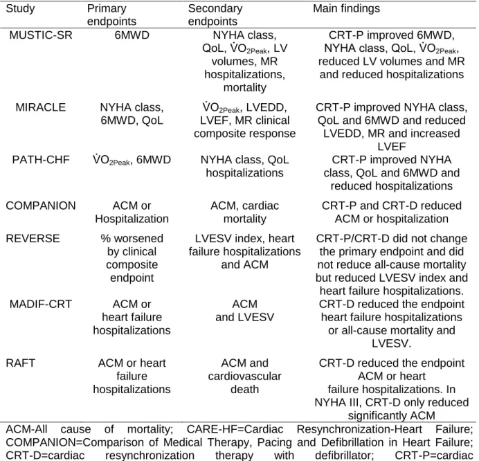
In the birth of CRT era, single centre trials were conducted, whereas limited by their external validity and statistical power, they provided useful information for later use in larger and multicentre trials (Leyva et al., 2014; Linde, Ellenbogen, & McAlister, 2012). According to a meta-analysis of CRT and selected RCT, until 2005 only nine clinical trials had been conducted and terminated (Linde, Ellenbogen & McAlister, 2012). Among these trials, analogous inclusion criteria could be observed. Depicted in Table 3, NYHA functional class (III/IV), depressed systolic LV function, LVEF< 35% and also QRS> 120ms with interventricular conduction disorder were inclusion criteria for these RCT. Importantly, most of the studies also reported improvements in all of these bus parameters. Similar primary end-points were also observed, although secondary end-points tend to vary from a strict range of echocardiographic measurements (e.g., LV systolic function, LVEF, LV reverse remodelling, LV volumes and Mitral regurgitation) (Table 4). Linde et al. (2012) refer that key studies on moderate to severe HF, as others on mild HF patients receiving CRT, were based on LVEF< 35% and QRS wider than 120ms. Jabbour et al. (2015) elaborated a meta-analysis were they pursue either or not some of the reported CRT benefits could be extended for narrow QRS patients? According to these authors, this extrapolation could be deceiving because poor study designs could have induced unwanted bias and conflicting results for CRT benefits in patients with narrow QRS. Conflicting evidence clearly complicates the complexity of synchrony and CRT. In addition, the indications for CRT are not limited to the measurement of LVEF, QRS complex, monitoring data, or even results of
15
electrophysiological studies, but further expanded to a wide range of cardiovascular signs and symptoms, disease states and physiological assessments (Russo et al., 2013).
Table 3. Inclusion criteria in selected randomized controlled studies
Study n NYHA class LVEF (%) LVEDD (mm) SR/AF QRS (ms) ICD MUSTIC-SR 58 III < 35% >60 SR >150 No MIRACLE 453 III, IV < 35% >55 SR >130 MUSTIC-AF 43 III < 35% >60 SR >200 PATH-CHF 41 III, IV < 35% NA SR >120 MIRACLE-ICD 369 III, IV < 35% >55 SR >130 Yes CONTAK- CD 227 II, IV < 35% NA SR >120 MIRACLE-ICD II 186 II < 35% >55 SR >130
PATH–CHF II 89 III, IV < 35% NA SR >120 Yes/No
COMPANION 1520 III, IV < 35% NA SR >120 CARE-HF 814 III, IV < 35% >30 IH SR >120 No CARE-HF EXTENSION 2006 813 III, IV < 35% >30 IH SR >120 REVERSE 2008 610 I, II < 40% >55 SR >120 Yes/No MADIF-CRT 1800 I, II < 30% NA SR >130 Yes
AF=atrial fibrillation; CARE-HF=Cardiac Resynchronization-Heart Failure; COMPANION=Comparison of Medical Therapy, Pacing and Defibrillation in Heart Failure; CONTAK-CD=CONTAK-Cardiac Defibrillator; CRT-D=cardiac resynchronization therapy with defibrillator; CRT-P=cardiac resynchronization therapy pacemaker; HF=heart failure; ICD=Implantable cardioverter-defibrillator; IH=indexed to the height; LVEDD=left ventricular end-diastolic diameter; LVEF=left ventricular ejection fraction; MADIT-CRT=Multicenter Automatic Defibrillator Implantation Trial with Cardiac Resynchronization Therapy; MIRACLE=Multicenter InSync Randomized Clinical Evaluation; MR=mitral regurgitation; MUSTIC=Multisite Stimulation in Cardiomyopathies; NA=Non-applicable; n=Number of patients; NYHA=New York Heart Association; PATH-CHF=Pacing Therapies in Congestive Heart Failure trial; REsynchronization reVErses Remodelling in Systolic left vEntricular dysfunction SR=sinus rhythm. *patients in atrial fibrillation
Adapted from Brignole et al.(2013)
Recent and main recommendations for CRT, from some of the most relevant American medical societies (e.g., ACCF, Heart Rhythm Society, AHA, Heart Failure Society
16
of America) and ESC, accomplishes a standardized approach that reflects the majority of clinical scenarios and permits individual expertize and technical appreciations of each individual case (Brignole et al., 2013; McMurray et al., 2012; Yancy et al., 2013).
Table 4. Endpoints, and main findings of some randomized clinical trials evaluating
CRT in sinus rhythm Study Primary endpoints Secondary endpoints Main findings
MUSTIC-SR 6MWD NYHA class,
Qo , V O2Peak, LV volumes, MR hospitalizations,
mortality
CRT-P improved 6MWD, NYHA class, Qo , V O2Peak, reduced LV volumes and MR
and reduced hospitalizations MIRACLE NYHA class,
6MWD, QoL
V O2Peak, LVEDD, LVEF, MR clinical composite response
CRT-P improved NYHA class, QoL and 6MWD and reduced
LVEDD, MR and increased LVEF
PATH-CHF V O2Peak, 6MWD NYHA class, QoL hospitalizations
CRT-P improved NYHA class, QoL and 6MWD and
reduced hospitalizations COMPANION ACM or Hospitalization ACM, cardiac mortality CRT-P and CRT-D reduced ACM or hospitalization REVERSE % worsened by clinical composite endpoint
LVESV index, heart failure hospitalizations
and ACM
CRT-P/CRT-D did not change the primary endpoint and did not reduce all-cause mortality but reduced LVESV index and
heart failure hospitalizations.
MADIF-CRT ACM or
heart failure hospitalizations
ACM and LVESV
CRT-D reduced the endpoint heart failure hospitalizations
or all-cause mortality and LVESV.
RAFT ACM or heart
failure hospitalizations
ACM and cardiovascular
death
CRT-D reduced the endpoint ACM or heart
failure hospitalizations. In NYHA III, CRT-D only reduced
significantly ACM
ACM-All cause of mortality; CARE-HF=Cardiac Resynchronization-Heart Failure; COMPANION=Comparison of Medical Therapy, Pacing and Defibrillation in Heart Failure; CRT-D=cardiac resynchronization therapy with defibrillator; CRT-P=cardiac resynchronization therapy pacemaker; LV= left ventricular; LVEDD=left ventricular enddiastolic dimension; LVEF=left ventricular ejection fraction; LVESV=left ventricular endsystolic volume; MADIT-CRT=Multicenter Automatic Defibrillator Implantation Trial with Cardiac Resynchronization Therapy; MIRACLE=Multicenter InSync Randomized Clinical Evaluation; MR=mitral regurgitation; MUSTIC=Multisite Stimulation in Cardiomyopathies; NYHA=New York Heart Association; PATH-CHF=Pacing Therapies in Congestive Heart Failure trial; QoL=quality-of-life score; RAFT= Resynchronization-Defibrillation for Ambulatory Heart Failure Trial; 6MWD=6-min walk distance.
17
It is not the scope of this project to analyse in depth the specific criteria for CRT implementation or the major differences approaching HF definition from medical societies, but instead we intent to identify the main points that lead to this significant clinical decision. Another significant issue is related with, the CRT patients eligibility for clinical trials may not be representative of those from the “real world” Finegold, Raphael, Levy, Whinnett, & Francis, 2013). Caution should also be taken into account because the assessment of patient clinical status relies upon individual clinical judgment. So, having this in consideration, we narrow the main criteria for CRT implementation to symptomatic HF and depressed LVEF, which can be objectively evaluated as: i) VEF≤ 35 ; ii) QRS duration≥ 120ms and morphology (i.e., LBBB); and finally, iii) NYHA functional class (Brignole et al., 2013; Poole, 2014; Russo et al., 2013; Vardas et al., 2007).
Importantly, CRT treatment is recommend in moderate to severe HF but more information is needed over its application in patients who differ from the aforesaid, specifically those with mild-to-moderate HF (i.e., class I-II), narrow QRS (i.e.,<120ms) and with right bundle branch block (Linde et al., 2012).
2.3 Autonomic function and resynchronization
In recent years, the autonomic nervous system (ANS) has gain attention because its importance in the natural history of HF. New non-invasive and valid ways to assess ANS have arisen through the study of heart rate variables (Piotrowicz, Baranowski, Piotrowska, & Zielinski 2009). The heart rate variability can be used as an index for parasympathetic reactivation post-exercise (Goldberger et al., 2006), while the heart rate recovery (HRR) is a prognostic variable and a good indicator of autonomic function (Arena, Myers, Abella, Peberdy, et al., 2010; Piotrowicz et al., 2009). The HRR has a prognostic and preventive value, and should be considered both in clinical and experimental fields (Arena, Guazzi, Myers, & Peberdy, 2006; Cahalin et al., 2013; Lipinski, Vetrovec, Gorelik, & Froelicher, 2005; Wu, 2014; Liu et al., 2014; Tang et al., 2009). In the scope of this document we will only focus on HRR.
AUTONOMIC NERVOUS SYSTEM MODULATION
The parasympathetic and the sympathetic independent structures, namely the nerve endings of both divisions, modulate the function of one another through complex interactions, varying HR accordingly distinctive adrenergic receptors selectivity stimulation. The HR is intimately dependent on the acetylcholine release in the synaptic cleft and the degree of interaction with muscarinic receptors in the heart (Mizuno et al., 2007).
18
Chakir et al. (2009) revealed CRT benefits beyond the restoration of electromechanical synchrony, and pointed out that calcium (Ca2+) transient improvement and depressed contractile function seem associated with cholinergic signalling receptors remodelling. The muscarinic receptors subtypes, can actually modulate, and be modulated, in their responsiveness, sensibility and specificity to particular amines. Coordinate sympathovagal control is essential for a normal hemodynamic function (Levy, 1984). Similar findings were achieved when vagus nerve stimulation and β-Blockers were used, which in turn seem to cause more norepinephrine release and reduced renin-angiotensin-aldosterone system effect (Zhang et al., 2009). Both of these adaptations improved autonomic control. In an analogous study, Kannenkeril et al. (2002) assessed parasympathetic effects on cardiac electrophysiology during moderate exercise and recovery, observing reduced sinus cycle length during recovery (p< .003), augmented ventricular effective refractory period (p< .005) and reduced Q-T interval (p< .02), all of these seemed to be associated with parasympathetic reactivation, sympathetic withdrawal or both. However, other findings suggest that discrepancy on sympathovagal tone could be observed in different levels of oxygen consumption (Saito & Nakamura, 1995), what could lead us to accept that exercise intensity could certainly be influencing these autonomic changes (Buchheit, Papelier, Laursen, & Ahmaidi, 2007).
EXERCISE AND AUTONOMIC FUNCTION
After finishing an exercise bout, parasympathetic reactivation occurs and sympathetic function decreases (Buchheit, Laursen, & Ahmaidi, 2007). Athletes tend to have higher vagal tone and more precise and accurate sympathetic modulation, an indicator and predictor of mortality (Prakash, 2012). The HRR at 1-minute (HRR1) reflects vagal reactivation, while the HRR at 3-minute (HRR3) seems to be mediated by sympathetic withdrawal (Okutucu et al., 2011). The autonomic balance and their major contributors interplay change continuously since the termination of an exercise bout and during the following recovery phases (Goldberger et al., 2006).
Still, controversial findings (Buchheit, Papelier, et al., 2007) question the initial fall in the post-exercise HR has a result, solely, of vagal reactivation, and argue that sympathetic withdrawal occurring immediately after exercise should also be taken into account. In accordance with these findings, others concluded the same for maximal and supramaximal exercise testing, which is similar to what was observed in the previous study, thereby, reinforcing a unknown role of the anaerobic metabolism influence (Oliveira, Mattos, Silva, Rezende, & Lima, 2013).
If these findings are important to understand the HRR dynamics, could we extrapolate this knowledge to a HF population? Or, vagal reactivation role on HR fall
post-19
exercise is reduced on low to moderate exercise intensity? It seems that in high intensity exercise, sympathetic and parasympathetic loops could also regulate the HRR (Yaylali et al., 2015). Also of note, and subject to scrutiny, are the different exercise intensities per se, or even the exercise mode, which could cause distinct central and peripheral fatigue (O'Leary, Morris, Collett, & Howells, 2015; Sahlin, Tonkonogi, & Soderlund, 1998), therefore contributing to different autonomic modulation.
The broad interpretation of the HRR, such as the difference between the HR at peak exercise and HR post-exercise, could to some extent be variable or even deceiving accordingly to individual scientific background or exercise approaches. For instances, Kubrychtova et al. (2009) used the HRR1 into the active recovery phase of the exercise test, while Lipinsky et al. (2005) used the HRR measured at 1, 2, 3, and 5-minute time points after treadmill testing. Although the HRR has been discussed mainly in the context of the ANS modulation and exercise, in good truth there is a far more complex domain to learn and explore. The ways adrenergic-cholinergic cross talk can exert their action and are influenced by other mechanisms, such as immune regulatory processes, clearly depicts the ANS capability in promoting or attenuating a response via similar paths (Chobanyan-Jurgens & Jordan, 2015; Levy, 1971; Mizuno, Tajima, Watanabe, & Kuratsune, 2014; Ogoh et al., 2005; Straburzynska, Wallace, & Potter, 1999). Other findings, associate autonomic imbalances and fetal genes activation with profound changes in cardiac structure and function (Paulus & Tschope, 2013). Others suggested that metaboreceptors, neurohumoral activation and baroreflex desensibilization may lead to sympathetic overactivation, resulting in HF vicious cycle of detrimental exercise capacity (Corra et al., 2014).
2.4 Exercise and resynchronization
Epidemiological evidence depicts physical activity and structured exercise importance for treatment and management of several chronic diseases (Bamman et al., 2014; Fleg et al., 2015; Kujala, 2006; Rosenthal & Dorsey, 2013). Exercise represents an independent protective role for the growing sociocultural problem of cardiometabolic diseases (Bamman et al., 2014; Powers, Smuder, Kavazis, & Quindry, 2014).
Conraads et al. (2007) compare the additive effect of exercise to the implantation of CRT, and verified that endurance training enhanced exercise tolerance in the intervention group versus the control for the V O2Peak (+40% versus +16%, p= .005), WattMax (+43% versus + 13%, p= .0005), and circulatory power (+74% versus + 32%, p= .01). Similarly, the HF-ACTION trial (Zeitler et al., 2015) enrolled a heterogeneous population of device patients, including CRT (n= 435), and allocation to usual care and exercise training. The three major findings were: first, the safety of the exercise; second, the improvement of
20
exercise capacity and QoL; lastlty, clinical events may be attenuated in patients with devices. According to Mayer et al. (2013) CRT falls under the category of ventricular assisted devices, which seems to benefit from exercise, but still lacks from a standardized exercise training programme. Independently of the specific programing of the pacemaker, some precautions should be made, namely: 1) the upper limit of the device which should be 20 beats per minute below the device intervention; 2) Graded exercise testing (GXT) is mandatory; 3) heart rate training zone and rate of perceived exertion should be measured at all times (Maeyer, Beckers, Vrints & Conraads, 2013).
If exercise training (ExT) in cardiac rehabilitation setting has class I indication for safety and effectiveness on HF functional status improvement, and class IIa recommendation for improvement of functional capacity, exercise duration, QoL and mortality, it is imperative to know the best form of exercise for this patients (Yancy, 2013). As new frontiers are reached new insides and discoveries happen. It´s obvious that old and new questions addressed to aerobic interval training (AIT) safety, efficacy, and physiological mechanisms should be made to an understanding of the functional improvement and survival rates it promotes, as well as to further bolster its clinical implementation. However, it seems clear its undeniable value, and therefore, more randomized controlled trials, with more statistical power should be conducted in order to clarify some of the concerns AIT may raise.
PHYSIOLOGICAL ADAPTATIONS TO EXERCISE PROTOCOLS
In recent years, novel approaches to exercise training and CRT have been done, either in the experimental or clinical field. AIT has been shown superior results over moderate continuous training (MCT) (Wisloff et al., 2007), and if there is biological plausibility for this superiority, it must be analysed, and also understand from which population, and which methodological approach this findings come from. Furthermore, it is of great relevance to differentiate myocardium adaptations arising from volume overload, from those arising from pressure overload, although this may be controversial and, further, raise new questions (Conraads & Beckers, 2010; Piepoli et al., 2011).
Myocardial architectural remodelling leads to alterations on the force-length curve relationship, and we can differentiate between eccentric and concentric remodelling, which in turn could lead, as already discussed in the HF continuum, to systolic or diastolic dysfunction respectively (Paulus & Tschope, 2013). Aerobic training can enhance exercise performance due to increased LV function (Haykowsky et al., 2013), cellular maladaptive hypertrophy regression (Gaasch & Zile, 2011), improved metabolic status (Bassett & Howley, 2000), mitochondrial adaptations (Hawley, Hargreaves, Joyner, & Zierath, 2014), improved cardiomyocyte contractility and also Ca2+ handling (Johnsen et al., 2013; Kemi et al., 2012).
21
Aerobic training also improves myocardium efficiency, leading to higher cardiac output and augmented peripheral oxygen consumption (Kemi & Wisloff, 2010). However, oxygen consumption and oxygen supply can´t be dissociated one from another. Oxygen consumption relies on the ability of the heart to contract efficiently, the ability to reach and maintain a high HR, and the relative wall tension, whereas oxygen supply depends on the arteriovenous difference and coronary blood flow. In CRT patients, ExT seems to improve energy metabolism (Hafstad et al., 2011), while pharmacological medicamentation could improve patient clinical status (Patwala et al., 2009).
As previously supported, we can assume that exercise intensity plays the key role in cardiac rehabilitation setting. The individual differences induced to individual cardiac myocytes are pivotal to the exercise induced cardioprotection (Powers et al., 2014). Exercise intensity relates with specific cellular metabolic pathways that are the foundation of either positive or adverse outcomes, through specific acute and chronic adaptations (Egan & Zierath, 2013). But another question emerges! How can we be precise and assume valid data while selecting exercise intensity? Intensity can be determined indirectly (i.e., formulas) and directly i e , HR, V O2). Other related variables are duration and exercise protocols. Aerobic training implies the presence of oxygen or more specifically oxidative mechanisms predominance for energy production (Egan & Zierath, 2013). Of note, aerobic interval training main energy system is oxidative, although the rate of oxygen utilization and therefore energy production is much higher than could be observed through, the also aerobically, continuous mode.
In a highly cited, and already considered a classic study from Trondheim team, Wisloff et al. (2007) AIT protocol was applied in patients undergoing optimal treatment for HF after myocardial infarction. In this controlled and randomized trial, the authors seek to understand differences between MCT and AIT in several parameters, ranging from reverse remodelling, exercise capacity and QoL. They conclude that AIT was superior to MCT in several parameters, including the V O2Peak (46% versus 14%, p< .001). CHF is clearly a disease of the elderly, and in this study, the included population had 88% of the patients with more than 65 years old, and 49% of the patients with more than 80 years old. The exercise protocol was maintained during the 12 weeks of the trial, in which it was revealed that, even in the elderly exercise is still able to induce adaptations.
Another study from Wisloff et al. (2001), and corroborated elsewhere (Kemi, Loennechen, Wisloff, & Ellingsen, 2002), concluded that exercising at 85/90% of the maximal oxygen uptake V O2Max) induces architectural and conformational transformations in cardiomyocytes, specifically inducing wider and long myocytes proteins, also termed as physiological hypertrophy. Exercise also seems to induce changes in Ca2+ transients, namely, faster systolic rise and diastolic decay. The magnitudes of these changes have a
22
direct and positive correlation with exercise intensity (Kemi et al., 2005). Kemi et al. (2005) observed a Ca2+ transient improvement of 40 on 85/90 of the V O2Max group, versus 20% for the moderate intensity. The same pattern was observed in mice and rats, as well as in humans, although some methodological and controversial issues could be raised (Helgerud et al., 2007).
Regarding the mode, most studies used treadmill walking (Fu et al., 2013; Iellamo et al., 2013; Wisloff et al., 2007), while others preferred concurrent training (Santa-Clara, Fernhall, Mendes, & Sardinha, 2002). However, dynamic exercise tends to put a volume overload in the myocardium (Erhman, 2010), while static exercise seems to depend more on neurogenic effect (Folland & Williams, 2007). The first mechanism is the basis for cardiac adaptation to aerobic exercise, while the second seems to focus mainly on peripheral mechanisms.
IN THE URGE OF A NEW EXERCISE METHODOLOGY
Several guidelines for exercise prescription in various populations, with specific intensities that promote optimal stimuli for expected adaptations are described elsewhere (Pescatello, 2013). These guidelines were born from robust scientific evidence supporting its value, but their practical application is often difficult and some pitfalls may be pointed out. Several training protocols (e.g., AIT, CMT), different modes (e.g., walking, cycling), different weekly frequency, different volume and intensity are proposed in literature (Erhman, 2010) as well as in several clinical trials (Fu et al., 2013; Rognmo et al., 2012; Wisloff et al., 2007). However, intensity is probably the most important variable, and apart from being independently manipulated, it also depends in the above-mentioned.
Nevertheless, some intriguing issues regarding exercise intensity can be raised, even more in the clinical domain. Specifically, we must question: i) the homogeneity of metabolic reactions among patients, which according to Arena et al. (2013) are heterogeneous among patients; ii) the methods accuracy and validity for evaluating metabolic reactions through gas analyses, which seems to be altered by several factors such as physiological and technical equipment variability (Bensimhon, 2008); iii) is the peak value e g , V O2, peak HR) the “true” maximal value for the tested variable? If not, can we measure the difference between them? iv) and most of all, is it possible to guarantee fixed and specific exercise intensity in the clinical domain? To address these concerns, we analyse Wisloff et al. (2007) work, in a well-designed and randomized controlled trial, and a reference work to the AIT apology. In this paper, and regarding exercise intensity, the authors write: “One of the weaknesses of
our study is that the usual care programme was not very well defined in means of exercise intensity” The authors also referred a variation of HR both between and within subjects.
23
confounding effect over the outcome. Fu et al. (2013) also adopted this strategy, which allows for distinguishing the periods of high intensity from the average periods and its effects over a specific outcome. Iellamo et al. (2012) adopted a training impulse method (TRIMP), alongside with complex algorithms to guarantee maximum control of possible confounders.
Percentages of the V O2Max and HR, as the hallmarks to set exercise training intensity, seem to yield heterogeneous metabolic and cardiocirculatory responses in different patients (Hofmann & Tschakert, 2011). Actually the same patient can respond differently upon different stressful and variable hormonal interactions (Hashimoto & Brooks, 2008) or even as a result of ExT chronic adaptation (Blazevich, 2006; Hambrecht et al., 2000). There has been proposed more accurate methods that rely upon submaximal markers, thresholds or turning points (Arena et al., 2012; Straburzynska-Migaj, Gwizdala, Siniawski, Ochotny, & Grajek, 2010). These are particularly important for those engaged on CRT (Conraads & Beckers, 2010). Could this actually be a turning point, or just a shifting to a more individualized and even more specific accurate method?
On a recent review over HF trials (Niederseer, Thaler, & Niebauer, 2013), it was observed a mismatch between the trials and the “real” world patients´ Spall et al 2007) went further and conducted a review of several RCT in which they seek to understand the nature and extent of exclusion criteria in certain patients´ population. Invariably, when assuming certain scientific methodology premises, we are also limiting the extent of our findings. Maybe one size fits all is not applicable in cardiac rehabilitation, moreover when regarding such a specific syndrome as HF (Hofmann & Tschakert, 2011), and with another complex variable such as CRT.
25
27
Methodology
3.1 Introduction
This chapter describes the methodology process of the study. Firstly, we identify our variables; secondly, our hypothesis; thirdly, the conceptual design of the study, where the patient population, as well as the inclusion and exclusion criteria are described; fourthly, the equipment and protocols of the assessments are overviewed in detail; and lastly, based on our data, we describe how our statistical treatment was conducted.
3.2 Variables in the study
Dependent variables. According to the following equipment and protocols
mentioned below, the present analysis considered the main variables of interest: the V O2Peak, the CPET duration, the resting exchange ratio (RER), the NYHA functional class, the systolic blood pressure and the diastolic blood pressure (rest and peak exercise), the HR (i.e., at rest, at peak exercise), the HR reserve and the HRR (i.e., 1-minute, 3-minute and 6-minute). Other variables were then computed based on the abovementioned.
Independent variables. ExT in the form of AIT was our independent variable.
3.3 Hypothesis
Main hypothesis. In 6 months, there will be differences between the intervention
and the control group on autonomic function.
Secondary hypothesis. In 6 months, there will be differences between the
intervention and the control group on exercise capacity, hemodynamics and chronotropic capacity.
3.4 Study design
The present analysis uses a longitudinal approach with two assessment time points: the baseline, before the cardiac implant (M1); and on the 6th month (M2), after the experimental therapy (i.e., ExT). Additionally, another moment was considered at the 3rd month (3Mo). All patients meeting inclusion criteria underwent the CPET in all three time points, and the data was collected. The 3Mo was utilized, when necessary, and in accordance with the CPET, to adjust exercise intensity. Aiming for the major outcome (i.e., autonomic function), the