Sanitary evaluation of large game hunted in
Idanha-a-Nova county.
Pilot study on evaluation of Sarcocystis spp. in muscular samples from large game harvested for human consumption.
Mestrado Integrado em Medicina Veterinária
Bruno Miguel Rafael Vinhas
Orientador:
Professora Doutora Maria Madalena Vieira-Pinto
Co-Orientador:
Dr. João Manuel Quirino Serejo Proença
Composição do Júri:
Presidente Doutor Nuno Francisco Fontes Santa Alegria, professor auxiliar da
Universidade de Trás-os-Montes e Alto Douro.
Vogais Doutora Alexandra Sofia Miguens Fidalgo Esteves, professora auxiliar com agregação da Universidade de Trás-os-Montes e Alto Douro.
Doutora Maria Madalena Vieira Pinto, professora auxiliar da
Universidade de Trás-os-Montes e Alto Douro
Doutora Ana Patrícia Lopes, professora auxiliar da Universidade de
Sanitary evaluation of large
game hunted in
Idanha-a-Nova County.
Pilot study on evaluation of Sarcocystis spp. in
muscular samples from large game harvested for
human consumption.
Nome do Candidato: Bruno Miguel Rafael Vinhas Orientador: Professora Doutora Maria Madalena Vieira-Pinto Co-Orientador: Dr. João Manuel Quirino Serejo Proença
Agradecimentos
Bem, depois de tão longa caminhada é difícil escolher um conjunto de palavras com um significado aproximado da intensidade vivida nestes últimos anos, que culminam com esta produção textual científica. De qualquer maneira algumas palavras têm que ser proferidas e sobretudo sentidas. Desejo apenas que não olhem para o texto seguinte como uma lista ordenada mas apenas como o livro do meu coração académico e os que me conhecem sabem que o meu coração tem espaço para todos os que quiserem lá entrar.
Particularizando, gostaria assim de começar pela minha Família, que é desde de sempre a grande razão deste caminho. Aos meus pais e o meu irmão expresso aqui a minha gratidão pelo apoio constante nos bons e maus momentos. Sem eles, percorrer este caminho não faria sentido. Depois à minha Família Transmontana, a Chica, o Fiúza, o Miguel e a Di, se sem a minha Família não faria sentido este caminho, não os encontrar a eles seria um fado de igual quebra neste percurso. Por isso, o meu muito obrigado aos quatro por estarem sempre lá. De seguida gostaria de mostrar o meu reconhecimento à Professora Madalena por ter embarcado na loucura da minha orientação. E quando digo loucura digo-o consciente, pois uma orientação já é por si difícil, mas quando se junta a incerteza do que esperar de uma pessoa face a este desafio é preciso realmente um pouco de loucura. Gostaria, também, de agradecer ao Dr. Serejo e família por me terem acolhido com tanto afeto e simpatia. Desde de logo tudo se tornou mais fácil, mesmo estando eu longe do meu mundo e encarando um novo mundo muito diferente do meu. Um apoio deste nível foi indispensável ao meu sucesso. Em especial ao Dr. Serejo quero agradecer, com entusiasmo, todos os conhecimentos transmitidos e acima de tudo o exemplo do que é realmente ser veterinário. É incalculável o valor desta passagem de saberes que não vêm nos livros e que de outra maneira poderiam perder-se no tempo. De seguida gostaria de agradecer à UTAD e em especial à Veterinária da UTAD. Sobretudo, mas não só, ao magnífico Corpo Docente que tem uma grande influência na rota escolhida por mim. O ambiente de amizade, cordialidade e acessibilidade é de certeza uma das razões da qualidade dos profissionais formados na UTAD. Este agradecimento estende-se ao ambiente académico único e à cidade que são sem dúvida a grande matéria-prima de toda a verdadeiro afeição gerado em quem vem estudar para esta Mui Nobre Bila. O meu muito obrigado. Outro capítulo importante desta aventura foi a AEMV-UTAD, a qual teve uma enorme responsabilidade na definição do meu ser atual. A todos os que partilharam esta experiência comigo fica aqui expresso o meu profundo e associativo reconhecimento por todas as experiências partilhadas, em especial para os que prosseguiram com os meus ideais. Por fim gostaria de agradecer, de forma Honrosa ao Laboratório de Histologia e Anatomia Patológica, sobretudo à Professora Isabel Pires pelo apoio técnico, particularmente na ilustração deste trabalho. Pois, as importantes fotos histológicas
muito embora estejam impressas com o meu nome são em grande parte sua responsabilidade. Tal como os fantásticos mapas de Idanha com os vários dados trabalhados neste estudo são obra do Professor Aranha, a quem desde de já agradeço todo tempo e perseverança. Por fim gostaria de agradecer pela ajuda no texto à Isabel e ao Miguel pelo tempo e paciência dispensados na leitura deste documento.
De uma forma geral, gostaria de agradecer a todos os que não nomeei anteriormente mas que de uma forma ou de outra contribuíram para esta feliz peripécia da minha vida.
“Agir, eis a inteligência verdadeira. Serei o que quiser. Mas tenho que querer o que for. O êxito está em ter êxito, e não em ter condições de êxito. Condições de palácio tem qualquer terra larga, mas onde estará o palácio se não o fizerem ali?” Fernando Pessoa
Abstract
In the last three decades, game hunting activity assumed a major role at social, cultural and economic level in several regions of Portugal. Large game hunting, with focus on red deer (Cervus elaphus) and wild boar (Sus scrofa), is a significant part of that hunting activity. The study area of this survey, Idanha-a-Nova County, is one of the most representative places of this hunting activity.
In this circumstances, the knowledge on the health of those animals become an essential matter, mainly if their meat is used for human consumption, being zoonotic agents the major target of veterinary attention. For that reason, presently in the Portuguese epidemiological risk area for tuberculosis in large game, it is mandatory the presence of a veterinarian in each driving hunt to do a sanitary evaluation in the field. The geographic area of this study is included in this risk area.
This survey occurred in hunting season 2011/2012 and had as main objectives the evaluation of game meat preparation conditions in the field and the Sarcocystis spp. occurrence in muscular samples collected from large game harvested for human consumption.
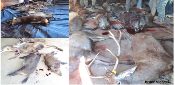
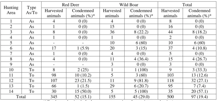
In order to fulfill the previous issues it were attended 24 drives hunting in 14 hunting areas in which were harvested 500 animals (345 red deer and 155 wild boars). From those, 52 red deer (15.1%) and 45 wild boars (29.0%) were declared unfit for human consumption. The main cause of condemnation observed was Tuberculosis compatible lesions, underlining the persistence and importance of this disease in large game hunted in Idanha-a-Nova County.
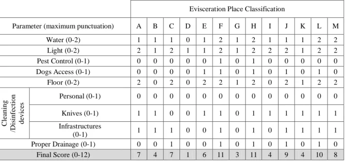
The evaluation of the game meat preparation conditions in the field, made on the evisceration place, gathered information from 3 main domains: structural requirements, hygienic requirements and transportation of game and carcasses, and by-products destination. The structural requirements domain was the one with the lowest score. In two cases, it was observed that it would be possible to overcome this problem if these Hunting Areas share the evisceration place with one geographically close with better classification. Within each domain several parameters were analyzed, being personal cleaning/disinfection devices, knives utilization, garment, footwear and mask the ones with the lowest punctuations, indicating the need for its improvement to ensure better protection of handlers and better hygiene of game meat.
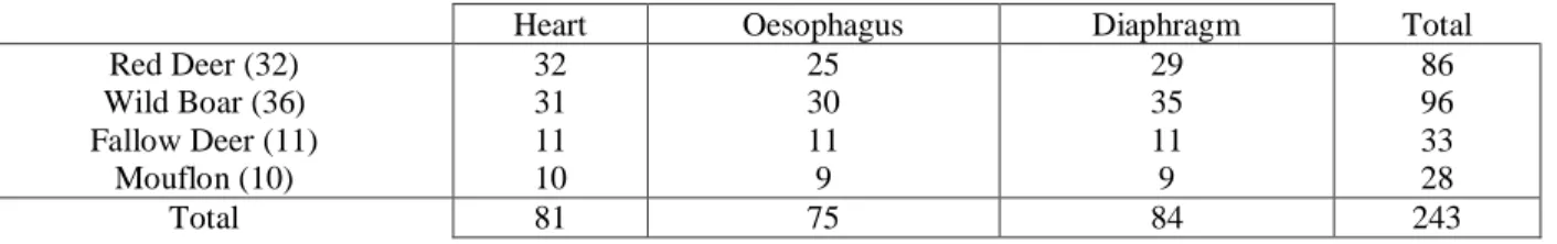
Through histological examination of the oesophagus, diaphragm, and heart, Sarcocystis spp. cysts were detected in 65.6% red deer, 38.9% wild boar, 100% fallow deer and mouflon. In red deer and wild boar oesophagus was the less often affected sample.
The applied diagnostic approach revealed high level of Sarcocystis spp. occurrence, underlining that life cycle and zoonotic potential should be further investigated and
indicates that several issues should be addressed when planning surveillance and prevention actions.
Resumo
Nas últimas três décadas a caça assumiu um papel importante a nível social, cultural e económico em várias regiões de Portugal. A caça maior, sobretudo veados (Cervus elaphus) e javali (Sus scrofa), é uma parte relevante dessa atividade. E a área de estudo deste trabalho, Idanha-a-Nova, é um dos concelhos mais representativos dessa importância.
Nestas circunstâncias, o conhecimento do estado sanitário destes animais torna-se uma questão essencial, sobretudo se para consumo humano, sendo os agentes zoonóticos o principal alvo da atenção veterinária. Por essa razão, na Área Epidemiológica de Risco para a Tuberculose dos Animais de Caça Maior é obrigatório a presença de um veterinário em cada montaria para efetuar a avaliação sanitária. A área geográfica deste estudo está incluída nesta área epidemiológica de risco.
Este estudo ocorreu na época venatória 2011/2012 e tinha como principais objetivos a avaliação das condições do local de evisceração e a ocorrência de Sarcocystis spp. em amostras musculares de animais de caça maior destinados ao consumo humano.
Para cumprir os objetivos foram acompanhadas 24 montarias, em 14 zonas de caça, nas quais foram caçados 500 animais (345 veados e 155 javalis). Destes animais, 52 veados (15.1%) e 45 javalis (29.0%) foram rejeitados para consumo. A principal causa da rejeição foram lesões compatíveis com Tuberculose, enfatizando a importância desta doença da caça maior em Idanha-a-Nova.
Para a avaliação das condições do local de evisceração foram recolhidas informações em 3 domínios: requisitos estruturais, requisitos higiénicos e transporte de animais caçados e carcaças e destino de subprodutos. O domínio dos requisitos estruturais foi o que teve o resultado mais baixo. Em dois casos foi observado que seria possível melhorar este resultado através da partilha, por parte das Zonas de Caça, do local de evisceração mais próximo e com melhor classificação. Cada domínio foi ainda dividido em parâmetros, sendo que os que obtiveram pontuações mais baixas foram: Dispositivos de Limpeza/Desinfeção Pessoal, Utilização das Facas, Vestuário, Calçado e Máscara. Este resultado indica a necessidade de melhorar estes parâmetros para assegurar uma maior proteção dos intervenientes e salubridade da carne de caça. Através do exame histológico de esófago, diafragma e coração foram detetados quistos de
Sarcocystis spp. em 65,6% dos veados, 38,9% dos javalis, 100% dos gamos e muflões. Tanto no
veado como no javali o esófago foi a amostra de tecido menos afetado.
Este exame revelou um nível elevado de ocorrência de Sarcocystis spp., reforçando a necessidade de investigar o ciclo de vida e o potencial zoonótico, indicando a necessidade de contemplar estas questões na elaboração de planos de vigilância e prevenção.
General Index:
1. Introduction ... 15
2. Bibliographic revision ... 17
2.1. Large Game Hunting in Portugal ... 17
2.1.1. Red deer (Cervus elaphus Linnaeus, 1758) ... 18
2.1.2. Wild boar (Sus scrofa Linnaeus, 1758) ... 19
2.2. Sarcocystis spp. ... 21
2.2.1. History and Taxonomy ... 21
2.2.2. Life Cycle ... 22
2.2.3. Specificity of hosts ... 27
2.2.4. Diagnostic ... 28
2.2.5. Species affecting red deer (Cervus elaphus) and wild boar (Sus scrofa)... 29
2.2.6. Sarcocystis as a Zoonotic Agent ... 31
3. Materials and Methods ... 35
3.1. Area of study ... 35
3.2. Game Sanitary Evaluation ... 38
3.3. Evaluation of game meat preparation conditions in the field ... 40
3.4. Analysis of the presence of Sarcocystis spp. in muscular samples of large game hunted in Idanha-a-Nova County ... 44
3.4.1. Sampling procedure ... 44
3.4.2. Histopathological Analysis ... 46
3.4.3. Statistical Analysis ... 46
3.5. GIS Methodology ... 48
4. Results and Discussion ... 49
5. Conclusion ... 71
6. Bibliography ... 73
Image Index:
IMAGE 1 - RED DEER GRAZING IN A FIELD IN IDANHA-A-NOVA COUNTY. ... 17
IMAGE 2 - RED DEER. ... 19
IMAGE 3 - WILD BOAR TRACES. ... 20
IMAGE 4 - SARCOCYSTIS SPP. LIFE CYCLE. ... 23
IMAGE 5 - WILD BOAR, OESOPHAGUS. IMAGE OF A SARCOCYSTIS SPP BRADYZOITE. IN TRANSVERSAL CUT. H&E. BAR=30µM. ... 24
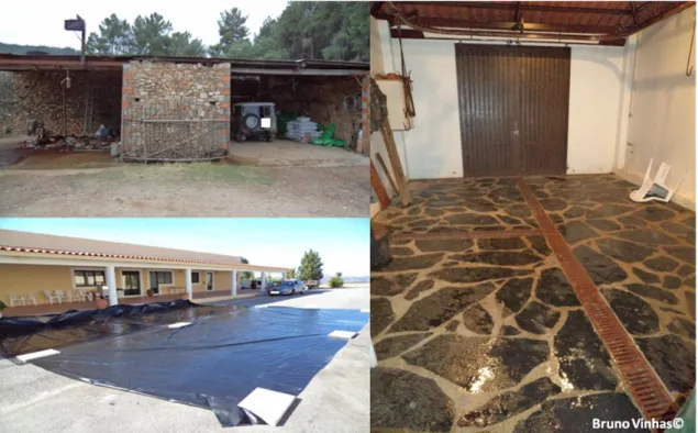
IMAGE 6 – REPRESENTATIVE IMAGE OF THE TWO LANDSCAPES FOUND IN IDANHA-A-NOVA. A NORTHEN MOUNTAINS. B SOUTH PLATEAU. ... 35
IMAGE 7 - LOCATION OF IDANHA-A-NOVA COUNTY IN THE PORTUGAL MAP.. ... 35
IMAGE 8 - EPIDEMIOLOGIC RISK AREA FOR TUBERCULOSIS MAP. ... 37
IMAGE 9 – SURVEYED HUNTING AREAS (HA) WITHIN IDANHA-A-NOVA COUNTY. ... 37
IMAGE 10 – IMAGE OF PARASITIC LARVAE OF OESTRIDS IN RED DEER. ... 39
IMAGE 11 - INSPECTION OF SEVERAL TISSUES. A – KIDNEY INCISION. B – HEART INCISION. C - SPLEEN INSPECTION. ... 39
IMAGE 12 – LYMPH NODES INSPECTION. A - MANDIBULAR LYMPH NODES INCISION. B - PRE-ESCUPULAR LYMPH NODES INCISION. ... 39
IMAGE 13 - LYMPH NODES INSPECTION. A - MESENTERIC LYMPH NODES. INCISION. B - INGUINAL LYMPH NODES INCISION... 40
IMAGE 14 - EXAMPLE OF THE THREE TYPES OF FLOOR FOUND IN THE STUDY. A – SOIL. B – PLASTIC PROTECTION. C - TILE. ... 41
IMAGE 15 - IMAGE OF THE SEVERAL EVISCERATION PLACES... 42
IMAGE 16 – IMAGE OF ABSENCE OF GAME HEAPING IN THE EVISCERATION PLACE. ... 43
IMAGE 17 - REPRESENTATIVE IMAGE OF THE HYGIENE REQUIREMENTS... 43
IMAGE 18 - IMAGE OF ANIMAL’S TRANSPORTATION TO THE COLLECTION SITE. ... 44
IMAGE 19 - REPRESENTATIVE IMAGE OF ANIMALS (A), CARCASS TRANSPORTATION (B) AND BY-PRODUCTS DESTINATION (C). ... 44
IMAGE 20 – REPRESENTATIVE IMAGE OF SAMPLES. A - SAMPLES READY TO FREEZE. B - CONTAINERS WHERE TISSUES WERE PRESERVED IN 10% FORMALDEHYDE. ... 45
IMAGE 21 – A - WILD BOAR HEART. EVIDENCE OF A HIGH NUMBER OF PARASITES OBSERVED. H&E. BAR=300µM. B - WILD BOAR, OESOPHAGUS. IMAGE OF A SARCOCYSTIS SPP. BRADYZOITE IN TRANSVERSAL CUT. H&E. BAR=30µM. ... 46
IMAGE 22 - HARVESTED ANIMALS IN EACH SURVEYED HUNTING AREA WITHIN IDANHA-A-NOVA COUNTY. ... 50
IMAGE 23 –TCL IMAGES. A – BLADDER. B – LIVER. C - MESENTERIC LN.. ... 50
IMAGE 24 - CONDEMNED CARCASSES. A - CONDEMNED CARCASS DUE TO DOG BITE. B - CONDEMNED CARCASS DUE TO EMACIATION. ... 51
IMAGE 25 – GEOGRAPHICAL DISTRIBUTION OF TB IN WILD BOAR HARVESTED WITHIN IDANHA-A-NOVA COUNTY. ... 52
IMAGE 26 – GEOGRAPHICAL DISTRIBUTION OF TB IN RED DEER HARVESTED WITHIN IDANHA-A-NOVA COUNTY ... 53
IMAGE 27 - A - IMAGE OF PEST CONTROL DEVICES. B - IMAGE OF A DOG WITH ACCESS TO BLOOD FROM GAME ANIMALS. ... 57
IMAGE 28 - IMAGE OF AN EVISCERATION PLACE WHERE IS EASY TO CONTROL DOG ACCESS DURING EVISCERATION AND SANITARY EVALUATION. ... 57
IMAGE 29 - REPRESENTATIVE IMAGE OF CARCASS TRANSPORTATION. A - CARCASSES INTENDED TO COMMERCIAL PROPOSE. B - CARCASSES INTENDED TO SELF-CONSUMPTION. ... 58
IMAGE 30 - REPRESENTATIVE IMAGE OF SEVERAL CLEANING AND DISINFECTION DEVICES. 58 IMAGE 31 – RED DEER LUNG. IMAGE OF TCL BEING CUT. ... 59
IMAGE 32 - REPRESENTATIVE IMAGE OF ONGOING EVISCERATION AND SANITARY EVALUATION... 59
IMAGE 33 - REPRESENTATIVE IMAGE OF EVISCERATION. A - INTERVENIENT DO NOT USE GLOVES NEITHER OWN KNIVES, FOOTWEAR OR GARMENT. B - EXAMPLE OF HOW TO BE DRESSED TO EVISCERATION. ... 59
IMAGE 34 - REPRESENTATIVE IMAGE OF DRAINAGE. ... 59
IMAGE 35 - REPRESENTATIVE IMAGE OF THE POSSIBLE BY-PRODUCTS DESTINATION. A - STORAGE OF BY-PRODUCTS TO CITY HALL SERVICES COLLECT. B - BURIAL. ... 60
IMAGE 36- REPRESENTATIVE IMAGE OF THE USE OF GLOVES. A - NOT WEARING GLOVES. B –
WEARING GLOVES. ... 60
IMAGE 37 – GEOGRAPHICAL DISTRIBUTION OF SARCOCYSTIS OCCURRENCE IN RED DEER HARVESTED WITHIN IDANHA-A-NOVA COUNTY. ... 64
IMAGE 38 – GEOGRAPHICAL DISTRIBUTION OF SARCOCYSTIS OCCURRENCE IN WILD BOAR HARVESTED IN IDANHA-A-NOVA COUNTY... 64
IMAGE 39 – REPRESENTATIVE IMAGE OF MEAT HARBORING SARCOCYSTIS SSP CYSTS. A - MOUFLON HEART. MUSCULAR TISSUE AND PURKINJE FIBERS WITH PARASITIC CYSTS. H&E. BAR=120µM. B – RED DEAR HEART. SARCOCYSTIS SPP. CYST IN A LONGITUDINAL CUT. H&E. BAR=30µM. ... 67
Table Index:
TABLE 1 - EXAMPLES OF SARCOCYSTIS SPECIES WITH RESPECTIVE INTERMEDIATE HOST AND DEFINITIVE HOST (YANG ET AL., 2002; UGGLA ET AL., 1990). ... 29TABLE 2 - PREVALENCE OF SARCOCYSTIS INFECTION IN RED DEER FOUND IN LITERATURE (IN MUSCULAR SAMPLES). ... 31
TABLE 3 - PREVALENCE OF SARCOCYSTIS INFECTION IN WILD BOAR FOUND IN LITERATURE. ... 31
TABLE 4 – NUMBER OF HUNTING AREAS AND CORRESPONDENT TYPE, SANITARY EVALUATION PLACE, COUNCIL, NUMBER OF DRIVING HUNTS AND HARVESTED ANIMALS. ... 38
TABLE 5 - HUNTING AREAS AND CORRESPONDENT ASSIGNMENT. ... 41
TABLE 6 - CLASSIFICATION OF THE STRUCTURAL REQUIREMENTS PARAMETERS PRESENT DURING GAME MEAT PREPARATION IN THE FIELD (MAXIMUM PUNCTUATION: 12). ... 41
TABLE 7 - CLASSIFICATION OF THE HYGIENIC REQUIREMENTS PARAMETERS PRESENT DURING GAME MEAT PREPARATION IN THE FIELD (MAXIMUM CLASSIFICATION: 12). ... 42
TABLE 8 - CLASSIFICATION OF THE PARAMETERS RELATED TO GAME AND CARCASS TRANSPORTATION AND TO BY-PRODUCTS DESTINATION (MAXIMUM CLASSIFICATION: 4). ... 43
TABLE 9 - NUMBERS OF COLLECTED MUSCLES BY SPECIE AND TISSUE. ... 45
TABLE 10 – TABLE EXAMPLE. ... 47
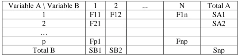
TABLE 11 - CONTINGENCY TABLE EXAMPLE. ... 47
TABLE 12 – K VALUES FOR C MAX CALCULATION. ... 48
TABLE 13 - NUMBER AND SPECIES OF HARVESTED ANIMALS IN EACH HUNTING AREA AND THE RESPECTIVE LEVEL OF CONDEMNED ANIMAL FOR HUMAN CONSUMPTION. ... 49
TABLE 14 - STRUCTURAL REQUIREMENTS SCORE FOR EACH EVISCERATION PLACE. ... 54
TABLE 15 - HYGIENIC REQUIREMENTS SCORE FOR EACH EVISTERATION PLACE. ... 55
TABLE 16- TRANSPORTATION OF GAME AND CARCASSES, AND BY-PRODUCTS DESTINATION SCORE FOR EACH EVISCERATION PLACE. ... 55
TABLE 17 - FINAL SCORE FOR EACH EVISCERATION PLACE. ... 55
TABLE 18 - CLASSIFICATION IN PERCENTAGE ... 56
TABLE 19 - SARCOCYSTIS SPP. FOUND IN EACH SAMPLED ANIMAL SPECIES. ... 62
TABLE 20 – SARCOCYSTIS SPP. OCCURRENCE ACCORDING TO THE AGE AND GENDER OF THE SAMPLED ANIMALS. ... 66
TABLE 21 - SARCOCYSTIS SPP. DISTRIBUTION AMONG THE ANALYSED MUSCLE SAMPLES COLLECTED FROM BOTH INFECTED ANIMALS SPECIES ... 66
TABLE 22 – PRESENCE OF SARCOCYSTIS SPP. IN MATCHED SAMPLES COLLECTED FROM INFECTED ANIMALS... 68
Abbreviation, Acronym and Symbol Index:
µm – micrometers
ANMP – National Association of Portuguese City Halls BD – By-products Destination
CT – Carcass Transportation DA – Dog Access
EFSA – European Food Safety Agency
ELISA – Enzyme-linked immunosorbent assay FW – Footwear
G – Garment g – Grams
GH - Game Heaping
GIS – Geographical Information Systems Gl – Gloves GU - Gloves Utilization h – Hour ha – Hectares HA – Hunting Area HE – hematoxylin-eozine
HPS – Human Hypereosinophilic Syndrome IFAT – Immunofluorescent Antybody test IHA – Indirect Hemagglutination test INE – National Institute of Statistics
IUCN – International Union for the Conservation of Nature Kg – Kilogram
km2 – square kilometres KnU – Knives Utilization M – Mask
M. – Micobacterium ºC – Centigrade OK – Own Knives
PAS – Periodic Acid-Schiff PC – Pest Control
PCDD – Personal Cleaning/Disinfection Devices PD – Proper Drainage
TB – Tuberculosis
Tcl – Tuberculosis compatible lesion USA – United States of America WK - Work Organization
1.
Introduction
In the last three decades, large game hunting activity assumed an increasing economic, social and cultural importance in several regions of Portugal, being the red deer and the wild boar the most important game species hunted.
These game species may be infected by several zoonotic agents requiring some veterinarian concerns and attention mainly if their meat is used for human consumption. For that reason, presently in the Portuguese epidemiological risk area for tuberculosis in large game, it is mandatory the presence of a veterinarian in each driving hunt to do a sanitary evaluation in the field. The geographic area of this study, Idanha-a-Nova County, is included in this risk area.
One of the zoonotic agents that may affect red deer and wild boar is Sarcocystis spp., a unicellular parasite that may be found in muscles of the infected animals. In Sarcocystis life cycle, humans may act as definitive host (gastrointestinal sarcocystosis) for S. suihominis and S.
hominis (Heydorn, 1977, cited by Mohammadi & Petri, 2006) or as an intermediate host
(muscular sarcocystosis) for a considerable number of species, some of them not yet determined (Mohammadi & Petri, 2006; EFSA, 2010; Esposito, D. H, et al,. 2012). In fact, according to several authors, it is necessary to make clear the impact in public health of Sarcocystis species to clarify their importance and the need of their monitoring (EFSA, 2010).
The lack of information of Sarcocystis spp. prevalence in large game species, the importance that Sarcocystis spp. may assume to the public health, the importance of large game as a source of meat for human consumption and the importance of sanitary evaluation of game species in the field, justify the development of this study which was carried in wild boars and red deer hunted in a Idanha-a-Nova County during the hunting season 2011/2012.
The main objectives of the present study include:
The characterization of the number of wild boar and red deer hunted in Idanha-a-Nova County and the main cause of condemnation of animals for human consumption;
The evaluation of game meat evisceration place;
Identification of the Sarcocystis spp. infection occurrence in red deer and wild boar hunted in Idanha-a-Nova County through histopathological analyses.
2.
Bibliographic revision
2.1. Large Game Hunting in Portugal
In the last three decades, large game hunting is increasing its importance on economical, social and cultural features of several regions of Portugal. To underline the increasing importance of large game hunting in Portugal is the size of hunting area, which was about 5 million ha in 2004. From those 5 million ha, 3.2 million ha (1/3 of Portugal) were divided for 1700 Association Hunting Area and 720 Tourist Hunting Area and 1.8 million ha dived for 600 of other types of hunting areas (Lopes et al., 2004). Other fact that emphasizes the increasing importance of large game hunting is the number of hunter’s licenses issued, which almost doubled from season 1999/2000 to season 2004/2005, (Vingada et al., 2010).
Hunting and game management are mainly regulated by Act 173/1999, of September 21st, which was updated to Act n. º 2/2011, of January 6th. To both activities there are several specific directives emitted to regulate specific cases. In accordance with these legal diplomas the large game species hunted in Portugal are the wild boar (Sus scrofa), the fallow deer (Cervus dama), the red deer (Cervus elaphus), the roe deer (Capreolus capreolus) and the mouflon (Ovis
ammon). These species can be hunted by sit and wait hunting, stalking, drive hunting, battue
hunting and spear hunting. While the red deer, the roe deer, and the wild boar are native species, the fallow deer and the mouflon were introduced, the first one centuries ago and the second one two decades ago (Lopes et al., 2004).
The present study was mainly focused on red deer (Cervus elaphus) (Image 1) and wild boar (Sus scrofa).
2.1.1. Red deer (Cervus elaphus Linnaeus, 1758)
Taxonomy: Kingdom-Animalia; Phylum-Chordata; Class-Mammalia; Order-Artiodactyla; Family-Cervidae; Genus-Cervus; Specie-C. elaphus.
The red deer (Image 2) is the largest wild ungulate hunted in Portugal. Its existence has suffered some ups and downs through history. While, in the medieval period, it was widespread in Portugal (Mendonça, 2003, cited by Vingada et al., 2010), in the last part of the nineteenth century, red deer was already on the edge of extinction (Bugalho, 2002, cited by Vingada et al., 2010). In the 1970’s red deer presented a reduced and spotted distribution confined to fenced areas (Tapada de Mafra, Torre Bela, Tapada de Vila Viçosa, Parâmio, Montesinho e Contenda) and was practically extinct in their natural habitat. Nonetheless, in the following decades, were made several deliberated attempts to restocking by releasing animals from those fenced herds (Vingada et al, 1997, cited by Vingada et al., 2010; Fonseca, 2004a, cited by Vingada et al., 2010). In addition, several animals dispersed naturally from Spain into Portuguese territory in the Montesinho Mountains, Contenda-Barrancos region, Tagus International and more recently Gerês Mountains (Vingada et al., 2010). Furthermore, in the border regions were taken a series of measures to improve habitat quality allowing red deer populations to settle permanently in our country. And, currently, this specie is widespread throughout Portugal, especially to south of the river Mondego, with emphasis on the populations of the International Tagus, Lousã, Alentejo (Moura, Mourão and Barrancos) and Silves. In the north of the country, it is evident the population of the north-eastern border (Lombada National Hunting Area). The expansion of red deer, seen in the last decades in Portugal, is for sure rare in the recent history of Western Europe. The totality of causes of this fact still to explain but is possible to mention some: the enormous plasticity and adaptation to different habitats; a progressive abandon of the rural lands, providing shelter; possible increase of the tolerance to man's presence; and a strong sense of territoriality, that doesn't allow great densities but forces the increase of the distribution area (Salazar, 2009).
In literature, is quoted that the number of red deer (Cervus elaphus) in Portugal should be approximately 15 000 to 20 000 animals and almost half of them are restricted to fenced areas. It was the importance of this specie in the large game that led to the promotion of red deer (Cervus
Because it is not possible to determine if the reintroduction of the animals were a success (different origins of animal from Spain, Scotland and Hungary) and because of the lack of studies on red deer genetics, it is generally accepted that Cervus elaphus hispanicus is the dominant subspecies in Portugal (Salazar, 2009).
Image 2 - Red deer.
2.1.2. Wild boar (Sus scrofa Linnaeus, 1758)
Taxonomy: Kingdom-Animalia; Phylum-Chordata; Class-Mammalia; Order-Artiodactyla; Family-Suidae; Genus-Sus; Species-S. scrofa.
Wild boar (Sus scrofa) is also one very important large game species hunted in Portugal. The distribution of this specie has also known several ups and downs through the Portuguese history. A serious decrease, in wild boar population, was prominent in the 19th century and in the beginning of the 20th century. During this period the population was constrained to mountainous areas contiguous to Spain and some Royal Hunting Grounds (Fonseca, 2004a, cited by Vingada
et al., 2010; Lopes et al., 2004). Taking into account the low density of populations, in 1967 the
wild boar hunting was prohibited outside enclosure areas (act 47 847, of August 14th in Vingada, J. et al. 2010). Also, in 1969, in the VIIth IUCN (International Union for the Conservation of Nature) Resource Technical Meeting (Fonseca, 2004a, cited by Vingada et al., 2010), the designation of endanger species was allocated to wild boar. The possible re-colonization by the clusters which remained next to the perimeter of Portugal, mainly those on south of Tagus Rive, were then responsible for the spreading out and the nowadays national distribution of wild boar populations (Lopes et al., 2004; Vingada et al., 2010). In the present days, wild boar populations are again widespread through all Portuguese territory, apart from the huge metropolitan and seashore areas (high human density), as according to the data reported by hunting areas
associations (Fonseca et al., 2004, cited by Vingada et al., 2010; Lopes et al., 2004). According to the official game statistics, in Idanha-a-Nova County were harvested 367, 708 and 817 (data only until 31-12-2009) wild boars in seasons 2006-07, 2007-08 and 2008-09 respectively (DGAV, 2010c).
2.2. Sarcocystis spp.
2.2.1. History and Taxonomy
The first report on Sarcocystis was made by Miescher in 1843, who described white threadlike cysts in striated muscles of a mouse (Mus musculus) (Dubey et al., 1989, cited by, Dahlgren, 2010). No scientific name was given at the time and in the following twenty years it was referred as “Miescher’s Tubules”. In 1865 similar structures were found in pig muscle by Kühn and named Synchrytium miescherianum (Dahlgren, 2010). Lankester in 1882 introduced the name Sarcocystis (Greek: sarkos - flesh, kystis - cyst) but only in 1899 the denomination
Sarcocystis meischeriana was proposed to identify this species (Dubey, 1989, cited by Fayer,
2004). Afterwards, for every intramuscular cyst found in a new host was proposed a new specie name. Throughout this time there was the controversy of which taxonomic group did it belong to (protozoa or fungi), on the account that sarcocyst stage was the only identified and that in several culture media there were seen hyphae and mycelia after a few days (result of contamination). In 1967, 124 year after the first report of Sarcocystis, there was a study using electronic microscopy on the bradyzoites, which revealed organelles like those seen on the apicomplexan protozoa in species such as Toxoplasma and Eimeria (Senaud, 1967, cited by Fayer, 2004). In1970, with the inoculation of bradyzoites from Sarcocystis of birds into cultured mammalian cells, there was development of sexual stages and oocysts (Fayer, 1970, cited by Fayer, 2004; Fayer, 1972, cited by Fayer, 2004). In 1972 the heteroxenous life was revealed when the life-cycles of several
Sarcocystis species of sheep, swine, and cattle were determined (Heydorn et al., 1972, cited by
Dahlgren, 2010; Rommel et al., 1972, cited by Dahlgren, 2010).
In the present, Sarcocystis is a unicellular parasite with the following classification:
Kingdom: Protozoa > Phylum: Apicomplexa > Class: Sporozoea > Subclass: Coccidia > Order: Eucoccidiorida > Suborder: Eimeriorina > Family: Sarcocystidae > Subfamily:
Sarcocystinae > Genera: Sarcocystis (Dalhgren, 2010).
Several coccidian species were revealed in the last two centuries, and its classification was mainly based on intermediate and definitive host species, life cycle and phenotypic characters, such as oocyst morphology (Tenter, 1995, cited by Tenter et al., 2002). For example, transmission studies using Sarcocystis cysts from three divergent morphological types found in cattle were fed to different potential definitive hosts: dogs, cats, and humans. These three genuses were infected and new species names were proposed: S. bovicanis, S. bovifelis, and S.
Bovihominis, respectively (Heydorn, et al., 1972 cited by Fayer, 2004; Rommel et al., 1972,
cited by Fayer, 2004; Rommel, et al., 1972, cited by Fayer, 2004). These species classification scheme, together with others misinterpretation took to a lightly misconstruction of the taxonomic classification within the genus Sarcocystis. In the following lines are listed those possible mistakes: 1 - the limited number of morphological characters, which were often misidentified or incompletely described; 2 - deficient awareness of the life-cycle of many Sarcocystis species; 3 - the fact that intermediate and definitive hosts are generally infected by more than one
Sarcocystis species; 4 - the fact that sarcocysts with similar morphology may appear in different
host species; 5 - and the imperfect acquaintance of the intermediate host range of a particular
Sarcocystis species (Dahlgren, 2010).
But with the application of molecular methods the classification of protozoa group is now seen in a different way and is possible that genetic information will sustain a new classification of Coccidia group in coming future. In fact, there have been a growing number of publications making the review and reclassification of the Apicomplexa group (Tenter, 1995, cited by Dahlgren, 2010; Tenter et al, 2002, cited by Dahlgren, 2010). Today, about 200 Sarcocystis species have been described from many species of reptiles, birds, and mammals from all over the world and some species have the definitive hosts determined but not all species have been assigned a name (Dahlgren, 2010).
2.2.2. Life Cycle
Sarcocysts spp. has an obligatory heteroxenous life cycle (Image 4), with a sexual stage
in enteroepithelial cells of the definitive host (predator), and asexual generation in the tissues of the intermediate host (prey) (Dubey et al., 1989, cited by Fayer, 2004; Kia, 2011).
Image 4 - Sarcocystis spp. life cycle (Source: http://dpd.cdc.gov/dpdx/html/imagelibrary/s-z/sarcocystosis/body_sarcocystosis_il5.htm, data: 22/02/2013 10:45).
2.2.2.1. Intermediate Host
The following description of the Sarcocysts spp. life cycle is based on studies of S. cruzi in cattle (Fayer, 1972, cited by Fayer, 2004; Fayer, 1982, cited by Fayer, 2004). Oocysts or free sporocysts from the definitive host are ingested by a susceptible intermediate host and pass to the small intestine. The sporocyst walls separate releasing four sporozoites. Motile sporozoites migrate through the gut epithelium entering, eventually, in endothelial cells on small arteries. Here they undergo the first of four asexual generations (called schizogony or merogony), producing numerous merozoites (cells morphologically similar to sporozoites and bradyzoites) about 15 to 16 days after ingestion of sporocysts. It is possible to see merozoites constituting the second generation in the peripheral blood 27 days after ingestion of sporocysts. The third asexual generation appears as multinucleate schizonts in capillaries. Merozoites from this generation enter muscle cells to form metrocytes (mother cells), and originate sarcocyst formation (Fayer, 2004). The tissue-cysts in the intermediate host are divided into compartments and contain two types of reproductive stages, metrocytes and bradyzoites (=cystozoites) (Dahlgren, 2010). At the beginning Sarcocysts is a unicellular bodie containing a single metrocyte. Through repeated
asexual multiplication, numerous metrocytes accumulate and the sarcocyst increases in size. Then sarcocysts mature from noninfectious metrocytes to infectious forms called bradyzoites (Greek: brady-slow, zoite-small animal) (Image 5).
Image 5 - Wild boar, oesophagus. Image of a Sarcocystis spp. bradyzoite in transversal cut. H&E. Bar=30µm.
The time that maturation takes varies between species and may take 2 months or more to sarcocysts become infectious for the definitive host. Mature sarcocysts may diverge in walls structures and thickness and villar protrusions size and organization. Sarcocysts may be found in all striated muscles of the body including the tongue, oesophagus, diaphragm, as well as cardiac muscle and, to a lesser extent, in smooth muscle. It is also possible to find sarcocysts, in lesser degree, in neural tissue (spinal cord, brain and Purkinje fibers of the heart). It seems that humans are accidental hosts when sarcocysts develop in striated muscles as there is little or no opportunity to maintain a life cycle (Fayer, 2004).
Considering the prepatent period, it varies with the Sarcocysts species. For example,
2.2.2.1.1.
Clinical Symptoms
In the intermediate host the clinical symptoms depend significantly on the infection occurrence and Sarcocystis species. For example, ingestion of less than 1 million sporocysts of S.
miescheriana generally causes subclinical illness in pig (Sus scrofa domesticus) (Dubey et al.,
1988) but ingestion of 50.000 sporocysts of S. suihominis cause illness in 100% of pigs and 50% of those fed with 1 million died (Dubey et al., 1988). Other factor is the animal condition. For example, according to Dubey et al. (1988) the ingestion of 50.000 S. Miesheriana sporocysts by a pregnant female is enough to cause abortion, become moribund or even die. Other study on mule deer (Odocoileus hemionus hemionus) with different sporocists dosage mentioned that all fawns orally inoculated became ill and 9 in 11 died (Hudkins, 1977) and relating the dosage levels with mortality rates we have : 1.0 x 10⁶ - 100%, 2.5 x 10⁵ -75% and 5.0 x 10⁴ - 75%. The intermediate host symptoms reported by several studies were anorexia, pyrexia, weakness, weight loss (Hudkins, 1977), less weight gain (Foreyt W. J., 1995), lethargy, anaemia, icterus, lymphadenopathy and abortion 2-5 weeks after infection (Dalghren, 2011). Being the main possible source of clinical signs the second generation of meront/schizont in endothelial cells of capillaries in most tissues and organs (Dalghren, 2011).
2.2.2.1.2.
Pathophysiology
In literature, it is possible to find several studies describing histological alterations caused by Sarcocystis in the intermediate host. Resuming, we may find mild to severe edema in all muscle tissues examined and several muscles with mild to moderate congestion and haemorrhages together with sarcocysts (Khatkar et al., 1993 cited by Avapal, 2003). In the kidneys, lungs, heart and liver we may see moderate to severe hemorrhages on serosal surfaces and we may also find serous atrophy of pericardial and perirenal fat reflecting starvation, anorexia and cachexia (Dubey et al, 1989 cited by Avapal, 2003; Thomson, 1989 cited by Avapal, 2003). In addition were also observed haemorrhages on pericardium, endocardium and on serosal and mucosal surface of intestine, (Avapal, 2003).
2.2.2.1.3.
Prophylaxis
Regarding prophylaxis there were made several studies. Those studies reported that immunity to one species of Sarcocystis does not seem to give rise to significant protection against another species (Fayer, 1984 cited by Baki, 2009; Munday, 1981 cited by Abdel-Baki, 2009) and can be limited in time (Dubey et al., 1988). Those studies also refer that animals surviving to a primary infection get protection against lethal or acute clinical disease in a secondary infection, month’s later (Dubey, 1981 cited by Abdel-Baki, 2009; Dubey, 1983 cited
by Baki, 2009; Leek et al., 1983 cited by Baki, 2009; Fayer, 1984 cited by Abdel-Baki, 2009; Ford,, 1985, cited by Abdel-Abdel-Baki, 2009; O'Donoghue, 1988, cited by Abdel-Baki, 2009). In this case the acquired immunity does not eliminate nor does it prevent further establishment of sarcocysts (O'Donoghue, 1988, cited by Abdel-Baki, 2009). Other fact quoted in literature is that sporozoites, as well as merozoites, are probably more immunogenic than bradyzoites (Lindsay et al., 1995). Furthermore, is referred that cell mediate immunity is probably more important than humoral immunity (Lindsay et al., 1995). Those studies also refer that the presence of Sarcocystis is not necessary for the maintenance of protective immunity (Lindsay et al., 1995). Referring now to chemoprophylaxis, there are three studies conducted in the USA mentioning that amprolium and salinomycin can diminish the symptoms of acute illness and act as a prophylactic measure (Fayer et al., 1975; Leek et al., 1980; Leek et al., 1983). Nevertheless, in one of the studies an additional group of five lambs were treated therapeutically with salinomycin beginning 21 days after sporocists inoculation. All five died from acute sarcocystosis (Leek et al., 1983).
2.2.2.2. Definitive Host
Concerning definitive host, the ingestion of meat with a sarcocyst by the definitive host, initiate this stage of the life cycle. In first place there must be the rupture or digestion of the sarcocyst wall which becomes bradyzoites motile. Bradyzoites leave the sarcocyst and penetrate the intestinal cells to develop into the male stadium called microgamete or the female stadium denominated macrogamete. In order to evolve for the next stage microgamete and macrogamete need to fuse. Subsequently the macrogamete initiates the maturation with the cytoplasm sequential development (sporogony) into an oocyst containing two sporocysts. Then the oocysts transpose to the intestinal lumen and appear in the fecal smears where may be find intact oocysts, only in the first few days of patency, or two adjacent sporocysts with the oocyst wall barely visible. Usually, the oocyst wall ruptures releasing sporocysts. This may be the only stadium observed in feces. Each sporocyst has four sporozoites which are the infective stage for susceptible intermediate hosts (Fayer, 2004).
2.2.2.2.2.
Prevention and Prophylaxis
There is no known and approved therapeutic prophylaxis or treatment for intestinal sarcocystosis, and avoid the ingestion of infected meat is the only measure recognized to be effective. (Fayer, 2004). For instance, in literature is mentioned a study with co-trimoxazole (Croft, 1994, cited by Fayer, 2004) or furazolidone (Mensa et al., 1999, cited by Fayer, 2004) but the efficacy of this two drugs remains to be demonstrated. And is also quoted that in Thailand six persons were submitted to surgical resection of the small intestine followed by antibiotic treatment. However this extremely aggressive treatment has not been applied in other cases (Bunyaratvej et al., 1982 cited by Fayer, 2004).
2.2.3. Specificity of hosts
Concerning the specificity of intermediate hosts, we may find many transmission studies, mostly in bovid, which indicate that there are some species without specificity and some with specificity. For example, S. hominis sporocysts infect cattle but not pigs while sporocysts from S.
suihominis infect pigs but not cattle (Damriyasa, 2004). On the other hand, S.cruzi from dogs
infect cattle (Bos taurus), water buffalo (Bubalus bubalis), and bison (Bison bison) (Fayer, 1982). Moreover, in a recent study, molecular data from Sarcocystis species of cattle and water buffalo stoutly suggested that the same Sarcocystis species infect both intermediate host species (Jehle et al., 2009, cited by Dahlghren, 2011). In other study, naturally infected cattle meat was fed to a human volunteer, who shed sporocysts. Then sporocysts were ingested by a water buffalo that was necropsied 119 days later. In the necropsy was found a larger number of
sarcocystis in skeletal muscles that were infective when ingested by two human volunteers
(Chen, 2003). This study proved that water buffalo can serve as intermediate host, as cattle, for
S. hominis (Damriyasa, 2004). This data induce us to think that Sarcocystis is genetically
predisposed to infect specific intermediate hosts or within closed related hosts (Solaymani-Mohammadi, et al., 2006). The same may happen to humans by ingesting sporocysts of predators of nonhuman primates. One study with a captive-born rhesus monkey, with an acute fulminant disease caused by Sarcocystis, report us that there is susceptibility to a primate have an acute infection although there is no apparent typical definitive host (Lane, et al., 1998 cited by Fayer, 2004). Other studies made in the 1990ies on Sarcocystis infections in exotic animals, born and raised in German zoos, indicated that these intermediate hosts had become infected by German Sarcocystis species, since they did not have the opportunity to become infected with the species usually occurring in its natural habitat (e.g., Stolte et al.. 1996, cited by Dahlghren, 2011;
Stolte et al., 1997 cited by Dahlghren, 2011). Furthermore, new intermediate hosts of S. neurona have continuously being found during the last decade (Elsheikha, 2009).
Analogous specificity can also be established for definitive hosts. For example, dogs and coyotes serve as definitive hosts for S. cruzi, but humans and cats do not (Leek, 1978 cited by Damriyasa, 2004). Humans, baboons, and rhesus monkeys can serve as definitive hosts for S.
hominis (Heydorn, 1976 cited by Damriyasa, 2004), and humans, chimpanzees, rhesus and
cynomolgus monkeys can serve as definitive hosts for S. suihominis (Fayer, 1979 cited by Damriyasa, 2004). No other definitive hosts have been identified for S. hominis or S. suihominis (Damriyasa, 2004). Other important fact reported in literature is that Sarcocystis uses either felids or canids, but not both (Huong et al., 1997, cited by Chen, 2011; Odening et al., 1995, cited by Chen, 2011; Odening et al., 1996, cited by Chen, 2011; Zuo et al., 1995 cited by Chen, 2011).
In resume, it is important to keep our mind open as every new study on this issue brings new facts. And studying on this issue is the only way to increase the knowledge and know for sure how each Sarcocystis cycle works.
2.2.4. Diagnostic
The first clinical traits to consider in diagnostic of sarcocistosis are reports of previous prevalence, if they exist, symptoms and ecopathology of affected animals (Tenter, 1995). This knowledge is an important complement to the laboratorial methods that may include a stool exam in the final host, direct observation of cysts in the carcass, histological examination, and digestion method and muscle squash, Enzyme-linked immunosorbent assay (ELISA), Indirect Hemagglutination test (IHA) and Immunofluorescent Antybody test (IFAT) in intermediate host.
Considering the stool exam, the best method is flotation based on high-density solutions rather than those based on formalin–ethyl acetate and other sedimentation methods (Saito Odening et al., 1998, cited by Solaymani-Mohammadi, 2006). In a stool smear it is possible to find three structures: 1 - intact oocysts (only in the first few days of patency) 2 - two adjacent sporocysts (oocyst wall may be visible or not) or 3 - individual sporocysts (sometimes the only
In slaughtered animals diagnose of Sarcocysts spp. may be done by the direct observation of cysts in the carcass if they are macroscopically visible. In these cases cysts appears as cream-colored, cylindrical cysts that resemble grains of rice running in parallel streaks through the muscle tissue (Friend, M. & et al. 1999).
But if the cysts have microscopic size, it can only be found through histological examination. In the histological examination can be used haematoxylin and eosin stain or periodic acid-Schiff (PAS) reaction. Nonetheless, a variability may be find in staining as the sarcocyst wall may be very thin or not clearly visible and in others the intensity of staining may not be sufficient to clearly determine that the wall is PAS positive (Fayer, 2004).
Other method, preferable if there is a large quantity of meat, is grinding it followed by an artificially digestion and centrifuge. After centrifuge the pellet can be stain or just examined microscopically for the presence of bradyzoites (Fayer, 2004; Hamidinejat, 2006). In the literature the digestion method is described as the gold standard for diagnoses of bradyzoites (Hamidinejat, 2006).
The same author also describes a method denominated muscle squash. In this method the collected muscle is cut in small pieces and strongly crushed between two slides. One of the slides is fixed with methanol and stained by giemsa, then examined with the optical microscope looking for bradyzoites (Hamidinejat, 2010).
It is also possible to use an ELISA test, with S. miescheriana as antigen, an IHA, with antigens from Sarcocystis gigantean and IFAT test for the detection of antibodies to S. cruzi bradyzoites (Moré et al., 2008).
2.2.5. Species affecting red deer (Cervus elaphus) and wild boar (Sus scrofa)
Sarcocystis spp. can be found worldwide and infect birds, poikilothermic animals and
mammals (Xinwen Chen, 2011) (Table 1).
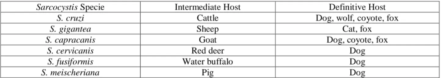
Table 1 - Examples of Sarcocystis species with respective Intermediate Host and Definitive Host (Yang et al., 2002; Uggla et al., 1990).
Sarcocystis Specie Intermediate Host Definitive Host
S. cruzi Cattle Dog, wolf, coyote, fox
S. gigantea Sheep Cat, fox
S. capracanis Goat Dog, coyote, fox
S. cervicanis Red deer Dog
S. fusiformis Water buffalo Dog
2.2.5.1. Red deer
The first report of Sarcocystis in a red deer (Cervus elaphus) was done by Hessling in 1854 (Drost et al., 1975, cited by Kutkienè, 2003). After that, major studies were performed in Europe and North America (Kutkienè, 2003) and eight species were described for red deer. In the following lines are described the eight species and its finding.
Previous to 1995 there were only thin-walled cysts described in red deer and the specie was named S. grueneri by Yakimoff and Sokoloff (1934) (Kutkienè, 2003). After, a study derived on the thin-walled cyst ultrastructure (Hernández-Rodríguez et al., 1981), this specie name changed to S. cervicanis (Kutkienè, 2003). Different studies derived in North America refer as S. waipiti to this specie (Dubey et al., 1989; Largerquist et al., 1993). In fact, there are some literatures whose authors describe S. gruneri, S. cervicanis and S. waipiti as single specie with low specificity to the intermediate host (Fayer et al., 1982; Matuschka, 1983; Balbo et al., 1988; Santini, 1997). Some of them even formulate the hypothesis that this type of cysts founded in cervids inhabiting all the Holarctic belong to the same species (Wesemeier et al., 1995 cited by Kutkienè, 2003). For these authors it was opportune to give this species the first name, S.
grueneri (Kutkienè, 2003).
Another Sarcocystis species reported in red deer was denominated as S. capreolicanis (Wesemeier et al., 1995 cited by Kutkienè, 2003). On the other hand, studies in North America refer to this species, which infect elk (Cervus canadensis), as S. sybillensis (Dubey et al., 1989; Largerquist et al., 1993).
Another species reported in literature was S. hofmanni (Wesemeier et al., 1995 cited by Kutkienè, 2003) also denominated as a Sarcocystis sp. similar to S. hofmanni (Stolte et al., 1996 cited by Dahlghren, 2009).
Several studies made in Norway described 5 more species found in red deer which were common to other cervids: S. hjorti (described for the first time and named in this study and also found in moose), S. tarandi (also in reindeer) S. rangiferi (also in reindeer), S. hardangeri (also in reindeer) and S. ovalis (also in moose) (Dalhgren, 2009).
Concerning the prevalence of Sarcocystis infection in red deer, the ones found in literature are resumed in the following table (Table 2).
Table 2 - Prevalence of Sarcocystis infection in red deer found in literature (in muscular samples).
Prevalence of Sarcocystis
infection (%) Country Reference
98 Hungary Kavai et al., 1976 cited by Goldová, 2008
63 Spain Navarrete et al, 1978 cited by Goldová, 2008
25 Czechoslovakia Lukesová et al., 1989 in Goldová, 2008
98.0 Germany Partenheimer-Hannemann, 199, cited by Goldová, 2008
90.2 Germany Spickschen et al., 2002 cite for Goldová, 2008
100 USA Largerquist et al., 1993 cited by William, 1995
94.3 Poland Tropilo et al., 2001 cited by Goldová, 2008
70.2 Lithuania Kutkiené, 2003
78.6 eastern Slovakia Goldová, 2008
50 eastern Slovakia Hvizdošová 2009
100 Norway Dalhgren, 2009
2.2.5.2. Sus scrofa
Referring to wild boar (sus scrofa), there are three known species that infect those animal specie: (1) S. miescheriana (synonym: Sarcocystis suicanis) (Kühn, 1865, cited by Tenter, 1995; Labbé, 1899, cited by Tenter, 1995; Erber, 1977 cited by Tenter, 1995), (2) S. porcifelis (Dubey, 1976 cited by Tenter, 1995) and (3) S. suihominis (Tadros et al., 1976 cited by Tenter, 1995; Heydorn, 1977 cited by Tenter, 1995) with final hosts being dog, cat and man, respectively.
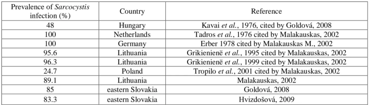
Concerning the prevalence of Sarcocystis infection in wild boar, the ones found in literature are resumed in the following table (Table 3).
Table 3 - Prevalence of Sarcocystis infection in wild boar found in literature.
Prevalence of Sarcocystis
infection (%) Country Reference
48 Hungary Kavai et al., 1976, cited by Goldová, 2008
100 Netherlands Tadros et al., 1976 cited by Malakauskas, 2002
100 Germany Erber 1978 cited by Malakauskas M., 2002
95.6 Lithuania Grikienienë et al., 1995 cited by Malakauskas, 2002
96.3 Lithuania Grikienienë et al., 1999 cited by Malakauskas, 2002
24.7 Poland Tropilo et al., 2001 cited by Malakauskas, 2002
89.1 Lithuania Malakauskas, 2002
85 eastern Slovakia Goldová, 2008
83.3 eastern Slovakia Hvizdošová, 2009
1.1.1. Sarcocystis as a Zoonotic Agent
In Sarcocystis world there are two known species that may involve humans as definite host (gastrointestinal sarcocystosis) in their life cycle: S. suihominis and S. hominis (Heydorn, 1977 cited by Mohammadi et al., 2006). Considering men as an intermediate host (Muscular
sarcocystosis) the number of species is not determined nor their name (Mohammadi et al., 2006; EFSA, 2010; Esposito, D. H, et al.2012).
1.1.1.1. Gastrointestinal Sarcocystosis and Prevention
Humans are a final host to S. suihominis and S. hominis. Generally, infections are self-limiting, of short duration, and often asymptomatic (Fayer, 2004), being, the main symptoms caused by these two species in man, gastrointestinal symptoms such as abdominal pain, nausea, vomiting and diarrhoea (Mohammadi et al., 2006). Anyway, it is referred in literature that S.
hominis may cause more stern symptoms like circulatory problems (tachycardia), drowsiness,
fatigue, anaemia and dyspnoea (Mohammadi et al., 2006; EFSA, 2010). S. hominis is also described in literature as the cause of human Hypereosinophilic Syndrome (HPS) as well as chronic diarrhoea (Nichpanit et al, 2010). Despite this vulgar form of infection in the definitive host, there are some recent studies reporting an alternative pathway. These studies distinguishes the vulgar non-invasive cycles from an invasive cycle, being S. fusiforms and S.meischeriana referred as possible species with the invasive cycle (Bunyaratvej et al., 2007). This alternative pathway may be the cause of an asexual phase in the definitive host, being a possible source of muscular sarcocystosis or chronic inflammation in the intestinal mucosa in man (Bunyaratvej et
al, 2007). So, as a recent report in Europe concludes, is necessary to make clear the impact in
public health of these Sarcocystis species to clarify it importance and the need of monitoring (EFSA, 2010). For that, additional studies are needed to clarify the Sarcocystis spp. cycles (Bunyaratvej et al, 2007).
Referring to prevention, avoiding the ingestion of cysts by the definitive host is the most effective way of prevent infection. When meat may be harbouring cysts, we should thoroughly frozen (-4ºC to -5ºC – 48h; 20ºC – 24h) or thoroughly cook (60ºC-20’; 70ºC-15’; 100ºC-5’) the meat to kill infectious bradyzoites (Fayer, 2004; Mohammadi et al., 2006). These measures will prevent the development of intestinal stages, where humans might serve as definitive hosts or host from erratic cycles (Fayer, 2004).
2004; EFSA, 2010). Southeast Asia is one of the most affected areas with human sarcocystosis (Bunyaratvej et al, 2007; Mohammadi et al., 2006; EFSA, 2010; Esposito, D. H, et al,. 2012), and a recent worldwide outbreak (Germany, France, Spain, Singapore, Belgium, Netherlands, Switzerland) of an acute muscular Sarcocystis-like illness affected 100 persons who had a recent travel to Tioman Islands in Malaysia. During the epidemiological study there were identified intramuscular cysts (Sarcocystis) in two of the involved persons in the outbreak (Esposito et al,. 2012). For that reason, the researchers believed that a Sarcocystis infection seems to be the most
likely cause of this outbreak (Esposito et al,. 2012). Before this outbreak, there were only about
92 cases of muscular sarcocystosis in humans reported worldwide (Fayer, 2004). In these cases humans are a dead end host (EFSA, 2010).
Referring to prevention, as it is hard to prevent and eliminate the animal’s infection, the most important to do is aware people to the health risk of each travel destination and the importance of appropriate hygiene and safe food and water consumption (Esposito et al,. 2012).
3.
Materials and Methods
3.1. Area of study
This survey was made in Idanha-a-Nova County which is located in southeast of central region of Portugal (1416.3 km2 and 10 147 habitants (AMNP, 2012) where occurs the transition of landscape from northern mountains to the south plateau of Portugal (Image 6).
Image 6 – Representative image of the two landscapes found in Idanha-a-Nova. A northen mountains. B South plateau.
Its limits are Penamacor County on north, Castelo Branco and Fundão Counties on west and Erges and Tejo Rivers on east and south (respectively), which corresponds to the Spain border (Extremadura). In the following image it is possible to observe the location of Idanha-a-Nova County in the Portugal map (Image 7).
Image 7 - Location of Idanha-a-Nova County in the Portugal map. (source: httpwww.hortasdidanha.ptsiteindex.phpas-terras, data: 12-03-2013, 11:10).
Idanha-a-Nova has a considerable rural area with approximately 50% of land dedicated to agriculture (43% dry agriculture, 9% watered agriculture, and 5% graze land), 30% are forested areas, mainly oaks, and 13% is shrub land and sparse vegetation. Domestic animal production (bovine and small ruminants, especially sheep), generally based on outdoor extensive production, has foremost significance in local economy.
Other important activity to local economy is hunting, emphasized by the continuous decreasing of the agriculture. In fact, this County is known as one of the best places to hunt in Portugal and large game hunting is the most significant part of it. Overall, there are 119816.39 ha of Hunting Areas (84.8% of the County) corresponding to 42 Association Hunting Areas with 51 702.65 ha, 31 Tourist Hunting Areas with 44 375.82 and 14 County Hunting Areas with 23 737.92 (DGAV, 2010a). Most of this land is shared by livestock and wild ungulates, which lead us to other important aspect to have in account; Idanha-a-Nova is in the Epidemiologic Risk Area for Bovine Tuberculosis (Image 8) in large game and has a high infection occurrence in wild boar and red deer (Santos et al., 2009; Vieira-Pinto et al, 2011; DGAV, 2010b; DGAV, 2010c). Additionally to the identification of the risk areas, in 2011, the Portuguese Veterinarian Authority published an internal law (edital n.º 1) describing mandatory rules that must be observed during each driving hunt organized in this Epidemiologic Risk Area for Bovine Tuberculosis. For example:
“…There must be a place to the evisceration of harvested animals…with proper conditions to carry out that task…”
There must be present a veterinary responsible by the initial examination of every harvested animal presented in the evisceration spot to come up with one of the following results:
Animals which present alterations that may suggest an health risk must go to byproducts or if the Hunting Area requests to a specific establishment for game meat preparation to a final decision been taken;
Animals which do not present alterations that may suggest a health risk go to self consumption or to a specific establishment for game meat preparation to be inspected and then placed in the market…”
Image 8 - Epidemiologic Risk Area for Bovine Tuberculosis map. (adapted from Edital nº1)
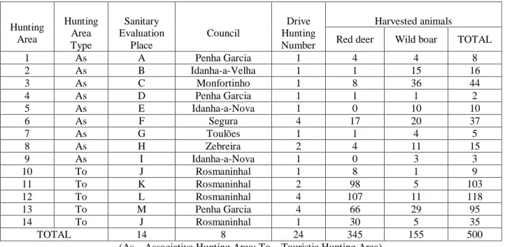
The present study was derived during the hunting season 2011-2012 from October to February, in 24 hunting campaigns (sampling plots) organized in 14 hunting areas from Idanha-a-Nova (Image 9, Table 4).
Table 4 – Number of hunting areas and correspondent type, sanitary evaluation place, council, number of driving hunts and harvested animals.
Hunting Area Hunting Area Type Sanitary Evaluation Place Council Drive Hunting Number Harvested animals
Red deer Wild boar TOTAL
1 As A Penha Garcia 1 4 4 8 2 As B Idanha-a-Velha 1 1 15 16 3 As C Monfortinho 1 8 36 44 4 As D Penha Garcia 1 1 1 2 5 As E Idanha-a-Nova 1 0 10 10 6 As F Segura 4 17 20 37 7 As G Toulões 1 1 4 5 8 As H Zebreira 2 4 11 15 9 As I Idanha-a-Nova 1 0 3 3 10 To J Rosmaninhal 1 8 1 9 11 To K Rosmaninhal 2 98 5 103 12 To L Rosmaninhal 4 107 11 118 13 To M Penha Garcia 4 66 29 95 14 To J Rosmaninhal 1 30 5 35 TOTAL 14 8 24 345 155 500
(As – Associative Hunting Area; To – Touristic Hunting Area)
3.2. Game Sanitary Evaluation
In each driven hunting action, the large game specimens hunted were collected and gathered in the evisceration place, where the veterinarian carried out the sanitary evaluation referred as initial examination of wild game on the spot. This procedure was based in the veterinary experience and in the Regulation (EC) 854/2004, which establish rules for the organization of official controls to animal origin products intended for human consumption. In brief, were pursued the following procedures:
1. questions to the hunters about abnormal behaviour;
2. carcass external analyses, that included evaluation of corporal conditions, wounds (shot, dog bite, others), secretions and joints palpation;
3. Carcass internal analyses, that included visual analyses of the carcass internal surface, looking for the generalized presence of tumours or abscesses, parasites (Image 10) and weird bodies not resulting from the hunting, incision of lymph nodes (ln.) (mandibular (Image 11A) ln. in wild boar and prescapular (Image 13B) and mesenteric ln. (Image
Image 10 – Image of parasitic larvae of oestrids in red deer.
Image 11 - Inspection of several tissues. A – Kidney incision. B – Heart incision. C - Spleen inspection.
Image 12 – Lymph nodes Inspection. A - mandibular lymph nodes incision. B - pre-escapular lymph nodes incision.
Image 13 - Lymph nodes Inspection. A - Mesenteric lymph nodes. incision. B - Inguinal lymph nodes incision.
According to the Edital Nº1, at the end of the procedure a sanitary decision was emitted as previously described. Then, the carcasses intended to commercial purpose were transported to a specific establishment for game meat preparation, where it was submitted to a sanitary inspection performed by the Official Veterinarian which included the Trichinella spp. test on wild boar. The carcasses intended for self consumption were taken by the hunters. In this context there were collected the data about the number of harvested animals and all the cases where meat was declared unfit for human consumption were registered.
3.3. Evaluation of game meat preparation conditions in the field
Additional to the number of harvested animals and the condemned cases declared unfit for human consumption, were collected data concerning the game meat preparation conditions in the field. This study was derived in 14 hunting areas in which were made 24 drives hunting. Due to the physical (geographical) difference between hunting areas and evisceration places (image 15), both were defined with specific assignments as presented in Table 5. In this topic was mainly gathered information related to evisceration, sanitary evaluation, elimination of animal by-products and derived products not intended for human consumption and carcass transportation. These data were grouped into three main domains:
Table 5 - Hunting areas and correspondent assignment.
Hunting Area 1 2 3 4 5 6 7 8 9 10 11 12 13 14
Corresponding Evisceration Place A B C D E F G H I J K L M J
In order to objectively rank each site/procedure used for game meat preparation in the field it was created a table with parameters to analyse and the punctuation for each analysed parameter, as it is described in the following tables.
Table 6 - Classification of the structural requirements parameters present during game meat preparation in the field (maximum punctuation: 12).
Parameter Classification
Water
0 - Absent
1 - Present – Insufficient Quantity or Availability
2 - Present in Suitable Conditions
Light
0 - Absent
1 - Present – Insufficient
2 - Present in Suitable Conditions
Pest Control 0 - Absent
1 - Present
Dogs Access 0 - Uncontrolled
1 - Controlled(1)
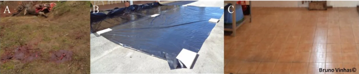
Floor (Image 14)(2)
0 - Soil
1 - Plastic Protection
2 - Floor (Tile or Concrete)
Cleaning/ Disinfection Devices Personal 0 - Absent 1 - Present Knives 0 - Absent 1 - Present Infrastructures 0 - Absent 1 - Present
Proper Drainage Runoff 0 - Without Drainage
1 - With Drainage
(1) It was considered controlled when sanitary evaluation place had a physical barrier; (2) Floor where the game meat sanitary evaluation was made.