University of Trás-os-Montes and Alto Douro
Advances in Canine Mammary Cancer: A Role
for Inflammatory Infiltrate in Tumor
Microenvironment
PhD Thesis in Veterinary Sciences
Maria Isabel da Silva Carvalho
Supervisor: Prof. Dr. Felisbina Luísa Pereira Guedes Queiroga Prof. Dr. Isabel Cristina Ribeiro Pires
University of Trás-os-Montes and Alto Douro
Advances in Canine Mammary Cancer: A Role
for Inflammatory Infiltrate in Tumor
Microenvironment
PhD Thesis in Veterinary Sciences
Supervisor: Prof. Dr. Felisbina Luísa Pereira Guedes Queiroga Prof. Dr. Isabel Cristina Ribeiro Pires
Jury composition:
Prof. Dr. Manuel João Rua Vilanova
Prof. Dr. Maria de Fátima Rodrigues Moutinho Gartner Prof. Dr. Maria de Fátima Monginho Baltazar
Prof. Dr. Maria dos Anjos Clemente Pires Prof. Dr. Adelina Maria Gaspar Gama Quaresma Prof. Dr. Felisbina Luísa Pereira Guedes Queiroga
Vila Real, 2017
V
I declare for all due purposes that the PhD thesis meets the technical and scientific standards required by the regulations of the University of Trás-os-Montes and Alto Douro. The presented doctrines are the exclusive responsibility of the author.
VII
This thesis was specifically prepared to obtain the PhD degree in Veterinary Sciences.
IX
Financial support provided by FCT (“Fundação para a Ciência e a Tecnologia”) and POPH-QREN/FSE (“Programa Operacional Potencial Humano-Quadro de Referência Estratégico Nacional/Fundo Social Europeu”), PhD Grant SFRH/BD/78771/2011.
XI
"Always do your best. What you plant now, you will harvest later."
XIII
To my parents for the dedication, To my brother for the complicity and friendship.
XV
Agradecimentos
Ao longo da realização deste trabalho muitas foram as pessoas que contribuíram com a sua amizade, compreensão e sabedoria. Assim sendo, a todos os que me acompanharam nesta jornada, quero aqui expressar o meu sincero reconhecimento:
À Universidade de Trás-os-Montes e Alto Douro (UTAD), na pessoa do senhor Reitor, Professor Doutor António Augusto Fontainhas Fernandes, pela disponibilização dos meios físicos para a realização desta tese.
À Professora Doutora Felisbina Luísa Pereira Guedes Queiroga, minha orientadora científica, por toda a dedicação e empenho que sempre teve comigo durante a elaboração deste trabalho. Quero agradecer, não só todas as oportunidades de aprendizagem e partilha de conhecimentos que me proporcionou, mas sobretudo todas as palavras de encorajamento e apoio. Obrigada por me mostrar, em todos os momentos, que mais importante que “dar o peixe é ensinar a pescar”. Obrigada pela amizade. Obrigada por tudo!
À Professora Doutora Isabel Cristina Ribeiro Pires, co-orientadora desta tese, pela ajuda imprescindível, pela mestria e amizade com que sempre me auxiliou durante todas as etapas deste trabalho. Pelas preciosas palavras de incentivo e, não menos importante, por todos os momentos de boa disposição.
À Professora Doutora Justina Maria Prada Oliveira por toda a disponibilidade, amabilidade e apoio prestados. Os seus conhecimentos e experiência foram, sem dúvida, um contributo essencial.
À Professora Doutora Maria dos Anjos Clemente Pires, enquanto responsável do Laboratório de Histologia e Anatomia Patológica da UTAD, por ter disponibilizado todos os meios solicitados.
XVI
A todos os funcionários do Laboratório de Histologia e Anatomia Patológica da UTAD pela simpatia e estímulo com que sempre me receberam. À Sra. Dona Lígia Lourenço, agradeço, adicionalmente, o excelente apoio técnico.
Às companheiras de laboratório Teresa Raposo e Helena Rodrigues gostaria de agradecer pela amizade e carinho em todos os momentos.
Ao Miguel Ribeiro e à Daniela Ferreira, pela amizade incondicional, por todo o apoio e cumplicidade nos bons e maus momentos. Obrigada por estarem sempre presentes.
À Anabela Garcia e à Susana Ferreira, amigas de todas as horas, pela amizade verdadeira, companheirismo, apoio e compreensão.
Aos meus pais e a toda a minha família agradeço o incentivo, os conselhos e principalmente todo o amor e carinho, sentimentos essenciais que acalentam o meu coração todos os dias.
Ao meu irmão, Ricardo Carvalho, por todo o amor e compreensão, por estar sempre ao meu lado em tudo e, essencialmente, pelo enorme coração que tem. Espero que sintas por mim o mesmo orgulho que eu sinto por ti!
Ao Duarte, porque "no teu colo cabem todos os meus medos", porque "até o pior da vida se acalma quando estou nos teus olhos".
Seria injusto não agradecer, mas mais injusto seria agradecer só a alguns, neste sentido, agradeço também a todos aqueles que, de uma forma direta ou indireta, me incentivaram e ajudaram no decorrer deste trabalho.
XVII
Acknowledgments
I am grateful to Professor Erika Jensen-Jarolim, head of Comparative Medicine at the interdisciplinary Messerli Research Institute, Vienna, which received me in her laboratory and allowed me to learn with all amazing investigators. I would like to thank also all the advices, share of knowledge and unfailing support.
I would like to thank Dr. Rodolfo Bianchini for being a mentor throughout my project in Vienna. Thank you so much all the scientific support, useful advices and constructive comments on my work.
I would like to thank the research technician Gerlinde Hofstetter, for the excellent scientific support at the laboratory in the Messerli Institute, her helpfulness and valuable experience.
I would like to thank Jelena Gotovina, Ina Hermann, Judit Fazekas, Stefanie Wagner, Stefan Gunther, Lukas Einhorn, Karin Hufnagl and all the other staff members at the laboratory of Prof. Erika Jensen-Jarolim for their kindness, friendship and spirit of cooperation.
XIX
Abstract
Cancer related inflammation is part of the major cancer hallmarks and has an important role in mammary carcinogenesis being involved in tumor aggressiveness and poor clinical outcome. In dogs only few studies have yet concentrated on the influence of immune cells in clinical outcome of dog mammary tumor patients. In this context, the present work was conducted with the main aim of up-to-date knowledge about the roles of the intertwined signaling pathways shared by T-lymphocytic/macrophage infiltrates and important tissue biomarkers in canine mammary tumors (CMT) progression, aggression and prognosis. Immunosuppression associated with tumor infiltrating T-lymphocytes (TILs) has been explained by the compartment of regulatory T-cells (Treg) that inhibit anti-tumor cytotoxic activities. Based on these evidences, first, we assessed, by immunohistochemistry, the characterization of intratumoral Treg cells, as well as some cytokines related by them: TGFβ and IL-35. Our results demonstrated that FoxP3 was present in tumors with more aggressive phenotypes: high histological grade of malignancy (HGM), presence of neoplastic intravascular emboli and presence of lymph node metastasis. Additionally, also showed that intratumoral FoxP3+ Treg cells were
associated with shorter overall survival (OS), both in univariate and multivariate analysis. Tregs can secrete inhibitory cytokines (TGFβ and IL-35), inducing tumors tolerance. Present work suggests a link between TGFβ and parameters of tumor aggressiveness, reflecting its involvement in CMT malignant transformation. Our data demonstrated also a positive correlation between intratumoral FoxP3, TGFβ levels, VEGF and CD31. Moreover tumors with abundant TGFβ and with concurrent high expression of TGFβ/FoxP3, FoxP3/VEGF, TGFβ/VEGF were associated with shorter OS time and the TGFβ/FoxP3 tumors class retained the association with worse survival in multivariate analysis, arising as an independent predictor of poor prognosis. IL-35 is a Treg cell-secreted cytokine that inhibits T-cells proliferation and function and to the best of our knowledge, this is the first study that investigates the role of IL-35 in CMT development and clinical outcome. Our results showed that IL-35 overexpression was significantly associated with advancement of tumor stage and unfavorable prognosis by univariate and multivariate survival analysis. Interestingly our findings indicate, for the first time in dog mammary tumors, the independent prognostic value of FoxP3+ Treg cells and
XX
TGFβ/FoxP3 tumors class. Additionally the IL-35 seems to be a new biomarker to predict the CMT clinical outcome.
For a more comprehensive approach about the role of immune cells in dog mammary carcinogenesis, we perform a wide study focused on the intertwined signaling pathways shared by T-lymphocytic/macrophage infiltrates and important tissue biomarkers in mammary tumor microenvironment. We focused on relevant cell biomarkers in mammary carcinogenesis, the COX-2, EGFR and c-kit which are often overexpressed or mutated in this type of tumor. Present data demonstrated a significant association of high COX-2 immunoexpression with CD3+ T-lymphocytes and MAC387 macrophages. Tumors with concurrent high COX-2/CD3 and high COX-2/MAC expression were associated with variables of tumor aggressiveness and shorter OS. Current results also demonstrated that the concurrent COX-2+/EGFR+ expression was associated with higher numbers of intratumoral CD3+ T-cells. Furthermore a significant association and a positive correlation between CD3+ T-lymphocytes and c-kit expression were observed. Tumors with high c-kit expression showed higher counts of CD3+ T-cells and were associated with high HGM, presence of neoplastic intravascular emboli, presence of lymph node metastasis, angiogenesis and shorter OS. The findings indicate that COX-2, EGFR and c-kit pathways are important not only for the remodeling of mammary tumor microenvironment but also could be a very important targets for tumor immunological therapy.
Finally, in an in vitro co-culturing experiment, was our aim to prove the potential bidirectional crosstalk between tumor cells and immune cells on COX-2 regulation. Our data showed that co-culturing of canine mammary carcinoma cell line Sh1b and canine peripheral blood mononuclear cells (PBMCs) induced a trend of COX-2 overexpression in mammary cancer cells. In turn, COX-2 expression by PBMCs, among them predominantly CD68+ macrophages, was significantly attenuated by co-culture with Sh1b. In accordance, co-culture with CD68+ differentiated THP1 (dTHP1) prompted an intracellular production of COX-2 in Sh1b cells. The intracellular COX-2 expression from dTHP1 decreased when they were treated with conditioned medium from cultured Sh1b cells. Present results represents a significant advance on understanding the possible role of COX-2 in inducing a cancer tolerogenic microenvironment in CMT namely through a cancer associated macrophages immunomodulation.
XXI
Overall present work demonstrated that, similarly to human breast cancer, also in CMT the inflammatory responses in mammary cancer sites are able to orchestrate hallmark-facilitating programs in tumor microenvironment.
Keywords: Canine mammary tumors; Macrophages; Prognosis; T-lymphocytes; Treg cells; Tumor microenvironment.
XXIII
Resumo
A inflamação faz parte dos principais “hallmarks” associados ao cancro e tem um papel relevante na carcinogénese mamária, relacionando-se com a agressividade tumoral e pior prognóstico. Nos tumores mamários dos canídeos (TMC) os estudos que se debruçam sobre o efeito das células imunitárias no prognóstico são escassos. Neste contexto, o presente trabalho foi realizado com o objetivo de fomentar o conhecimento sobre o papel das vias de sinalização compartilhadas pelo infiltrado de linfócitos T/macrófagos e importantes biomarcadores tumorais na progressão, agressividade e prognóstico destes tumores.
A imunossupressão associada ao infiltrado de linfócitos T no tumor tem sido explicada pela capacidade inibitória das células T reguladoras (Treg) que impedem as atividades citotóxicas anti-tumorais. Com base nestas evidências, caracterizámos por imunohistoquímica as células Treg intratumorais (FoxP3+), bem como algumas citoquinas relacionadas com elas: TGFβ e IL-35. Os nossos resultados demonstraram que a maior imunorreactividade para o FoxP3 está presente em tumores com fenótipos mais agressivos: elevado grau histológico de malignidade (GHM), presença de embolos intravasculares e metástases nos linfonodos. Adicionalmente, mostrámos que as células Treg FoxP3+ intratumorais estão associadas com uma menor sobrevida total (ST), por
análise univariada e multivariada. As células Treg secretam citoquinas inibidoras (TGFβ e IL-35), induzindo a tolerância aos tumores. Os nossos resultados sugerem uma associação entre o TGFβ e parâmetros de agressividade tumoral, refletindo o seu envolvimento na transformação maligna. Demonstraram também uma correlação positiva entre o FoxP3 intratumoral, os níveis de TGFβ, VEGF e CD31. Além disso, tumores com abundante TGFβ e com elevada expressão simultânea de TGFβ/FoxP3, FoxP3/VEGF, TGFβ/VEGF foram associados ao menor tempo de ST e a classe de tumoresTGFβ/FoxP3 elevados revelou-se como um fator independente de mau prognóstico na análise multivariada. A IL-35 é uma citoquina secretada pelas células Treg que inibe a proliferação e função das células T. Demonstrámos que a sobre-expressão da IL-35 está associada com o estádio clínico mais avançado e com um prognóstico desfavorável na análise univariada e multivariada. Foi possível constatar, pela 1ª vez nos TMC, o valor prognóstico independente das células FoxP3+ Treg e da classe de tumores TGFβ/FoxP3
XXIV
elevados. A IL-35 evidenciou-se ainda como um novo biomarcador capaz de prever a evolução clínica dos TMC.
No seguimento deste trabalho, realizámos um amplo estudo que incidiu sobre as vias de sinalização compartilhadas pelo infiltrado de linfócitos T/macrófagos e importantes biomarcadores tumorais no microambiente dos TMC. Com esse propósito estudámos biomarcadores celulares importantes na carcinogénese mamária (COX-2, EGFR e c-kit), que estão frequentemente sobre-expressos ou mutados nos TMC. Constatámos uma associação significativa da imunoexpressão elevada da COX-2 com os linfócitos T CD3+ e macrófagos MAC387. Tumores com elevada expressão de COX-2/CD3 e COX-2/MAC estão associados com variáveis de agressividade tumoral e menor ST. Demonstrámos ainda que a expressão simultânea de COX-2+/EGFR+ está associada com maior número de células T CD3+ intratumorais e foi observada uma associação significativa e uma correlação positiva entre os linfócitos T CD3+ e a expressão de c-kit. Tumores com alta expressão de c-kit apresentaram maior número de células T CD3+ estando associados a um GHM elevado, presença de êmbolos intravasculares, metástases nos linfonodos e menor ST. Os resultados demonstram que as vias de sinalização que envolvem a COX-2, o EGFR e o c-kit são importantes, não só para a remodelação do microambiente do tumor mamário, mas também como alvos para a terapia imunológica anti-tumoral.
Para complementar o trabalho, foi realizada uma experiência de co-cultura in vitro onde se pretendia demonstrar a potencial participação bidirecional das células tumorais e células imunitárias na regulação da COX-2. Os resultados mostraram que a co-cultura entre a linha celular de carcinoma mamário de cão Sh1b e as células mononucleares de sangue periférico de cão (PBMCs) induziram uma tendência para a sobre-expressão de COX-2 nas células cancerígenas. Por sua vez, a expressão de COX-2 pelos PBMCs, entre eles predominantemente macrófagos CD68+, foi significativamente atenuada pela co-cultura com Sh1b. Em conformidade, a co-co-cultura com CD68+ THP1 diferenciada (dTHP1) provocou um aumento da produção intracelular de COX-2 pelas células Sh1b. A expressão intracelular de COX-2 pela dTHP1 diminuiu quando estas células foram tratadas com meio condicionado das células Sh1b. Estes resultados representam um avanço significativo na compreensão do papel da COX-2 na remodelação do microambiente tumoral nos TMC, nomeadamente através da imunomodulação dos macrófagos associados ao tumor.
XXV
Globalmente, o presente trabalho demonstrou que, à semelhança do cancro de mama da mulher, também nos TMC as células inflamatórias no microambiente do tumor são capazes de orquestrar programas facilitadores da progressão tumoral.
Palavras-chave: Tumores mamários dos canídeos; Macrófagos; Prognóstico; Linfócitos T; células Treg; Microambiente tumoral.
XXVII
Thesis Outline
Cancer related inflammation is part of the major cancer hallmarks and has an important role in mammary carcinogenesis being involved in tumor aggressiveness and poor clinical outcome. In dogs only few studies have yet concentrated on the influence of immune cells in clinical outcome of dog mammary tumor patients. In this context, the present work was conducted with the main aim of up-to-date knowledge about the roles of the intertwined signaling pathways shared by T-lymphocytic/macrophage infiltrates and important tissue biomarkers in canine mammary tumors (CMT) progression, aggression and prognosis.
The present thesis was divided into five chapters.The main motivation that conducted this work was the need to better understanding the influence of immune cells in dog mammary carcinogenesis and to establish new prognostic factors and molecular therapeutic targets that could be of value in the treatment of CMT.
Chapter I is a general introduction on the state of the art about CMT pathophysiology. Additionally it was describes the intertwined signaling pathways shared by T-lymphocytic/macrophage infiltrates and important tissue biomarkers in both human and dog mammary carcinogenesis.
Chapter II encompass original data focusing on the characterization of intratumoral FoxP3 regulatory T-lymphocytes, TGFβ and IL-35 immunoexpression and attempt to clarify the role of these molecular markers in CMT aggressiveness and prognosis. Chapter III includes original data assessing the relationship between T-lymphocytic/macrophage infiltrate and emergent molecular targets. There is a growing list of signaling molecules released by inflammatory cells that serve as effectors of their tumor-promoting actions. This chapter emphasizes the important association of intratumoral T-lymphocytes/macrophages with COX-2, EGFR and c-kit that seems to contribute to the amplification of the tumoral inflammatory state related with cancer progression and worse clinical outcome.
Chapter IV focuses on the determination, in an in vitro co-culturing study, of the bidirectional COX-2 regulation between cancer cells and monocytes/macrophages in CMT. This original data represent a significant advance on understanding the possible
XXVIII
role of COX-2 in inducing a cancer tolerogenic microenvironment namely through cancer associated macrophages immunomodulation.
XXIX
Publications and Communications
The author of this thesis declares to have actively participated in the elaboration and execution of experimental work that led to the results presented, whichoriginated several publications in international scientific journals with Referee, as well as oral and poster communications in national and international meetings.
Publications in international scientific journals with Referee
Maria Isabel Carvalho, Isabel Pires, Justina Prada, Felisbina Luísa Queiroga (2014). A Role for T-Lymphocytes in Human Breast Cancer and in Canine Mammary Tumors. BioMed Research International. doi:
10.1155/2014/130894
Maria Isabel Carvalho, Isabel Pires, Justina Prada, Adriano Fernandes Ferreira, Felisbina Luísa Queiroga (2015). Positive Interplay between CD3+
T-lymphocytes and Concurrent COX-2/EGFR expression in Canine Malignant Mammary Tumors. Anticancer Research. 35(5): 2915-2920. doi: 10.4142/jvs.2015.16.2.225
Maria Isabel Carvalho, Isabel Pires, Marlene Dias, Justina Prada, Hugo Gregório, Luís Lobo, Felisbina Luísa Queiroga (2015). Intratumoral CD3+ T-lymphocytes
immunoexpression and its association with c-kit, Angiogenesis, and Overall Survival in Malignant Canine Mammary Tumors. Analytical Cellular Pathology. Article ID 920409. doi: 10.1155/2015/920409
Maria Isabel Carvalho, Isabel Pires, Justina Prada, Teresa Raposo, Hugo Gregório, Luís Lobo, Felisbina Luísa Queiroga (2016). High COX-2 expression is associated with increased Angiogenesis, Proliferation and Tumoral Inflammatory Infiltrate in Canine Malignant Mammary Tumors: a Multivariate Survival Study. Veterinary and Comparative Oncology. doi: 10.1111/vco.12206.
Maria Isabel Carvalho, Isabel Pires, Justina Prada; Hugo Gregório; Luís Lobo; Felisbina Luísa Queiroga (2016). Intratumoral FoxP3 expression is associated with Angiogenesis and Prognosis in Malignant Canine Mammary Tumors.
XXX
Veterinary Immunology and Immunopathology. 178: 1-9. doi:
10.1016/j.vetimm.2016.06.006
Maria Isabel Carvalho, Ricardo Silva-Carvalho, Isabel Pires, Justina Prada, Rodolfo Bianchini, Erika Jensen-Jarolim, Felisbina Luísa Queiroga (2016). A Comparative Approach of Tumor Associated Inflammation in Mammary Cancer between Humans and Dogs. BioMed Research International. doi: 10.1155/2016/4917387
Maria Isabel Carvalho, Isabel Pires, Justina Prada, Carla Pinto, Hugo Gregório, Luís Lobo, Felisbina Luísa Queiroga. Crosstalk between TGFβ, FoxP3 expression and Angiogenesis in Malignant Canine Mammary Tumors: association with Clinicopathological Parameters and Prognosis. Submitted manuscript
Maria Isabel Carvalho, Rodolfo Bianchini, Judit Fazekas, Ina Herrmann, Irene Flickinger, Johann G. Thalhammer, Isabel Pires, Erika Jensen-Jarolim, Felisbina Luísa Queiroga. Bidirectional Regulation of COX-2 expression between Cancer Cells and Macrophages. Submitted manuscript
Maria Isabel Carvalho, Isabel Pires, Justina Prada, Hugo Gregório, Carla Pinto, Felisbina Luísa Queiroga. Assessing the Interleukin 35 immunoexpression in Malignant Canine Mammary Tumors: association with Clinicopathological Parameters and Prognosis. Submitted manuscript
Book chapters
Maria Isabel Carvalho, Teresa Raposo, Helena Rodrigues, Isabel Pires, Justina Prada, Felisbina L. Queiroga (2014). Neoplastic Diseases of the Canine Mammary Gland. In Book: Mammary Glands: Anatomy, Development and Diseases, Edmund B. Rucker (Ed), Nova Science Publishers, Inc, NY. ISBN: 978-1-62948-856-1 eBook
Oral communications
Maria Isabel Carvalho, Isabel Pires, Justina Prada, Felisbina Luísa Queiroga. The interplay between CD3+ T-lymphocytes and Concurrent COX-2/EGFR
XXXI
expression in Canine Malignant Mammary Tumors. 9th International Conference of Anticancer Research, 6-10 October, 2014, Sithonia, Greece.
Poster communications
Maria Isabel Carvalho, Isabel Pires, Justina Prada, Teresa Raposo, Helena Rodrigues, Felisbina Luísa Queiroga. High COX-2 expression is associated with increased Angiogenesis, Proliferation and Tumoral Inflammatory Infiltrate in Canine Mammary Tumors. European Society of Veterinary Oncology Congress, 22-24 May, 2014, Vienna, Austria.
Maria Isabel Carvalho, Marlene Dias, Isabel Pires, Justina Prada, Felisbina Luísa Queiroga. Intratumoral CD3+ T-lymphocytes immunoexpression and its
association with c-kit and Angiogenesis in Malignant Canine Mammary Tumors: a Multivariate Survival Analysis. European College of Veterinary Internal Medicine-Companion Animals, ECVIM-CA Congress, 10-12 September, 2015, Lisbon, Portugal.
Maria Isabel Carvalho, Isabel Pires, Justina Prada, Felisbina Luísa Queiroga. Intratumoral FoxP3 expression in Malignant Canine Mammary Tumors: its association with Clinicopathological Parameters, Angiogenesis and Prognosis. European College of Veterinary Internal Medicine-Companion Animals, ECVIM-CA Congress, 10-12 September, 2015, Lisbon, Portugal.
Awarded works
ESVONC Best Poster Communication
Maria Isabel Carvalho, Isabel Pires, Justina Prada, Felisbina Luísa Queiroga. Intratumoral FoxP3 expression in Malignant Canine Mammary Tumors: its association with Clinicopathological Parameters, Angiogenesis and Prognosis. European College of Veterinary Internal Medicine-Companion Animals, ECVIM-CA Congress, 10-12 September, 2015, Lisbon, Portugal.
XXXIII
Table of Contents
Agradecimentos ... XV Acknowledgments ... XVII Abstract ... XIX Resumo ... XXIII Thesis Outline ... XXVII Publications and Communications ... XXIX Table of Contents ... XXXIII List of Figures and Tables ... XLI List of Abbreviations ... XLVII Chapter I - General Introduction ... 1 I.1 Neoplastic Diseases of the Canine Mammary Gland ... 3 Mammary Gland Development ... 3 Mammary Gland Anatomy and Blood Supply ... 3 Lymph Drainage of the Mammary Gland ... 4 Canine Mammary Tumors (CMT) ... 5 A General Approach ... 5 Predisposing Factors ... 5 Aetiology ... 6 Diagnosis and Clinical Management ... 8 Histological Classification of Canine Mammary Tumors: World Health Organization Guidelines ... 10 Prognostic Factors ... 12 Treatment ... 13 References ... 15 I.2 A Role for T-lymphocytes in Human Breast Cancer and in Canine Mammary Tumors ... 25XXXIV
Chronic Inflammation and Cancer ... 25 Adaptive Immunity and Cancer Development: A Role for T-Lymphocytes ... 26 T-Lymphocytes and Human Breast Cancer: Friends or Foes? ... 28 T-Lymphocyte Infiltrate in Canine Mammary Tumors ... 29 References ... 38 I.3 A Comparative Approach of Tumor Associated Inflammation in Mammary Cancer between Humans and Dogs ... 45
Inflammatory Cells and Sustained Angiogenesis... 47 Inflammatory Cells, Tissue Invasion and Metastasis ... 49 Inflammatory Cells and Cancer Cell Biomarkers ... 51 Inflammatory infiltrates and tumor COX-2 expression ... 51 Inflammatory infiltrates and receptor tyrosine kinases (RTKs) ... 54 Concluding remarks ... 56 References ... 59 Chapter II – The Role of FoxP3 Regulatory T-Lymphocytes, TGFβ and IL-35 in Canine Mammary Carcinogenesis ... 71 II.1 Intratumoral FoxP3 expression is associated with Angiogenesis and Prognosis in Malignant Canine Mammary Tumors ... 73
Abstract ... 73 Introduction ... 75 Materials and Methods ... 76 Study population and sample collection ... 76 Histopathological examination ... 77 Immunohistochemistry ... 77 FoxP3, CD31 and VEGF staining evaluation ... 78 Follow-up study ... 78 Statistical analysis ... 79 Results ... 79
XXXV
Clinicopathological data ... 79 Expression of FoxP3, CD31 and VEGF in malignant CMT ... 79 Associations of FoxP3, CD31 and VEGF immunostaining with clinicopathological features ... 80 Correlation between FoxP3 and CD31 immunoexpression ... 80 Association of intratumoral FoxP3 and MVD with levels of VEGF expression in malignant CMT ... 80 FoxP3, CD31 and VEGF associations with OS ... 84 Discussion ... 86 Conclusion ... 88 References ... 88 II.2 Crosstalk between TGFβ, FoxP3 and Angiogenesis in Malignant Canine Mammary Tumors: association with Clinicopathological Parameters and Prognosis 95
Abstract ... 95 Introduction ... 97 Materials and Methods ... 98 Patient selection, sample collection, clinicopathological characteristics, treatment and follow up ... 98 Histopathological examination ... 99 Immunohistochemistry ... 99 TGFβ, FoxP3, VEGF and CD31 staining evaluation ... 100 Statistical analysis ... 100 Results ... 101 Clinicopathological data ... 101 Expression of TGFβ, FoxP3, VEGF and CD31 in malignant CMT ... 101 Associations of TGFβ immunostaining with clinicopathological features ... 102 Correlation between TGFβ, FoxP3, VEGF and CD31 immunoexpression ... 102
XXXVI
Association of TGFβ/VEGF class with intratumoral FoxP3 and MVD in malignant CMT ... 102 Relationship of TGFβ/FoxP3, TGFβ/VEGF and TGFβ/CD31 classes with clinicopathological variables of tumor aggressiveness ... 104 Follow-up study ... 105 Discussion ... 105 Conclusion ... 109 References ... 112 II.3 Assessing the Interleukin 35 immunoexpression in Malignant Canine Mammary Tumors: association with Clinicopathological Parameters and Prognosis ... 117
Abstract ... 117 Introduction ... 119 Materials and Methods ... 120 Patient selection and tissue sample collection ... 120 Histopathological examination ... 120 Immunohistochemistry ... 121 IL-35 staining evaluation ... 121 Follow-up study ... 121 Statistical analysis ... 122 Results ... 122 Clinicopathological data ... 122 Expression of IL-35 in malignant CMT ... 123 Associations of IL-35 immunostaining with clinicopathological variables ... 124 Follow-up study ... 124 Discussion ... 125 Conclusion ... 127 References ... 130
XXXVII
Chapter III – Relationship of T-Lymphocytes and Macrophages with Emergent Molecular Targets ...... 133
III.1 High COX-2 expression is associated with increased Angiogenesis, Proliferation and Tumoral Inflammatory Infiltrate in Canine Malignant Mammary Tumors: a Multivariate Survival Study ... 135 Abstract ... 135 Introduction ... 137 Materials and Methods ... 138 Tissue samples ... 138 Immunohistochemical analysis ... 138 Quantification of immunoreactivity ... 139 Follow-up data ... 140 Statistical analysis ... 140 Results ... 141 Tumors ... 141 COX-2; CD31; VEGF; Ki-67; CD3+ T-lymphocytes and MAC387 macrophages immunostaining ... 141 Associations between COX-2; CD31; VEGF; Ki-67; CD3+ T-lymphocytes and MAC387 macrophages ... 142 Associations of clinicopathological features with COX-2, CD31, VEGF, Ki-67, CD3+ T-lymphocytes and MAC387 macrophages immunostaining ... 142
Relationship of COX-2/CD31; COX-2/VEGF; COX-2/Ki-67; COX-2/CD3 and COX-2/MAC groups with clinicopathological variables of tumor aggressiveness ... 143 Follow-up study ... 143 Discussion ... 146 Conclusion ... 149 References ... 155
XXXVIII
III.2 Positive Interplay between CD3+ T-lymphocytes and Concurrent COX-2/EGFR
expression in Canine Malignant Mammary Tumors ... 159 Abstract ... 159 Introduction ... 161 Materials and Methods ... 162 Tissue samples ... 162 Immunohistochemical analysis ... 162 Quantification of immunolabelling ... 163 Statistical analysis ... 163 Results ... 164 Tumors ... 164 CD3+ T-lymphocytes, COX-2 and EGFR immunostaining ... 164 Relationship of CD3+ T-lymphocytes with clinicopathological variables ... 164 Correlation between COX-2 and EGFR and relationship of COX-2/EGFR groups with clinicopathological variables. ... 166 CD3+ T-lymphocytes and COX-2/EGFR groups associations ... 166 Correlation between CD3+ T-lymphocytes and COX-2/EGFR groups ... 167
Discussion ... 168 Conclusion ... 169 References ... 170 III.3 Intratumoral CD3+ T-lymphocytes immunoexpression and its association with
c-kit, Angiogenesis and Overall Survival in Malignant Canine Mammary Tumors ... 175 Abstract ... 175 Introduction ... 177 Materials and Methods ... 178 Mammary tumors and clinicopathological variables ... 178 Immunohistochemical technique ... 178 Evaluation of immunoreactivity ... 179
XXXIX
Clinical follow-up ... 179 Statistical analysis ... 179 Results ... 180 Expression of CD3+ T-lymphocytes, c-kit, VEGF and CD31 in CMT ... 180 CD3+ T-lymphocytes, c-kit, VEGF and CD31 correlations ... 182 CD3+ T-lymphocytes, c-kit, CD31 and VEGF expression associations ... 182 Relationship of CD3/c-kit, CD3/VEGF and c-kit/VEGF with clinicopathological variables of tumor aggressiveness ... 183 Comparison between CD3/c-kit, CD3/VEGF, c-kit/VEGF and OS ... 184 Discussion ... 184 Conclusion ... 187 References ... 190 Chapter IV – The Potential Crosstalk between Tumor Cells and Immune Cells on COX-2 Regulation: an in vitro Co-Culturing Experiment ... 195 IV.1 Bidirectional Regulation of COX-2 expression between Cancer Cells and Macrophages ... 197
Abstract ... 197 Introduction ... 199 Material and Methods... 200 Cell culture ... 200 Collection of tumor cell line cultured supernatants ... 200 THP1 differentiation in response to PMA and treatment with cancer cell cultured supernatants ... 200 Isolation of dog peripheral blood mononuclear cells (PBMCs) ... 201 Co-culture experimental conditions ... 201 Assessment of CD68, CD3 and intracellular COX-2 by Flow Cytometry ... 201 Statistical analysis ... 202 Results ... 202
XL
Co-culturing canine cancer Sh1b and canine PBMCs from tumor patients: effects on COX-2 ... 202 Co-culturing cancer cell lines and human macrophages dTHP1: effects on COX-2 ... 203 Effects of cancer cell supernatants on intracellular COX-2 expression by dTHP1 ... 204 Discussion ... 205 Conclusion ... 207 References ... 207 Chapter V - General Discussion and Concluding Remarks ... 211 References ... 218
XLI
List of Figures and Tables
Chapter I
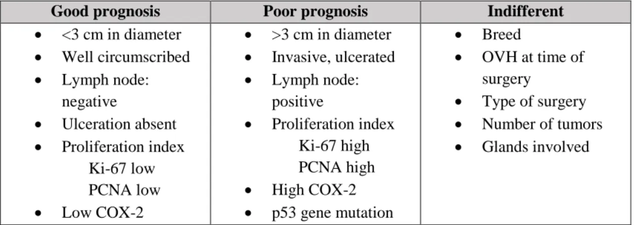
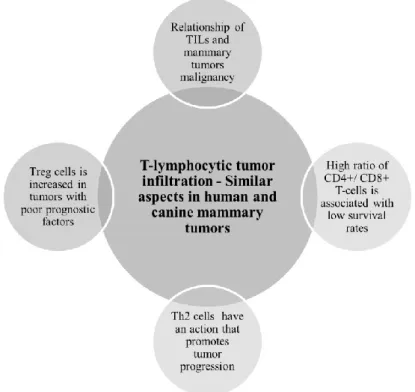
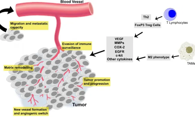
Figure 1 - Prognosis of malignant CMT. ... 11 Figure 2 - Similarities between human breast cancer and canine mammary tumors regarding tumor T-lymphocyte infiltration... 32 Figure 3 - Cancer related inflammation has an important role in mammary carcinogenesis, contributing to tumor evasion of immune surveillance, matrix remodeling and angiogenic switch, acquisition of metastatic capacity and tumor proliferation and progression. ... 46
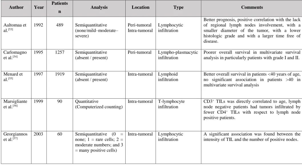
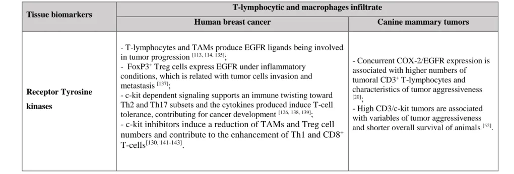
Table 1 - Lymphatic drainage of normal and neoplastic canine mammary gland. ... 4 Table 2 - Canine mammary tumor staging - modified WHO. ... 9 Table 3 - Histological Classification – WHO. ... 11 Table 4 - Summary of CMT prognostic factors. ... 14 Table 5 - Studies of a generalized lymphocytic infiltration in human breast cancer. .... 33 Table 6 - Studies of T-lymphocytic infiltrate in Canine mammary tumors. ... 37 Table 7 - Relationship between T-lymphocytic and macrophages infiltrate and tissue biomarkers in human and dog mammary tumors. ... 57
Chapter II
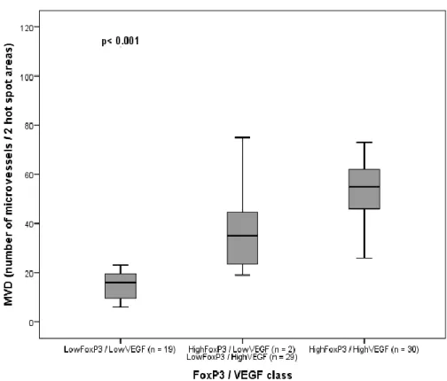
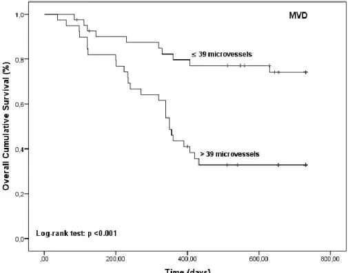
Figure 1 – FoxP3 T-cells distributed according to the VEGF class low or high and respective value of statistical significance for the ANOVA test. ... 83 Figure 2 - Association of MVD (number of microvessels) distributed according to the FoxP3/VEGF class and respective value of statistical significance for the ANOVA test. ... 83 Figure 3 - Kaplan-Meier OS curves comparing FoxP3 T-cells categories in 80 dogs with malignant mammary tumors. ... 84 Figure 4 - Kaplan-Meier OS curves comparing MVD categories in 80 dogs with malignant mammary tumors. ... 84 Figure 5 - Kaplan-Meier OS curves comparing VEGF categories in 80 dogs with malignant mammary tumors. ... 85
XLII
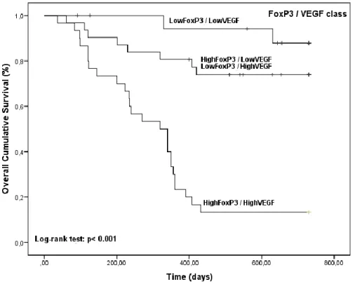
Figure 6 - Kaplan-Meier OS curves comparing FoxP3/VEGF categories in 80 dogs with malignant mammary tumors. ... 85 Figure 7 – (A) FoxP3+ expression in the nuclei of lymphoid cells infiltrating a
tubulopapillary carcinoma, bar = 50 µm; (B) Tubulopapillary carcinoma with high expression of TGFβ, note the most evident diffuse cytoplasmic staining, with prominence of the cytoplasmic membrane, bar = 100 µm. ... 101 Figure 8 - FoxP3 T-cells distributed according to the TGFβ/VEGF class and respective value of statistical significance for the ANOVA test. ... 104 Figure 9 - Association of MVD (number of microvessels) distributed according to the TGFβ/VEGF class and respective value of statistical significance for the ANOVA test. ... 105 Figure 10 - Kaplan-Meier OS curves comparing TGFβ class in 67 dogs with malignant mammary tumors. ... 106 Figure 11 - Kaplan-Meier OS curves comparing TGFβ/FoxP3 class in 67 dogs with malignant mammary tumors. ... 106 Figure 12 - Kaplan-Meier OS curves comparing TGFβ/VEGF class in 67 dogs with malignant mammary tumors. ... 107 Figure 13 - Kaplan-Meier OS curves comparing TGFβ/CD31 class in 67 dogs with malignant mammary tumors. ... 107 Figure 14 – Immunoreactivity for IL-35 in (A) solid carcinoma, note the predominant staining in the epithelial cells in a diffuse manner, bar = 200 μm; (B) carcinosarcoma, note the intense positivity in the mesenchymal component, bar = 100 μm; (C) complex carcinoma. The myoepithelial area was negative or revealed a weak focal positivity, bar = 500 μm and (D) neoplastic intravascular emboli. The labelling intensity of IL-35 in primary tumor and neoplastic intravascular emboli was similar, bar = 100 μm. ... 123 Figure 15 - Kaplan-Meier OS curves comparing IL-35 class in 72 dogs with malignant mammary tumors. ... 125
Table 1 - Relationship between intratumoral FoxP3 T-cells, MVD by CD31 (average number of microvessels) and clinicopathological parameters in malignant CMT. ... 81 Table 2 - Relationship between VEGF class and clinicopathological parameters in malignant CMT. ... 82
XLIII
Table 3 - Association between considered molecular markers and overall survival times. ... 86 Table 4 - Relationship between TGFβ class and clinicopathological parameters in malignant canine mammary tumors. ... 103 Table 5 - Relationship of TGFβ/FoxP3, TGFβ/VEGF and TGFβ/CD31classes with clinicopathological variables of tumor aggressiveness. ... 110 Table 6 - Association between considered molecular markers classes and overall survival. ... 111 Table 7 - Relationship between IL-35 expression and clinicopathological parameters in malignant CMT. ... 128 Table 8 - Relationship of clinicopathological variables and IL-35 expression with overall survival. ... 129
Chapter III
Figure 1 - Tubulopapillary carcinoma with high expression of COX-2 (A); VEGF (B) and CD31 (C). Solid carcinoma with low expression of COX-2 (D); VEGF (E) and CD31 (F). ... 143 Figure 2 - Kaplan-Meier overall survival curves comparing COX-2 categories in 109 dogs with malignant mammary tumors. Female dogs with highCox-2 had significantly shorter overall survival intervals. ... 144 Figure 3 - Kaplan-Meier overall survival curves comparing COX-2/CD31 group categories in 109 dogs with malignant mammary tumors. Female dogs with highCox-2/highCD31 tumors had significantly shorter overall survival intervals. ... 144 Figure 4 - Kaplan-Meier overall survival curves comparing COX-2/VEGF group categories in 109 dogs with malignant mammary tumors. Female dogs with highCox-2/highVEGF tumors had significantly shorter overall survival intervals. ... 145 Figure 5 - Kaplan-Meier overall survival curves comparing COX-2/Ki-67 group categories in 109 dogs with malignant mammary tumors. Female dogs with highCox-2/highKi-67 tumors had significantly shorter overall survival intervals. ... 145 Figure 6 - Kaplan-Meier overall survival curves comparing COX-2/CD3 group categories in 109 dogs with malignant mammary tumors. Female dogs with highCox-2/highCD3 tumors had significantly shorter overall survival intervals. ... 146
XLIV
Figure 7 - Kaplan-Meier overall survival curves comparing COX-2/MAC group categories in 109 dogs with malignant mammary tumors. Female dogs with highCox-2/highMAC tumors had significantly shorter overall survival intervals. ... 146 Figure 8 - Association of CD3+ T-lymphocytes and COX-2/EGFR groups in malignant canine mammary tumors. ... 166 Figure 9 - Immunoreactivity for c-kit in tubulopapillary carcinoma; note the simultaneous cytoplasmic and membranous staining; bar =50 𝜇m. ... 180 Figure 10 - Immunoreactivity for c-kit in carcinosarcoma; bar =50 𝜇m. ... 181 Figure 11 - Immunoreactivity for c-kit in adnexal non-tumoral mammary gland; bar = 100 𝜇m. ... 181 Figure 12 - Association of CD3+ T-lymphocytes and c-kit in malignant canine mammary tumors. ... 182 Figure 13 - Association of MVD and CD3/VEGF groups in malignant canine mammary tumors. ... 183 Figure 14 - Association of MVD and c-kit/VEGF groups in malignant canine mammary tumors. ... 183 Figure 15 - Kaplan-Meier overall survival curves comparing CD3/c-kit categories in 80 dogs with malignant mammary tumors. ... 184 Figure 16 - Kaplan-Meier overall survival curves comparing CD3/VEGF categories in 80 dogs with malignant mammary tumors. ... 185 Figure 17 - Kaplan-Meier overall survival curves comparing c-kit/VEGF categories in 80 dogs with malignant mammary tumors. ... 185
Table 1 - Associations of clinicopathological features with COX-2, CD31 and VEGF immunostaining. ... 150 Table 2 - Associations of clinicopathological features with Ki-67, CD3+T-lymphocytes
and macrophages immunostaining. ... 151 Table 3 - Relationship of COX-2/CD31; COX-2/VEGF; COX-2/Ki-67; COX-2/CD3 and COX-2/MAC groups with clinicopathological variables of tumor aggressiveness. .... 152 Table 4 - Association between considered molecular markers groups and overall survival. ... 154 Table 5 - Relationship between tumoral CD3+ T-lymphocytes and clinicopathological parameters. ... 165
XLV
Table 6 - Relationship of COX-2/EGFR groups with clinicopathological variables. . 167 Table 7 - Relationship of CD3/c-kit, CD3/VEGF and c-kit/VEGF groups with clinicopathological variables of tumor aggressiveness ... 188 Table 8 - Association between considered molecular markers groups and overall survival. ... 189
Chapter IV
Figure 1 - Effects of co-culture of PBMCs and canine cancer cells on intracellular COX-2 expression. (A) Graphs show the overlay of flow cytometric expression of intracellular COX-2 by Sh1b mammary cancer cells (gray filled histogram) or Sh1b cancer cells in co-culture with canine PBMCs (black solid line). Co-co-cultured Sh1b mammary cancer cells with canine PBMCs showed a trend of overexpression of COX-2 in comparison with SH1b alone. The co-culture between Sh1b mammary cancer cells and canine PBMCs results in (B) a decreased expression of intracellular COX-2 by the PBMCs (*** p = 0.0002), (C) induced a decreased expression of intracellular COX-2 specifically in the fraction of CD68+CD3- cells within the PBMCs (* p = 0.0215), but (D) did not induce alterations in COX-2 expression in the CD68-CD3+ cells. Graphs show the mean of 5 independent experiments +/- SD. P-values were calculated by two tail paired Student t-test: ns= not significant, *p<0.05, **p<0.01, ***p<0.001). ... 203 Figure 2 - The effects of co-culture between human macrophage cell line and canine or human cancer cells lines on COX-2 expression by the cancer cells. Co-cultured dTHP1 with canine mammary cancer cell Sh1b (A), result in overexpression of intracellular COX-2; with human mammary cancer cell BT474 (B), results in a decreased intracellular production of COX-2; and with human colon cancer cell HT29 (C), result in overexpression of intracellular COX-2. Graphs show the overlay of flow cytometric expression of intracellular COX-2 by cancer cells (gray filled histogram) or cancer cells in co-culture (black solid line). ... 204 Figure 3 - Effects of soluble factors derived from cultured cancer cells on intracellular COX-2 expression by macrophages. dTHP1 macrophages were cultured in medium alone (white column) or treated with 50% v/v conditioned medium from cultured canine mammary cancer cells Sh1b (black; * p = 0.0257) or human colon cancer cells HT29 (gray; ** p = 0.0028) for 48h. Graph shows the z-score of intracellular COX-2 MFI of 2
XLVI
independent experiments +/- SD. P-values were calculated by one-way ANOVA with Bonferroni’s multiple comparisons post test: *p<0.05, **p<0.01. ... 205
XLVII
List of Abbreviations
A
AKT or PKB, Protein kinase B ANOVA, Analysis of variance AP-1, Activator protein 1 B
BRCA1, Breast cancer susceptibility gene-1 BT474, Human breast carcinoma cell line C
cAMP, Cyclic adenosine monophosphate CCL2/MCP-1, Monocyte chemotactic protein -1 CCL8 or MCP-2, Monocyte chemotactic protein 2 CCL18, Chemokine (C-C motif) ligand 18
CCR, C-C chemokine receptor
CD31, Platelet Endothelial Cell Adhesion Molecule 1- PECAM 1 CD3, Cluster of differentiation 3
CD4, Cluster of differentiation 4 CD8, Cluster of differentiation 8 CK5, Cytokeratin 5
c-kit, Tyrosine-protein kinase Kit CMT, Canine mammary tumors COX-2, Cyclooxygenase 2
CSF1, Macrophage colony-stimulating factor 1 D
DCs, Dendritic cells
DFS, Disease-free survival
DMEM, Dulbecco’s modified Eagle’s medium
E
XLVIII
EGFR, Epidermal growth factor receptor (ErbB1 or HER-1) EMT, Epithelial-to-mesenchymal transition
Ephs, Ephrin receptors ER, Estrogen receptor F
FCS, Fetal calf serum
FGF, Fibroblast growth factor FoxP3, Forkhead box P3 G
G-CSF, Granulocyte colony-stimulating factor GH, Growth hormone
H
HBSS, Hank’s Balanced Salt Solution
HER-2/neu, Human epidermal growth factor receptor 2 (ErbB2) HGF/SF, Hepatocyte growth factor/scatter factor receptor HGM, Histological grade of malignancy
HIF-1α, Hypoxia-inducible factor 1α HT29, Human colon cancer cell line I
IFN-γ, Interferon gamma
IGF-1, Insulin-like growth factor 1 IL, Interleukin
M
MAPK/ERK1/2, Mitogen activated protein kinase M-CSF, Macrophage colony-stimulating factor MDSCs, Myeloid-derived suppressor cells MEK p44/42, Mitogen-activated protein kinase MHC, Major histocompatibility complex MIF, Macrophage migration inhibitory factor MMP, Matrix metalloproteinase
XLIX N
n, Number of observations NFκB, Nuclear factor kappa-B NK, Natural killer
O
OS, Overall survival OVH, Ovariohysterectomy P
PBMCs, Peripheral blood mononuclear cells PBS, Phosphate-Buffered Saline
PCNA, Proliferating cell nuclear antigen PDGF, Platelet-derived growth factor PGE2, Prostaglandin E2
PI3K, Phosphatidylinositol-4,5-bisphosphate 3-kinase PMA, Phorbol myristate acetate
PRL, Prolactin
p53, Tumor suppressor p53
p63, Transformation-related protein 63 R
Ras, Protein superfamily of small GTPases RPMI, Roswell Park Memorial Institute medium RTKs, Receptor tyrosine kinases
S
SCF, Stem cell factor SD, Standard deviation SE, Standard error
Sh1b, Canine mammary carcinoma cell line
STAT3, Signal transducer and activator of transcription 3 T
TAMs, Tumor associated macrophages TCR, T-cell receptor
L
TGFβR, Transforming growth factor beta receptor Th1, T-helper 1 lymphocytes
Th2, T-helper 2 lymphocytes Th17, T-helper 17 lymphocytes THP1, Human monocytic cell line
TILs, Tumor-infiltrating T-lymphocytes TKIs, Tyrosine kinase inhibitors
TNFα, Tumor necrosis factor alpha Tregs, Regulatory T-lymphocytes U
uPA, Urokinase-type plasminogen activator V
VEGF, Vascular endothelial growth factor VEGFR, Vascular endothelial growth factor receptor
W
WHO, World Health Organization Others
Chapter I - General Introduction
Book Chapter:
Neoplastic Diseases of the Canine Mammary Gland.
In Book: Mammary Glands: Anatomy, Development and Diseases, Edmund B. Rucker. Maria Isabel Carvalho, Teresa Raposo, Helena Rodrigues, Isabel Pires, Justina Prada, Felisbina Luísa Queiroga.
(Ed), Nova Science Publishers, Inc, NY, 2014 (ISBN: 978-1-62948-856-1 eBook)
Review Articles:
A Role for T-lymphocytes in Human Breast Cancer and in Canine Mammary Tumors. Maria Isabel Carvalho, Isabel Pires, Justina Prada, Felisbina Luísa Queiroga.
BioMed Research International, 2014
(doi: 10.1155/2014/130894)
A comparative approach of Tumor Associated Inflammation in Mammary Cancer between Humans and Dogs.
Maria Isabel Carvalho, Ricardo Silva-Carvalho, Isabel Piresa, Justina Prada, Rodolfo Bianchini, Erika Jensen-Jarolim, Felisbina Luísa Queiroga.
BioMed Research International, 2016
Chapter I – General Introduction
3
I.1 Neoplastic Diseases of the Canine Mammary Gland
The development of the mammary gland is a puzzling phenomenon and the research on this field has been focused mostly on the carcinogenesis, with a less goal-oriented concern in basic histology. Canine mammary tumors (CMT) represent a serious problem in veterinary medicine [1]. Clinically, these tumors share many similar features with human breast cancer in terms of histopathology, biological behavior, hormone dependence, risk factors and genetic alterations. These similarities have led to a proposed canine model for the study of breast cancer in women [2-5].
Mammary Gland Development
The mammary gland is a modified apocrine sweat gland present in the subcutaneous connective tissue, containing a nipple and consisting of a network of ducts surrounded by a fibrovascular and adipocyte-rich stroma. Mammary development can first be recognized during the embryonic stages by the appearance of 2 ventral ridges of ectoderm, which cover the mesodermic regions from the axillary to the inguinal area. The ectodermal cells migrate along each milk line and coalesce in a complex interaction with the mesenchymal cells to form a placode, which eventually becomes an individual mammary gland [6]. At birth, the mammary gland is not fully developed. Its development continues at puberty, and throughout reproductive life the gland undergoes a morphogenetic process controlled by its hormonal environment. This involves complex interactions between ovarian steroid and polypeptide hormones and growth factors produced in successive estrous cycles. The last stages of development of the mammary gland occur in the adult female only during pregnancy, with the proliferation of the ductal tissue, differentiation into milk-producing acini, secretion of milk by the acinar cells, and, at the end of lactation, involution of the secretory component of the gland with preservation of the ductal structures [6, 7].
Mammary Gland Anatomy and Blood Supply
Most dogs develop 5 pairs of mammary glands, although 4 or 6 pairs have been found in a few animals. There are 2 thoracic (M1 and M2), 2 abdominal (M3 and M4), and 1 inguinal (M5) pair of mammary glands [8-10]. The blood supply in canine mammary glands derives from the sternal branches of the internal and lateral thoracic artery, intercostal, the cranial and caudal superficial epigastric arteries, deep cranial epigastric,
Chapter I – General Introduction
4
abdominal segmented and deep iliac circumflex. Venous drainage of the glands is similar to the arterial supply, though small veins may cross the midline between the right and left mammary glands [8, 11].
Lymph Drainage of the Mammary Gland
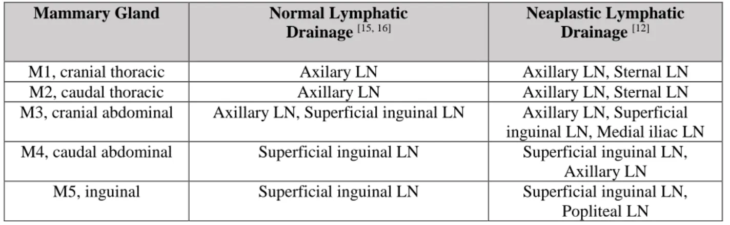
By convention, most malignant epithelial neoplasms (carcinomas) metastasize via the lymphatic system, whereas malignant mesenchymal neoplasms (sarcomas) metastasize via capillaries and veins. Patsikas and colleagues have methodically examined the lymph drainage of CMT located in each of the mammary glands [12] (Table 1).The lymph drainage of the fourth and fifth neoplastic mammary glands is directed to the ipsilateral superficial inguinal lymph nodes as sentinel lymph nodes of the fourth and fifth healthy mammary glands, whereas the two most cranial neoplastic mammary glands generally drain to the axillary lymph nodes. These authors also strength the clinician’s notion that mammary carcinomas occurring in the inguinal (M5) gland may show retrograde metastasis via the lymphatic plexus in the subcutis of the inner thigh and to the popliteal lymph nodes. Retrograde metastatic spread is a rare event, but mammary carcinoma metastasis to the vagina have been reported in bitches [13, 14]. In normal healthy dogs, lymphatic fluids drain into the ipsilateral lymph nodes. There is no drainage to the contralateral gland or lymph node, but drainage may be altered in cases of mammary neoplasia [6].
Table 1 - Lymphatic drainage of normal and neoplastic canine mammary gland.
Lymphatic vessels can also cross the abdominal midline and in doing so pass through the abdominal and thoracic walls. The axillary lymph node collects lymph from thoracic mammary glands, while the superficial inguinal lymph node collects lymph from the
Mammary Gland Normal Lymphatic
Drainage [15, 16]
Neaplastic Lymphatic Drainage [12]
M1, cranial thoracic Axilary LN Axillary LN, Sternal LN
M2, caudal thoracic Axillary LN Axillary LN, Sternal LN
M3, cranial abdominal Axillary LN, Superficial inguinal LN Axillary LN, Superficial inguinal LN, Medial iliac LN M4, caudal abdominal Superficial inguinal LN Superficial inguinal LN,
Axillary LN
M5, inguinal Superficial inguinal LN Superficial inguinal LN,
Popliteal LN
Chapter I – General Introduction
5
abdominal mammary glands. Generally, the lymphatic drainage of the third mammary gland occurs cranially to the axillary lymph node, but deviation is possible to the inguinal drainage region [8, 11]. The dual drainage system of this mammary gland is explained by the formation of plexiform lymphatic connections in between the abdominal and caudal thoracic mammary glands [12, 15, 16].
Canine Mammary Tumors (CMT)
A General Approach
The investigations of mammary tumors in dog are of particular interest because they constitute an excellent model for the study of breast cancer in women. In fact mammary tumors are very common in the population of domestic dogs and show remarkable clinical, histopathological, and even genetic and molecular similarities with women breast cancer [4].
The recent sequencing of the canine genome and the evidence of its similarity to the human genome has emphasized the dog as an attractive alternative model for cancer research [17, 18]. In addition, dogs have a considerable body size, genetic variability similar to humans, and develop spontaneous tumors in the context of a natural immune system. Moreover, unlike laboratory animals, these species share the same environment and are exposed to the same carcinogens; however, they develop tumors in shorter time, due to their shorter longevity compared with the humans [4, 19-22]. In the future it’s possible that this comparative oncology can play a role in translational medicine, making early clinical applications from knowledge obtained from this model [23].
In bitches, mammary tumors are the most common type of cancer, with a high incidence in geographic regions where ovariohysterectomy is not commonly practiced [24]. When both sexes are considered, breast tumors are the most common non-cutaneous tumors [25]. As in humans, there is a marked sexual predisposition, males have a risk of developing breast tumors equal or less than 1% [26, 27]. Studies conducted over the past decade reveal that in dogs diagnosed with cancer, 56% have mammary tumors [28] and the incidence rate is 205/100,000 cases per year [29].
Predisposing Factors
The incidence of CMT increases with age [30, 31], with middle-aged and older bitches mostly affected [32]. The mean age of incidence is approximately 10 years; however, the
Chapter I – General Introduction
6
risk of mammary tumors is increased in dogs over 6 years of age [33, 34]. Animals younger than 4 years are rarely affected by this condition [27]. The existence of a racial predisposition is a matter of disagreement among authors. Different breeds have been identified as predisposed to the development of these tumors: English springer spaniel, Doberman, Boxer [30], and also Cocker Spaniel, Dachshund [35] and Bouvier Bernois [36]. One study performed in 101 bitches reveals a lower incidence and a smaller dimension of malignant mammary tumors in small breeds [37]. This divergence between authors is possibly caused by the different contribution of each breed to the sample size under study. According to some researchers, obesity and type of diet influence the risk of developing mammary tumors in dogs, similar to what happens in humans [38]. In women, particularly during menopause, obesity is associated with increased development of breast cancer [24, 31, 39, 40] and epidemiological studies have shown that consumption of high fat diets is positively correlated with mortality rates caused by this type of cancer [41]. The low prevalence of mammary tumors in herbivores leaves us tempted to assume that a diet low in fat and high in vegetable fiber is beneficial. This is contrary to what happens in dogs, who feed mainly on meat (high fat and a tiny amount of fiber) and have a high prevalence of mammary cancer [42]. In dogs, obesity at a young age, as well as a diet based mostly on red meat and homemade food, appear to be factors potentially associated with a higher risk to develop mammary tumors and dysplasias [43].
Aetiology
The development of a neoplasia is a complex process resulting from various carcinogenic factors, such as ionizing radiation, chemical compounds and oncogenic viruses. The impact of several endogenous factors, genetic, immune and hormonal, is of irrefutable importance and deserves special attention [27]. Hormonal factors have been thoroughly studied and play a fundamental role in canine mammary carcinogenesis [44-46]. The development and growth of the mammary gland is influenced by ovarian hormones (e.g. estrogen and progesterone) and the functional contribution of these hormones has been acknowledged in various physiologic and pathologic situations [42]. As for the genesis of canine mammary gland neoplasia, the role of these hormones has been well established [42, 47, 48]. The first observation of the hormonal contribution in mammary carcinogenesis arose in the late 70s, when the effect of administering progestogen inhibitors of the oestrous cycle was first described [49]. This hypothesis is further supported by evidence of the protective effect of ovariohysterectomy (OVH) in the
Chapter I – General Introduction
7
development of mammary tumors, especially when it occurs at an early age, since the source of hormones is surgically removed [27, 31, 44]. It has been reported that mammary tumor incidence rises sharply whenever the OVH is performed following the second oestrous cycle. When compared with female dogs spayed before the first oestrous cycle, those spayed after the first and second oestrous cycle present an 8% and 26% increased risk of developing mammary tumors, respectively [50].
Analysis of steroid hormone participation in the acquisition of malignancy has shown a loss of estrogen receptors during the neoplastic progression, which suggests a hormone independence at stages of higher tumor aggressiveness [51]. However, conducting an OVH as a therapeutic measure after the initiation of cancer progression does not seem to have a significant beneficial effect in the course of the neoplastic disease [52]. In fact, CMT appear to be clearly dependent on steroid hormones (estrogen and progesterone) and therefore, the expression of estrogen and progesterone receptors constitutes additional evidence of the endocrine component in mammary neoplasia [53, 54].
Presently, other hormones are also known to be involved in mammary carcinogenesis. During CMT development, changes are found in the production of pituitary hormones, prolactin (PRL) [45] and growth hormone (GH), have a specific function in mammary tumorigenesis [46]. Malignant CMTs produce greater amounts of prolactin compared with benign CMT, and these also produce more prolactin that normal mammary glands. Prolactin and steroid hormones are produced in the mammary gland, functioning in an autocrine-paracrine mode as local growth factors, favouring the progression of malignant mammary tumors [45]. Concerning GH, excessive production induced by endogenous or exogenous progestagens might promote mammary tumorigenesis while stimulating the proliferation of susceptible mammary epithelium cells [48]. GH tissue levels in malignant CMT were also astoundingly augmented in relation to benign CMT [46]. Over the last decade, some studies point out the existence of other factors intervening in canine mammary carcinogenesis, such as insulin-like growth factor I (IGF-1) [46] epidermal growth factor (EGF) [55] and epidermal growth factor receptor (EGFR) [56].
Besides endocrine factors, genetic mutations are crucial in the development and progression of tumors. The first to occur is the initiation step, an initial lesion that will evolve under the influence of promoting factors until the later stages of invasion and metastasis unfold. Among several genes participating in mammary carcinogenesis, p53, p27 and c-erB-2 or HER2/neu are worth mentioning for the research conducted throughout the last decade [57-59]. The p53 gene is a major regulator of tumor
Chapter I – General Introduction
8
suppression, inducing the apoptosis of cells bearing damaged, irreparable DNA such as neoplastic cells. p53 gene mutations have been reported in 30-35% of CMT [57, 60, 61]. Not only do malignant and metastatic tumors bear p53 mutations, but also benign CMT, which might indicate this is an early event in mammary carcinogenesis [62].
The p27 gene, an inhibitor of cyclin dependent kinases, hampers the transition to the cell cycle replicative phase. Primary carcinomas and lymph node metastasis have a reduced expression of p27 mRNA, suggesting a preponderant role in tumorigenesis [58].
HER2/neu is an oncogene frequently mutated in human breast cancers, as the encoded receptor is targeted in clinical application by the drug Herceptin. The involvement of this gene as a prognostic marker in CMT is yet to be well established, but the expression of HER2/neu in tumor biopsies, coupled with other molecular markers (e.g. estrogen receptors, p63 and CK5) is useful in determining a molecular phenotype which, similarly to what has been observed in the human counterpart, presents a relationship with a worse prognosis [59]. The environmental aetiology has already been suggested, but hitherto not proven true. It is possible that anti-parasitic drugs containing pyrethroid substances accumulate in the peri-mammary adipose tissue and sensitize epithelial cells to proliferation [63].
Diagnosis and Clinical Management
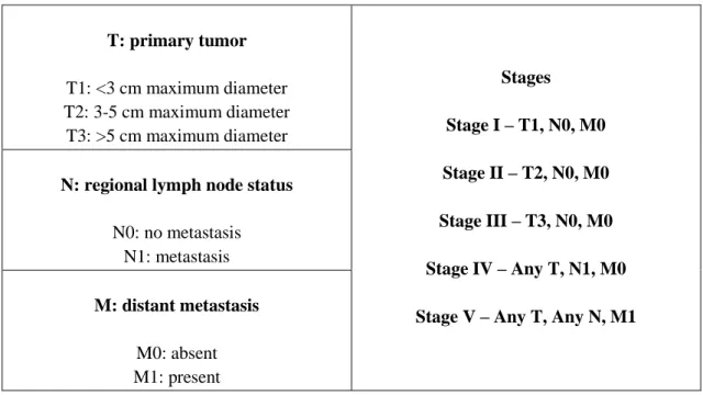
CMTs may present as solitary or multiple masses. The caudal mammary glands are more affected than cranial glands, probably due to their larger size. Clinical signs that indicate a malignant lesion include the observation of the rapid growth of a mass not circumscribed, invasiveness, adherence to the skin or surrounding tissues, and ulceration. However the absence of these signs does not mean that it is excluded as malignant lesion [34]. Similarly, large lesions can merely mean that the search for veterinary care was delayed [24]. Mammary tumors are staged according to the World Health Organization (WHO) TNM system or a modification of this system [64, 65] (Table 2).
To stage the mammary gland tumors in a dog, information regarding tumor size, lymph node status, and presence of metastasis needs to be collected and verified. The largest malignant tumor in dogs with more than one tumor should be considered in the stage assignment. The lymph nodes should be identified, and if they are clinically enlarged, fine-needle aspirates for cytological evaluation must be performed. Dogs with malignant mammary tumors would also be evaluated for metastatic disease. Three-view thoracic radiographs remain the standard diagnostic method for evaluating veterinary patients for
Chapter I – General Introduction
9
the presence of metastasis [6, 24], since the lungs are the most common site for distant metastasis [66, 67]. However, additional staging tests, including abdominal ultrasound, skeletal radiographs, or other imaging modalities, may be indicated if clinical examination identifies other sites suspicious for metastasis. On physical examination, clinical signs suggestive of breast cancer are easily identified; however, definitive diagnosis of CMT is reached only after a histopathological study of the sample obtained by biopsy. The cytology by fine needle aspiration, despite being a minimally invasive methodology and easy to perform, is not reliable in the diagnosis of mammary tumors in female dogs due to the high variability and pleomorphism that the same histological tumor can present. Despite the limitations in diagnostic, cytology is a good method to dispose of some differential diagnoses and to determine the nodal disease [68].
Table 2 - Canine mammary tumor staging - modified WHO.
T: primary tumor T1: <3 cm maximum diameter T2: 3-5 cm maximum diameter T3: >5 cm maximum diameter Stages Stage I – T1, N0, M0 Stage II – T2, N0, M0 Stage III – T3, N0, M0 Stage IV – Any T, N1, M0 Stage V – Any T, Any N, M1 N: regional lymph node status
N0: no metastasis N1: metastasis
M: distant metastasis
M0: absent M1: present
For neoplastic lesions of the mammary gland in dogs, the classification recommended by the World Health Organization (WHO), published in 1999, is commonly accepted [69]. The breast neoplasms are divided into benign or malignant, and they may have their origin in epithelial, myoepithelial or mesenchymal tissue. Most mammary gland tumors are of epithelial origin. Some, however, can have mixed histology consisting of both epithelial and myoepithelial tissue, with areas of cartilage and bone, and a few tumors are of purely mesenchymal origin [24]. The term inflammatory carcinoma is not included in WHO classification and is a distinct clinical entity in dogs, characterized histologically by the presence of emboli in dermal lymphatic vessels which is responsible for the severe