Luís Manuel Redondo Raposo
Mestre em Biotecnologia (Engª Bioquímica)
Transcriptome and proteome profiling of
canine mammary tumors: dog as a genetic
model for unraveling mammary cancer
molecular signatures
Dissertação para obtenção do Grau de Doutor em Biologia, variante Genética molecular
Orientador: Doutora Maria Alexandra Núncio de Carvalho Ramos Fernandes, Professora Auxiliar, Faculdade de Ciências e Tecnologia
da Universidade Nova de Lisboa
Co-orientador: Doutor Armando José Latourrette de Oliveira Pombeiro, Professor Catedrático, Instituto Superior técnico da Universidade de Lisboa
Júri:
Presidente: Doutor Luís Manuel Camarinha de Matos Arguentes Doutor Fernando António da Costa Ferreira Doutor António José de Freitas Duarte Vogais: Doutor Pedro Miguel Ribeiro Viana Baptista
Doutora Maria Alexandra Núncio de Carvalho Ramos Fernandes
Transcriptome and proteome profiling of canine mammary tumors: dog as a genetic model for unrevealing mammary cancer molecular signatures
Copyright © Luís Manuel Redondo Raposo, Faculdade de Ciências e Tecnologia, Universidade Nova de Lisboa.
i
Agradecimentos
Quero agradecer do fundo do coração à Professora Doutora Alexandra Fernandes por me ter aceite como seu aluno de doutoramento, por me ter feito embarcar numa experiência fantástica e ser a primeira responsável por tudo o que aprendi. Muito obrigado pela sua valiosíssima orientação científica e humana. Muito obrigado professora, por me ter apoiado, ajudado sempre que foi preciso e chorado, também, a meu lado. Obrigado também pelos risos que partilhamos e pelas pequenas e grandes conquistas que tivemos no nosso dia-a-dia.
Agradeço também ao Professor Doutor Armando Pombeiro por ter ser meu co-orientador e por todo o conhecimento que partilhou, a disponibilidade, amabilidade e cuidado que teve para comigo.
Quero agradecer ao Centro de Química Estrutural do IST, ao Departamento de Ciências da Vida da Faculdade de Ciências e Tecnologia da Universidade Nova de Lisboa por me terem acolhido e dado todas as condições para realizar o meu doutoramento. Agradeço também à Fundação para a Ciência e Tecnologia pela bolsa SFRH/BD/70202/2010 que me possibilitou realizar este doutoramento.
Agradeço ao Professor Doutor Pedro Baptista por ter partilhado comigo sabedoria, amizade e de se ter tornado, informal e desinteressadamente, um co-orientador meu que muito ajudou nesta jornada.
Agradeço também aos doutores Pedro Faísca e Jorge Correia e por toda a ajuda imprescindível na avaliação histopatológica dos tumores mamários e no acesso que nos deram a amostras. Agradeço também aos doutores Joaquim Henriques e Gonçalo Pereira por me terem permitido recolher tumores de pacientes.
ii
Quero agradecer enternecidamente a todos os colegas e amigos que partilharam o laboratório comigo desde a Lusófona até ao DCV e com os quais tive a oportunidade de aprender e dividir emoções.
Agradeço àqueles que são a minha Alma, o meu Ser. Ao meu Pai José, que me ensinou o que é coragem, à minha Mãe Júlia que me ensinou a amar sem reservas, ao meu Irmão Nuno, o menino,
rapaz e Homem que me faz querer sempre ser melhor, à minha Irmã “emprestada” Marta que me
mostra como ser menino apesar de ser já não o ser no corpo.
iii
Abstract
Within this work, two canine mammary tumors (CMT) derived cell lines named FR37-CMT and FR10-CMT were immortalized and characterized. These cell lines are good models for the development of new therapeutic compounds and novel strategies for the management of CMTs and for the study of CMT resistance to chemotherapy. FR37-CMT cell line is also is a good model for the study of epithelial-to-mesenchymal transition (EMT) while the FR10-CMT cell line can be considered a model for the study of malignant CMTs with repression of EMT.
Two new organometallic compounds, TS262 and TS265, have been tested in FR37-CMT and FR10-CMT cell lines. In both cell lines, the IC50 for TS262 and TS265 were significantly lower
than the displayed by cisplatin and doxorubicin (1.05µM and 1.39µM respectively for FR37-CMT and 0.55µM and 0.80µM respectively for FR10-CMT) and are thus good therapeutic possibilities for the treatment of CMTs.
iv TS265 (nanoTS265) into FR37-CMT and FR10-CMT cells, enhancing the cytotoxicity displayed by the free compounds.
A quantitative proteomic analysis was also performed in the FR37-CMT cells exposed to TS262 and TS265 in order to obtain more insights into the mechanisms of toxicity and the responses of the cells induced by these compounds.
Using principal component analysis (PCA) of gene expression, it was possible to distinguish metastatic from non-metastatic grade III CMTs using our set of 21 biomarkers (10 miRNAs and 11 mRNAs). The differential expression of miR-155, DICER1 and ESR1 between metastatic and non-metastatic grade III CMTs highlighted the importance of these genes in the metastatic transition of CMTs.
Keywords: Canine mammary tumors; Epithelial to mesenchymal transition; chemotherapeutic compounds and nanotechnology; Quantitative proteomics; FR37-CMT; FR10-CMT
v
Resumo
No trabalho desenvolvido para esta tese, foram imortalizadas e caracterizadas duas novas linhas celulares obtidas a partir de tumores mamários caninos (TMCs), FR37-CMT e FR10-CMT. Estas linhas são óptimos modelos para o desenvolvimento de novos compostos e estratégias terapêuticas para o tratamento de TMCs e para o estudo da resistência à quimioterapia exibida por estes tumores. A linha FR37-CMT é também um óptimo modelo para o estudo da transição epitelial-mesenquimatosa (TEM) enquanto que a linha FR10-CMT pode ser considerada um modelo para o estudo para o estudo de TMCs malignos com repressão de TEM.
Dois novos compostos metálicos, T262 e TS265, foram testados nas linhas celulares FR37-CMT e FR10-CMT. Em ambas as linhas, o IC50 para TS262 e TS265 é significativamente
mais baixo do que o exibido pela cisplatina e doxorubicina (1.05µM e 1.39µM, respectivamente, para FR37-CMT e 0.55µM e 0.80µM, respectivamente, para FR10-CMT) e são assim boas possibilidades terapêuticas para o tratamento de TMCs.
vi TS262 (nanoTS262) e TS265 (nanoTS265) para o interior das células das linhas FR37-CMT e FR10-CMT, aumentando a citotoxidade exibida pelos compostos livres.
Foi efectuada uma análise de proteómica quantitativa em células da linha FR37-CMT expostas a TS262 e TS265 de maneira a aprofundar o conhecimento dos mecanismos de toxicidade e resposta celular induzidas por estes compostos.
Foi possível distinguir TMC metastáticos de não metastáticos utilizando análise de componentes principais (ACP) da expressão genética de um conjunto de 21 biomarcadores (10 miRNAs e 10 mRNAS). A diferença de expressão de miR-155, DICER1 e ESR1 entre os TMCs metastáticos e não metásticos sublinhou a importância destes genes na transição metastática dos TMCs.
vii
Table of Contents
AGRADECIMENTOS ... I
ABSTRACT ... III
RESUMO ...V
TABLE OF CONTENTS ... VII
FIGURE INDEX ... XIII
TABLE INDEX ... XV
LIST OF ABBREVIATIONS ... XVII
I. BIBLIOGRAPHIC REVIEW IN CANINE MAMMARY TUMORS ... 1
I.1. THESIS MOTIVATION ... 1
I.2. INCIDENCE ... 2
I.3. CMTRISK FACTORS ... 3
I.3.1. Age 3 I.3.2. Diet 3 I.3.3. Hormonal dependency ... 4
I.3.4. Breed dependency ... 5
I.3.5. Germline mutations that predispose for CMTs ... 5
I.4. CMT CLASSIFICATION ... 6
I.5. CMTETIOLOGY AND CLINICAL PROGNOSTIC FACTORS ... 8
I.6. CANINE MAMMARY CARCINOGENESIS ... 11
viii
I.6.2. Major genomic alterations involved in canine mammary carcinogenesis
revealed by global expression studies ...14
I.6.3. Oncogenes and tumor suppressor genes implicated in canine mammary carcinogenesis ...17
I.6.4. Influence of tumor microenvironment in CMT growth, migration and invasion 18 I.7. CMT TREATMENT ... 22
I.7.1. Cytotoxic chemotherapy ...22
I.7.2. Hormonal treatment ...23
I.7.3. Non-steroidal anti-inflammatory drugs (NSAID) ...24
I.7.4. Receptor Tyrosine Kinase (RTK) Inhibitor, SU11654 (Toceranib) ...25
I.7.5. Desmopressin and p62 vaccine...25
I.7.6. Therapeutic compounds tested in vitro for the treatment of CMTs ...25
II. IMMORTALIZATION AND CHARACTERIZATION OF A NEW CANINE MAMMARY TUMOUR CELL LINE FR37-CMT ... 27
II.1. ABSTRACT ... 29
II.2. KEYWORDS ... 29
II.3. INTRODUCTION ... 31
II.4. MATERIALS AND METHODS ... 32
II.4.1. Sample collection ...32
II.4.2. Establishment of immortalized CMT primary cell line ...32
II.4.3. Tumour sample preparation for histopathology and immunohistochemistry ...33
II.4.4. Chromosome preparations from FR37-CMT cell line ...33
II.4.5. Determination of the doubling time of FR37-CMT cell line ...34
II.4.6. Clonogenic assays: Soft agar colony formation and collagen colony assays 35 II.4.7. Growth of FR37-CMT cell line on the top of a Fibroblast cell monolayer 35 II.4.8. Wound healing assay ...35
II.4.9. Tumorigenicity of FR37-CMT cell lines in NOD-SCID mice ...36
II.4.10. DNA extraction from FR37-CMT cell line and from tumour xenografts cells monolayers ...37
II.4.11. PCR for cOR9S13 and PRCD canine genes ...37
II.4.12. RNA extraction ...38
II.4.13. Quantitative PCR (RT-qPCR) ...38
II.4.14. Total protein extraction ...40
ix
II.4.17. Cell viability assays in presence of cisplatin and doxorubicin ...42
II.4.18. Statistical analysis ...42
II.5. RESULTS ... 43
II.5.1. Immortalization of FR37-CMT cell line ...43
II.5.2. Loss of contact inhibition and invasion ability ...45
II.5.3. Tumorigenicity of FR37-CMT in NOD-SCID mice ...48
II.5.4. Molecular characterization of FR37-CMT cell line ...49
II.5.5. Effect of cisplatin and doxorubicin in the cell viability of FR37-CMT cell line 52 II.6. DISCUSSION ... 53
II.7. CONCLUSIONS ... 58
III. TARGETING CANINE MAMMARY TUMORS VIA GOLD NANOPARTICLES FUNCTIONALIZED WITH PROMISING CO(II) AND ZN(II) COMPOUNDS 59 III.1. ABSTRACT ... 61
III.2. KEYWORDS: ... 62
III.3. INTRODUCTION ... 63
III.4. MATERIALS AND METHODS ... 65
III.4.1. Compounds 65 III.4.2. Gold nanoparticles synthesis and assembly of Au-nanoconjugates ...65
III.4.3. FR37-CMT cell culture ...67
III.4.4. Cell viability assays ...67
III.4.5. Wound healing assay ...67
III.4.6. Statistical analysis ...68
III.5. RESULTS ... 68
III.5.1. Synthesis of Gold nanoconjugates ...68
III.5.2. Effects of TS262 and TS265 on cell viability ...70
III.5.3. Effect of TS262 and TS265 on the migration of FR37-CMT cells evaluated by wound healing assay ...71
III.5.4. Effect of NanoTS262 and NanoTS265 on FR37-CMT cell line ...72
III.6. DISCUSSION ... 73
III.7. CONCLUSIONS ... 75
IV. PROTEOMIC STUDY OF FR37-CMT CELL LINE EXPOSED TO CO(II) AND ZN(II) COMPOUNDS ... 77
IV.1. ABSTRACT ... 79
IV.2. KEYWORDS: ... 79
IV.3. INTRODUCTION ... 81
IV.4. MATERIALS AND METHODS ... 82
x
IV.4.2. Two-Dimensional Electrophoresis (2-DE)...82
IV.4.3. In-gel digestion and MALDI-TOF mass spectrometry analysis ...83
IV.5. RESULTS AND DISCUSSION ... 84
IV.6. CONCLUSIONS ... 86
V. IMMORTALIZATION, CHARACTERIZATION AND TOLERANCE TO PROMISING GOLD NANOPARTICLES FUNCTIONALIZED WITH CO(II) AND ZN(II) COMPOUNDS OF A NOVEL CANINE MAMMARY TUMOR CELL LINE FR10-CMT ... 89
V.1. ABSTRACT ... 91
V.2. KEYWORDS ... 92
V.3. INTRODUCTION ... 93
V.4. MATERIALS AND METHODS ... 94
V.4.1. Sample collection ...94
V.4.2. Establishment of CMT primary cell line ...94
V.4.3. Chromosome preparations from FR10-CMT cell line ...95
V.4.4. Determination of the doubling time of FR10-CMT cell line ...96
V.4.5. Clonogenic assays ...96
V.4.6. Growth of FR10-CMT cell line on top of a Fibroblast cell monolayer ...97
V.4.7. Tumorigenicity of FR10-CMT cell lines in NOD-SCID mice and tumor sample preparation for histopathology/immunohistochemistry ...97
V.4.8. RNA extraction ...98
V.4.9. Quantitative PCR (RT-qPCR). ...99
V.4.10. Total protein extraction ... 100
V.4.11. Western blot 100 V.4.12. Chemotherapeutic compounds ... 101
V.4.13. Cell viability assays in presence of cisplatin and doxorubicin ... 101
V.4.14. Statistical analysis ... 102
V.5. RESULTS AND DISCUSSION ... 102
V.5.1. Establishment of FR10-CMT cell line ... 102
V.5.2. Loss of contact inhibition and invasion ability of FR10-CMT ... 104
V.5.3. Tumorigenicity of FR-10 Cells in Nod/SCID mice ... 106
V.5.4. Molecular characterization of FR10-CMT cell line ... 108
V.5.5. Effect of cisplatin and doxorubicin in the cell viability of FR10-CMT cell line 112 V.5.6. Effect of TS262 and TS265 in the cell viability of FR10-CMT cell line .. 113
V.5.7. Effect of NanoTS262 and NanoTS265 in the cell viability of FR10-CMT cell line 114 V.6. DISCUSSION ... 115
xi
VI. MOLECULAR TYPING OF GRADE III CANINE MAMMARY TUMORS
CAN DISTINGUISH METASTATIC FROM NON-METASTATIC TUMORS ...121
VI.1. ABSTRACT ... 123
VI.2. KEYWORDS ... 123
VI.3. INTRODUCTION ... 125
VI.4. MATERIALS AND METHODS ... 125
VI.4.1. Sample collection ... 125
VI.4.2. Analysis of mRNA and miRNA expression ... 126
VI.5. RESULTS AND DISCUSSION ... 128
VI.6. CONCLUSIONS ... 131
VII. CONCLUDING REMARKS AND PERSPECTIVES ...133
REFERENCES ...141
xiii
Figure Index
FIGURE I.1–WORK PLAN HIGHLIGHTING MAJOR SCOPES OF THIS THESIS. ... 1
FIGURE I.2–CLASSIFICATION OF CMTS ACCORDING TO THE 1999WHO GUIDELINES. ... 7
FIGURE I.3–BIOGENESIS OF MIRNAS ... 21
FIGURE II.1-REPRESENTATIVE IMAGES OF THE ORIGINAL TUMOUR FROM WHICH FR37-CMT CELL LINE WAS ORIGINATED ... 43
FIGURE II.2-REPRESENTATIVE IMAGES OF ADHERENT FR37-CMT CELLS ... 44
FIGURE II.3-CHROMOSOME PREPARATIONS OF THE FR37-CMT CELL LINE ... 45
FIGURE II.4-PCR RESULTS FOR PRCD AND COR9S13 CANINE GENE AMPLIFICATION ... 45
FIGURE II.5-REPRESENTATIVE IMAGES OF THE SOFT AGAR ASSAY ... 46
FIGURE II.6-REPRESENTATIVE IMAGES OF THE COLLAGEN ASSAY ... 46
FIGURE II.7-REPRESENTATIVE IMAGES OF FR37-CMT GROWTH ON THE TOP OF A HUMAN FIBROBLASTS MONOLAYER ... 47
FIGURE II.8-REPRESENTATIVE IMAGES OF THE WOUND HEALING ASSAY ... 47
FIGURE II.9-REPRESENTATIVE IMAGES OF A TUMOR XENOGRAFT ... 48
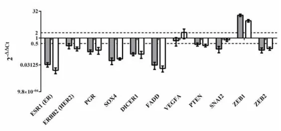
FIGURE II.10-RELATIVE EXPRESSION OF GENES INVOLVED IN BREAST AND MAMMARY TUMORIGENESIS IN THE ORIGINAL TUMOUR AND IN THE FR37-CMT CELL LINE ... 50
FIGURE II.11-RELATIVE EXPRESSION OF MIRNAS INVOLVED IN BREAST AND MAMMARY TUMORIGENESIS IN THE ORIGINAL TUMOR AND IN THE FR37-CMT CELL LINE ... 51
FIGURE II.12-PROTEINS EXPRESSED IN FR37-CMT AND MCF-7 CELL LINES ... 52
FIGURE II.13-CELL VIABILITY OF FR37-CMT CELL LINE AFTER 48 H OF EXPOSURE TO DIFFERENT CONCENTRATIONS OF CISPLATIN AND DOXORUBICIN ... 53
FIGURE II.14-EMT MEDIATED BY THE RAS-ERK1/2-SNAI2 SIGNALLING PATHWAY. ... 55
FIGURE III.1-GOLD NANOPARTICLES AS NANOVECTORIZATION SYSTEMS FOR THE DELIVERY OF TS262 AND TS265 IN FR37-CMT CELL LINE. ... 64
FIGURE III.2-PHYSICOCHEMICAL CHARACTERIZATION OF AUNP CONSTRUCTS. ... 69
xiv
FIGURE III.4-VIABILITY OF FR37-CMT CELLS AFTER 48 H OF EXPOSURE TO DIFFERENT CONCENTRATIONS OF
TS262 AND TS265 ... 70
FIGURE III.5-WOUND HEALING ASSAY OF FR37-CMT CELLS EXPOSED TO 1.5X IC50 CONCENTRATIONS OF TS262 (1.6 µM) AND TS265(2.0 µM) ... 71
FIGURE III.6–CELL VIABILITY OF FR37-CMT CELL LINE AFTER 48 H EXPOSURE TO AUNPS@PEG@BSA, FREE TS262 AND TS265 NANOTS262 AND NANOTS265 ... 73
FIGURE IV.1–REPRESENTATIVE PROTEIN PATTERNS OF FR37-CMT ... 84
FIGURE IV.2-VENN DIAGRAM DEMONSTRATING THE NUMBER OF SPOTS THAT ARE IN COMMON AMONG THE 3 ANALYZED SAMPLES CELLS AND DOUGHNUT DIAGRAM RESUMING THE FOLD VARIATIONS BETWEEN TS262 AND TS265 ... 85
FIGURE IV.3-:PRINCIPAL COMPONENT ANALYSIS OF THE PROTEIN FOLD OBTAINED IN FR37-CMT CELLS ... 86
FIGURE V.1-REPRESENTATIVE IMAGES OF ADHERENT FR10-CMT CELLS ... 103
FIGURE V.2-REPRESENTATIVE IMAGE OF CHROMOSOMES PREPARATION OF FR10-CMT CELLS ... 103
FIGURE V.3-REPRESENTATIVE IMAGES OF THE TIME COURSE OF CLONOGENIC ASSAYS FOR FR10-CMT CELL CULTURE ... 104
FIGURE V.4-REPRESENTATIVE IMAGES OF FR10-CMT CELL LINE GROWTH ON TOP OF A HUMAN FIBROBLAST MONOLAYER ... 105
FIGURE V.5-REPRESENTATIVE IMAGES OF WOUND HEALING ASSAY ... 106
FIGURE V.6–REPRESENTATIVE IMAGES OF A MOUSE TUMOR XENOGRAFT ... 107
FIGURE V.7-RELATIVE EXPRESSION OF GENES INVOLVED IN BREAST AND MAMMARY TUMORIGENESIS IN THE TUMOR THAT ORIGINATED THE CELL LINE ... 109
FIGURE V.8-RELATIVE EXPRESSION OF MIRNAS INVOLVED IN BREAST AND MAMMARY TUMORIGENESIS IN THE ORIGINAL TUMOR AND THE FR10-CMT CELL LINE ... 110
FIGURE V.9-PROTEINS EXPRESSED IN FR10-CMT AND MCF-7 CELL LINES ... 111
FIGURE V.10-CELL VIABILITY OF FR10-CMT CELL LINE AFTER 48 H OF EXPOSURE TO DIFFERENT CONCENTRATIONS OF CISPLATIN AND DOXORUBICIN. ... 113
FIGURE V.11-CELL VIABILITY OF FR10-CMT CELL LINE AFTER 48 H OF EXPOSURE TO DIFFERENT CONCENTRATIONS OF TS262 AND TS265 ... 114
FIGURE V.12–CELL VIABILITY OF FR10-CMT CELL LINE AFTER 48 H EXPOSURE TO AUNPS@PEG@BSA, FREE TS262 AND TS265, NANOTS262 AND NANOTS265 ... 115
xv
Table Index
TABLE I.1–THE ELSTON AND ELLIS GRADING SYSTEM ... 8
TABLE I.2–TNM(TUMOR-NODE-METASTASIS) STAGING SYSTEM ... 9
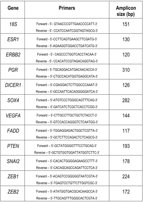
TABLE II.1-SEQUENCES OF THE PRIMERS USED FOR CANINE MRNA QUANTIFICATION. ... 39
TABLE II.2-AMPLIFICATION CONDITIONS USED FOR CANINE MRNA QUANTIFICATION. ... 40
TABLE VI.1-SUMMARY OF CANINE MAMMARY TUMORS INFORMATION ... 126
TABLE VI.2-PRIMER SEQUENCES AND AMPLICON SIZES USED FOR CANINE MRNA QUANTIFICATION BY RT-PCR.127 TABLE VI.3-AMPLIFICATION CONDITIONS USED FOR CANINE MRNA QUANTIFICATION. ... 128
xvii
List of Abbreviations
2-DE – Two-dimensional electrophoresis 5-AzaC - 5-Azacytidine
BCRP - breast cancer resistance protein BSA – Bovine serum albumin
CAFs - carcinoma-associated fibroblasts CDKN1B - cyclin-dependent kinase inhibitor 1B CDKN2A - Cyclin Dependent Kinase Inhibitor 2A
CMT – Canine mammary tumors CSC – Cancer stem cell
DDAVP - 1-deamino-8-d-arginine vasopressin DION - 1,10-phenanthroline-5,6-dione
DLS – Dynamic light scattering DFS – Disease free survival DNMT – DNA methyltransferase
DMEM - Dulbecco’s Modified Eagle’s Medium
DMEM-FBS-PenStrep – DMEM supplemented with 10% fetal bovine serum, and a mixture of penicillin (100 U/mL) and streptomycin (100 mg/mL)
EGF – Epithelial growth factor
EGFR – Epidermal growth factor receptor EMT – Epithelial to mesenchymal transition ESS – English Springer Spaniels
xviii FBS – Fetal Bovine Serum
FGF - fibroblast growth factor GH – Growth hormone
GHR – Growth hormone receptor
GnRH – Gonadotropin releasing hormone superagonist GWAS – Genome wide association study
HBC – Human breast cancer HE – Hematoxylin and eosin
HER2 – Human epidermal growth factor receptor 2 IBC – Inflammatory breast carcinoma
ICMT – inflammatory canine mammary tumor
ICP-MS - Inductively Coupled Plasma Mass Spectrometry IEF – Isoelectric electrophoresis
IGF-I – Insulin-like growth factor I IHC - Immunohistochemistry
IMC – Inflammatory mammary carcinoma INSR – Insulin receptor gene
HIF I – Hypoxia induced factor I MDR – Multidrug resistance
MES - 2-(N-morpholino)ethanesulfonic acid miRNAs – micro RNAs
MMPs – matrix metalloproteinases
MRP1 - multidrug resistance-associated protein 1 MVD – Microvessel density
NanoTS262 – gold nanoparticle system composed of polyethylene-glycol, bovine-serum-albumin and TS262
xix NSAID – Non-steroidal anti-inflammatory drugs
OS – Overall survival
PBS – Phosphate buffer saline PCA – Principal component analysis PEG - Polyethylene glycol
PGR – Progesterone receptor
PI3K/AKT - Phosphatidylinositol-4,5-bisphosphate 3-kinase/AKT serine/threonine kinase 1 PTEN – Phosphatase and tensin homolog
RT – Room temperature
SDS-PAGE - Sodium dodecyl sulfate polyacrilamide gel electrophoresis siRNA – Small interference RNA
SPR - surface plasmon resonance TAM – Tumor-associated macrophages
TBST – Tris buffered saline with 0.1% (v/v) Tween 20 TEM – Transmission electron microscopy
TGF-β - tumor growth factor beta VEGF - Vascular epithelial growth factor WHO – World health organization
1
I.
Bibliographic review in Canine Mammary
Tumors
1
I.1.
Thesis Motivation
Canine mammary tumors (CMTs) are the most frequent neoplasms diagnosed in non-spayed female dogs.From these, 50% of all CMTs are malignant, and mastectomy (and ovariohysterectomy in intact dogs) is the only broadly accepted treatment since chemotherapy with tamoxifen, doxorubicin and docetaxel, e.g., has not been proven effective against CMTs.
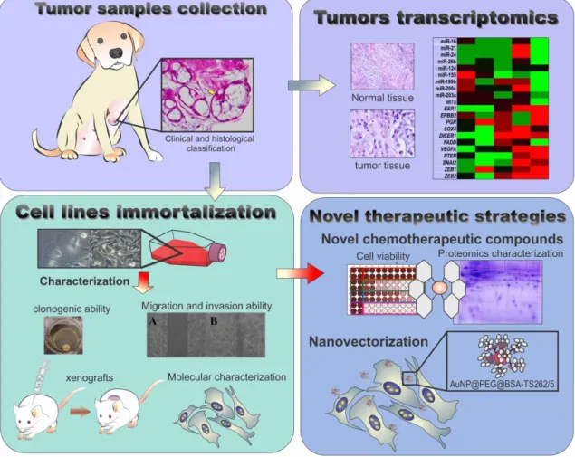
In Figure I.1, the outline of the project is presented.
Figure I.1 – Work plan highlighting major scopes of this thesis. CMTs will collected and processed for
2 Our aim was to establish new, well characterized CMT cell lines derived from the primary tumors by spontaneous immortalization. Relevant information on the CMTs collected during this work is described in table A.1 in the appendix. The molecular characterization of the cell lines will allow the identification molecular events and pathways responsible for their metastatic potential and cancer resistance.
The acquisition of metastatic potential has been linked to epithelial to mesenchymal transition (EMT) in human breast cancer (HBC). Based on the information available on HBC and in CMT models, we aimed to select panels of microRNAs (miRNAs) and mRNAs linked to this transition in order to identify molecular profiles linked to the metastatic transition in the original CMTs tissues and in the immortalized cell lines. At the same time, these new cell lines will serve as models for the identification of novel compounds with chemotherapeutic potential. For the most promising compounds their mechanism of action and molecular targets will also be explored using a quantitative proteomic analysis.
I.2.
Incidence
Mammary cancer is the most common neoplasia of the intact female dog and the second most frequent when both sexes are considered. In the Alameda county, California, USA the incidence of mammary cancer, was 252 per 100 000 female intact dogs per year.1 The reported
incidence of CMTs in the United Kingdom is 205 per 100 000 dogs per year,2 while in Sweden is
111 per 10 000 female dogs per year.3 The numbers described in the USA and the UK are similar
3 Tumor Registry reported that 56% of the diagnosed tumors were of the mammary gland while only 1.9% of male dogs were affected.5 These numbers are consistent with the findings in
Switzerland, where the country’s canine cancer registry revealed that 20.5% of all tumors in both
sexes were mammary tumors.6 The differences of incidence between sexes are consistent with
previous reports.7 Among the histopathological characterized CMTs, approximately 50% of the
tumors are malignant.8-10
I.3.
CMT Risk factors
I.3.1.Age
The majority of CMT cases are reported in dogs with 4 years of age or more.1-3, 8 The
average age of onset for benign CMT was been reported to be 8.5 years while for malignant CMT it has been reported to be 9.5 years.11 However, the average age of onset for overall CMTs has
also been reported to be 7.3 and 8.0 years.3, 12 Despite the differences in age of onset reported, it
is largely described that the incidence of CMT peaks at ages 9-12,1-4, 9 which is also the case
when all types of canine cancer are considered.5, 6, 13
I.3.2.Diet
Nutritional factors have been implicated in CMT onset. A study in pet dogs in the United States found that among spayed dogs, the risk of developing mammary cancer was reduced if the dogs were thin at 9 to 12 months of age.14 A study in Spain made a similar observation, that
4 observed that the consumption of homemade meals, when compared to the intake of commercial foods was also significantly associated with a higher incidence of CMTs.15 These authors also
associated the consumption of beef and pork with increased risk of the dogs developing mammary tumors.15
I.3.3.Hormonal dependency
The data collected from different canine tumor registries point that CMTs are rare in male dogs and frequent in intact female dogs.1, 4, 6, 7, 9 Furthermore, the incidence of CMTs is greatly
reduced in spayed female dogs when compared to intact female dogs.1, 9, 16 These observations
confirm that the onset of CMTs is dependent of sexual hormones. This also may be the reason for the increased incidence of CMT in Sweden (5 times higher) when compared to USA and UK, since in Scandinavia pet dogs are not routinely spayed.3
The relative risk of acquiring malignant tumors in female dogs spayed before the first estrus is 0.5%.16 If the dogs are spayed after the first or second estrus the relative risk rises to 8% or
26%, respectively.16 The risk for benign tumor decreases even when neutering is performed later
in the life of the dog but this does not reduce the risk of acquiring malignant tumors.17 A protective
effect of spaying is also observed when the animals are sterilized within two years after the detection of the malignant tumors.18, 19 The protective effect of early pregnancy observed in
women has not been demonstrated in dogs.16
5 frequent development of benign tumors, in dogs treated with progestins.21-24 A report by Geil et
al. 1977 reported that combinations that treatment of dogs with combinations of progestins with
estrogens led to the development of malignant mammary tumors in the animals tested.25
I.3.4.Breed dependency
Purebreds have been shown to be more predisposed to CMTs than crossbreds.1 It was
calculated a 7-fold higher risk for the purebred animals when compared with cross breed dogs.1
In particular, several spaniel breeds, the poodle, the terrier and dachshund breeds seem to be more predisposed to the condition.7, 13, 26-28 The differences in risk between breeds and between
purebreds and crossbreeds have been suggested to be due to genetic heritable components such as the ones observed in human breast cancer (see chapter I.3.5).28 Two Japanese studies
reported a correlation between the size of the dogs and mammary cancer: smaller dog breeds have a higher incidence of CMTs when compared with large dog breeds.13, 29
I.3.5.Germline mutations that predispose for CMTs
The BRCA1 and BRCA2 genes are known to increase the risk of HBC.30 Studying English
Springer Spaniels (ESS) one of the breeds with the higher risk of developing CMTs, showed that this risk was also associated with germline mutations in the canine BRCA1 and BRCA2 genes.31
Until now, several polymorphisms and insertion/deletion mutations have been identified in canine BRCA1 and BRCA2 genes that may be implicated in CMT predisposition.32-35
6
estrogen receptor α protein, ERα, were also implicated in the increased risk of the ESS breed to
develop CMTs.36
A germline deletion in the canine p53 gene, TP53, (exons 3 to 7) was described as being associated with the development of CMT in a boxer female dog.37
A genome wide association study (GWAS) performed in ESS dogs demonstrated an association between a deletion in chromosome 11, spanning the CDK5RAP2 gene, with the predisposition of the breed to CMTs.38
I.4.
CMT classification
7
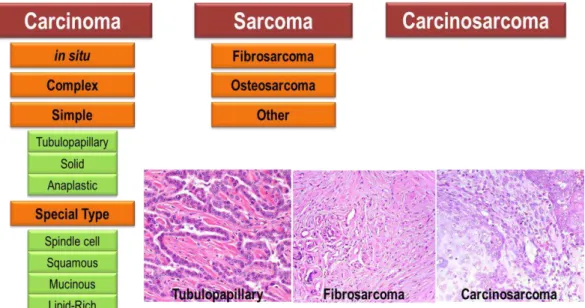
Figure I.2 –Classification of CMTs according to the 1999 WHO guidelines. Carcinomas are classified as in
situ (non-infiltrating), complex (with involvement of myoepithelial cells), simple and mixed (with involvement of mesenchymal cells). Less frequent types of carcinoma include spindle-cell, squamous, mucinous or lipid-rich carcinomas. Canine mammary sarcomas are classified as fibrosarcoma, osteosarcomas or other types of rare mammary tumors such as chondrosarcomas or liposarcomas. Carcinosarcoma is a rare type of mammary mixed tumor in which both the epithelial and mesenchymal components of the tumor are malignant. Images exemplifying a carcinoma, a sarcoma and a carcinosarcoma are also depicted.
Epithelial carcinomas are the most common form of malignant CMTs.8, 39 The most rare types
of malignant CMT are carcinosarcoma and carcinoma or sarcoma in benign tumor.39, 40 Sarcomas
such as chondrosarcomas and liposarcomas can also found be found in the mammary gland of dogs but are uncommon.39, 40
Besides man, dog is the unique animal species in which spontaneous inflammatory mammary carcinoma (IMC) has been reported.41 Both human inflammatory breast carcinoma
(IBC) and canine IMC are considered the most malignant type of mammary cancer with an extremely poor prognosis and is still poorly characterized in dogs.41 IMC and HBC cannot be
8
I.5.
CMT Etiology and clinical prognostic factors
CMTs progress from the benign to the malignant form.8, 11, 39 CMTs usually present
themselves in older intact female dogs, with 1 or, in approx. 70% of the cases, multiple tumors in one or both mammary chains and are predominantly located in the caudal abdominal and inguinal mammary glands.8, 11 When metastases occur, the primary sites are the regional lymph
nodes and the lungs due to the anatomy of the canine lymphatic system.42-44 The risk of dogs
developing primary CMTs is 32% greater in patients with previous benign or malignant CMTs and 58% if the CMTs are located in the ipsilateral side of the mammary gland.11, 45
The Elston and Ellis grading system is widely used in veterinary for the grading of CMTs, with slight adaptations (Table I.1).40, 46-49
Table I.1 – The Elston and Ellis grading system evaluates three morphological features of mammary
9 This method has been demonstrated as having prognostic value since the two year survival after surgery of dogs is higher in grade I CMTs, lesser in grade II and the smallest overall survival was found in the patients with grade III CMTs.47 Also the TNM staging system, which uses
information on tumor size (T), the presence of metastases at the local lymph nodes (N) and at other organs (M), and classifies CMTs in clinical stages I (initial stage) to V (final stage), has been shown to possess prognostic value (Table I.2).19 It was demonstrated that patients with
tumors greater than 5 cm in diameter and with clinical stages IV or V are associated with the worse two year survival.19
Table I.2 –TNM (Tumor-node-metastasis) staging system as defined by the WHO for CMTs. The staging
system considers the size of the primary tumor (T), the presence of metastasis at the regional lymph nodes (N) and the presence of metastasis in distant organs (M). Patients with stage I CMTs have higher overall survival time and on the contrary, patients with tumors larger than 5 cm or with metastasis at the lymph nodes (stage IV) or distant metastasis (stage V) have a poor overall survival time.
HBC have been classified in 5 molecular subtypes associated with different clinical outcomes: luminal A, luminal B, HER2-overexpressed, basal-like and normal-like.50-53 Luminal A
HBCs have the best prognosis and basal-like HBCs have the worst prognosis.53 Luminal B HBC
10 classification has been increasingly adopted in HBC, due to their proved prognostic value and the discriminative value for the use of targeted therapy for ER and HER2 expressing tumors.54
In CMT, identification of the molecular subtype of tumors has been also attempted by immunohistochemistry (IHC).55-61 However, four independent transcriptomic studies performed
did not confirm the existence of these (or other) molecular subtypes.62-65 Despite this limitation,
several authors assigned molecular classifications to the various CMTs with different conclusions on the prognostic value of the identification probably due to the different histopathologic markers used in the studies.56-58, 61 Also, the assumption made in all works, that luminal B mammary
tumors are HER2 positive and that luminal A CMTs are HER negative do not correlate with HBC, in which 14% of luminal A are HER2 positive and 24% of luminal B tumors are HER2 positive.54
Interestingly, Gama et al. 2008 observed a significant statistical correlation between poor survival, grade III and CMTs assigned to the basal-like phenotype.57 However, in a study by Sassi
et al. 2010, the majority of grade III CMTs were luminal B while tumors classified as basal-like
were grade I and no observable prognostic value was attributed to the molecular classification.58
A third study by Im et al. 2014 confirmed a correlation between poor prognosis, grade III and basal-like CMTs.56 Another IHC study used ERα, HER2 and CAV-1 staining for molecular
phenotyping, and Shinoda et al. 2014 found a significant correlation between the level of positive
staining, ERα localization, HER2 and CAV-1 staining and the behavior and prognosis of the tumor.61 However, the authors did not confirm a correlation between CMTs classified into the 5
11
I.6.
Canine mammary carcinogenesis
At the moment, there is a small understanding on which specific genetic alterations contributes to the progression of CMTs. However, major changes in the cellular metabolism of the mammary gland may contribute to the formation and development of CMTs.
I.6.1.Role of steroid hormones and growth factors in the
initiation and malignant progression in mammary
carcinogenesis
As depicted in I.3.3, epidemiology of CMTs shows a clear connection between sexual steroid hormones and the initiation of mammary carcinogenesis.
Estrogens and progesterone levels also appear to be essential for the acquisition of
malignant behavior in CMTs. Serum levels of progesterone, 17β-estradiol, androstenedione, dehydroepiandrosterone, testosterone, estrone sulfate, prolactin, growth hormone (GH) and of insulin-like growth factor I (IGF-I) were higher in the serum and in the tumor tissues of female dogs with malignant tumors compared with controls or with dogs with benign tumors.66-70 Higher
levels of epithelial growth factor (EGF) were also found in the tissue of dogs with malignant CMTs.71 The levels of prolactin, GH, IGF-I and EGF were significantly correlated with the levels of
17β-estradiol and progesterone.66, 67, 71
It has also been established that patient dogs with higher serum and mammary tissue
12 Administration of progestins to female dogs was shown to increase the secretion of GH in the mammary gland.72-74 Also, mammary cells producing GH where shown to be positive for the
progesterone receptor (PGR) by IHC.75 Parallel to the increase in GH production, a rise in the
blood levels of IGF-I and IGF-II has been shown to occur, which stimulate mammary cell proliferation.66, 76 Likewise, a synergy has been establish between 17β-estradiol and IGF-I
although the systemic or local origin of the IGF-1 has not been established by this study.66 It is
possible to speculate that GH/IHF-I stimulate the proliferation of mammary stem cells as a first step in the process of mammary carcinogenesis in progesterone dependent CMTs.66, 76 A recent
study observed an association between increased levels of aromatase, poorly differentiated (grade III) CMTs and overweight and obese dogs.77 The authors also found a correlation between
the expression of aromatase, leptin and IGF-I and the presence of ERα and PGR in the CMTs studied.77
These findings may justify that a faster growth of CMTs is probably due to the higher expression of aromatase and associated conversion of cholesterol into steroid hormones in the tumors or in the adjacent mammary tissue.77 The autocrine/paracrine production at the mammary
gland of GH, IGF-I, EGF and prolactin has been described.66, 70, 74-76, 78-80 It is possible that tumor
formation and malignant transformation depend on cellular division and growth induced by the increased expression of steroid hormones and, consequently, of the above mentioned growth factors by CMTs and/or by the adjacent normal mammary cells. In vitro, it was shown that knock-down of the growth hormone receptor (GHR) in CMT-U27 cell line (derived from canine mammary carcinoma) reduced the percentage of cells dividing, by down regulating the ERK1/2 signaling pathway, and increased the percentage of cells in apoptosis.81
13 expression is even more reduced in metastasis.82-90 Transcriptomic studies also corroborate
these findings: mRNA levels of ESR1 and PGR genes are decreased in metastatic CMTs.64, 65
The mRNA levels of Insulin receptor gene (INSR) were also found to be downregulated in metastatic CMTs.91
The ErbB protein family or epidermal growth factor receptor (EGFR) family is a family of four structurally related receptor tyrosine kinases, EGFR (ErbB1), HER-2 (ErbB2), ErbB3 and ErbB4.92 IHC has shown that EGFR is overexpressed in almost 50% (40.7% to 55.7%) of the
malignant CMTs analyzed while it is overexpressed in approximately 18.5% (17.4 to 19.6%) of benign CMTs.93-96 This overexpression has been positively correlated with tumor necrosis,
histological grade of malignancy (and in particular one of its features, mitotic index) and clinical stage (and with two of its features, tumor size and presence of metastasis at the regional lymph nodes).93, 95 Furthermore, RNA based studies show another aspect of the expression of EGF and
EGFR genes in metastatic CMTs: a downregulation of both in metastatic CMTs when compared
to normal mammary gland and non-metastatic CMTs.64, 97
IHC of CMTs for canine HER-2 receptor revealed that approximately 16% (12.5% to 19.1%) of malignant CMTs are overexpressing HER-2 while the benign CMTs are reported to express from 0% to 8.6%.96, 98-100. These values are in the same range as seen for HBC.98
Remarkably, HER-2 genomic amplification, which is present in 85% to 90% of the HBC HER-2 overexpressing tumors, was not found in the HER-2 positive CMTs.98
Discrepancies between the above summarized values of HER-2 expression are possible to observe in three other papers. A paper by Rungsipipat et al. 1999 reported that 50% of benign CMT overexpress HER-2 receptor as evaluated by IHC100 which is very different from the
14 of HER-2 which was not used in the other works.96, 98-100 Two other studies by Hsu et al. 2009
and Ressel et al. 2013, reported that 29.7% and 28.65% of malignant CMTs, respectively, had overexpression of HER-2.101, 102 The discrepancy between these studies and the previously
reported values for malignant CMTs is due to the interpretation of the Herceptest results. The authors of both papers consider tumors HER-2 positive when graded with the score of 2+, though the samples should only be considered positive with a score of 3+ in order to avoid equivocal results.98 This discrepancy in the analysis of IHC results is possible to observe even in works
from the same research group, e.g. Dutra et al. 2004 and Bertagnolli et al. 2011. In Dutra et al. 2004 the authors also assumed that CMT samples were positive when scored 2+ and reported an average of 34.4% of HER-2 positive results.99 For the purpose of this thesis, it was possible to
review the results available within that paper allowing the calculation of the percentage of HER-2 positive samples scored 3+ (12.5%). This number is similar to the 14.8% of HER-2 positive CMTs reported made in Bertagnolli et al. 2011 (from the same group) in which it was considered that only CMTs scored 3+ would be considered positive.96
I.6.2.Major genomic alterations involved in canine mammary
carcinogenesis revealed by global expression studies
Genomic copy number alterations are a main genomic event present in both HBC and CMTs.103, 104 Flow cytometry studies have shown that genomic instability increases from benign
to malignant CMTs as measured by DNA aneuploidy; 15% to 25% of benign CMTs display aneuploidy while 50% to 60% malignant tumors have shown to be have either an increase or a decrease in total amount of DNA.105-107 Variations in gene copy number have been reported in
15 CHEK2, COL9A3, CYP2E1, PTEN, MAP2K4, RB1 and TP53 genes, while gain in copy number
has been described for MYC, KIT and PFDN5 genes.103, 104
The importance of several signaling pathways in both HBC and CMT was highlighted in a microarray analysis of mRNA expression in mammary tumors from humans and dogs by Uva et al. 2009.63 Analysis of gene expression data revealed a great degree of similarity in the affected
signaling networks in both types of cancer.63 The acquisition of malignancy in CMTs was
accompanied by molecular signatures compatible with loss of phosphatase and tensin homolog (PTEN) function, hypoxia induced factor I (HIF I) activation [both signatures compatible with activation of the Phosphatidylinositol-4,5-bisphosphate 3-kinase/AKT serine/threonine kinase 1 (PI3K/AKT) signaling pathway], immune response expression signature, interferon signaling, activation of KRAS signaling pathway and expression loss of negative regulators of
Wingless-type MMTV integration site family member (WNT)/β-catenin and MAPK signaling pathways.63 A
recent work by Yu et al. (ahead of print) confirmed the importance of the role of WNT/β-catenin signaling in CMTs.108 In malignant CMTs it was found a significant upregulation of DKK1, SFRP1,
FZD3, CTNNB1, and LEF1 which correlated with higher levels of β-catenin and LEF1 protein in tumor cells.108
Analyzing malignant CMTs with different malignancy grades, Pawlowski et al. 2013 were able to find a significant overlap between mRNA expression patterns and histological grade III.62
The grade III CMTs were shown to have an increased expression of genes involved in inflammation and cytokine signaling, which can indicate that these events occur later in the canine mammary carcinogenesis.109
16 this work an overall decrease in the expression of membrane receptors, such as ESR1, PGR, EGFR, FGFR1, GHR, PDGFR, TGFDR, etc. which would indicate that metastatic CMTs are no
longer dependent of steroid hormones or growth factors to proliferate.64, 65
The expression changes of three genes (overexpression of BMP2 and DERL1 genes and downregulation of LTBP4 gene) were proposed to be discriminative of malignancy in CMTs.97
Proteomic studies were able to identify different expression patterns between benign, malignant and metastatic CMTs.110, 111 The benign expression profile consisted in the
overexpression of β-actin, keratin 8, keratin 17 , keratin 19 and phosphoglycerate mutase 1 proteins and downregulation of Calumenin, fibrinogen beta chain, fibrinogen gamma chain and siderophilin in non-malignant CMTs.110 The malignant expression profile involved the
overexpression of eukaryotic translation initiation factor 4A3, creatine kinase B, tropomyosin 1 α
and 14-3-3-ζ proteins and the downregulation of Gelsolin and peptidase D proteins in all malignant CMTs.110 In the metastatic expression profile it was possible to observe the
17
I.6.3. Oncogenes and tumor suppressor genes implicated in
canine mammary carcinogenesis
As already stated in chapter I.3.5, germline mutations in the canine genes BRCA1 and BRCA2 genes are connected with the increased risk of CMTs onset and progression.31-35 The
loss of BRCA1 nuclear localization was associated with ERα negative, PGR negative and triple
negative CMTs, and with the increased abundance of a high proliferation marker, Ki67, in malignant CMTs when compared to normal mammary gland and with dysplasia lesions.112, 113
The loss of nuclear localization of BRCA1 was accompanied by the ectopic localization of the protein in the cytoplasm of tumor cells.112, 113 The levels of BRCA2 mRNA were found to be
reduced in primary CMTs when compared to normal mammary tissue.114 In another study,
conflicting results were reported.115 BRCA2 gene was up-regulated in 50% of the malignant
CMTs analyzed and in 50% of the lymph nodes metastases of CMTs.115 At the same time, the
expression levels of RAD51 mRNA were found to be up-regulated in 60% of the primary CMTs and in 80% of the lymph nodes CMTs present in the study.115
As mentioned in chapter I.3.5, a germline deletion in canine TP53 gene was correlated with increased risk of dogs acquiring CMTs,37 however, somatic mutations in this gene have been
mainly encountered in malignant CMTs, suggesting that p53 has a similar role in CMTs and in HBC in the induction of malignancy in mammary cancer.37, 116-121
The Cyclin Dependent Kinase Inhibitor 1A, CDKN1A, mRNA levels for p21CIP1 were found to
be increased in all malignant CMTs tested, whereas only 40% of the benign CMTs and 40% of metastases CMTs tested displayed overexpression of CDKN1A mRNA.122 This may be an
18 The CKN1B cyclin-dependent kinase inhibitor 1B (CDKN1B) gene which codes for p27KIP1
was found to downregulated in 40% of the benign CMTs, 90% of the malignant CMTs and 80% of the metastatic CMTs metastases tested.122 By IHC, it was possible to identify nuclear p27KIP1 in
90% of benign of benign CMTs and in approximately 20% of malignant CMTs.123 Taken together
these observations seem to indicate that loss of p27KIP1 ability to inhibit cell cycle progression
may be associated with malignant progression in CMTs.122, 123
As previously mentioned in I.6.2, the loss of PTEN copy number and function are important events in canine mammary carcinogenesis. 63, 103, 104 The levels of PTEN mRNA were shown to
be 10 fold reduced in malignant CMTs and 100-fold reduced in metastatic CMTs when compared to normal mammary cells.124 However, only 33% of malignant CMTs had loss of PTEN protein, as
evaluated by IHC.125
Genetic alterations in genes involved in apoptotic pathways have been demonstrated to be crucial for the development of cancer.126 CMTs have been demonstrated to possess increased
expression of anti-apoptotic proteins such as Bcl-2, Bcl-X and survivin and a significant decreased expression of pro-apoptotic proteins such as Bax, Caspase 8 and Caspase 3.127, 128
I.6.4. Influence of tumor microenvironment in CMT growth,
migration and invasion
As tumors grow, the masses of cells need more nutrients from the microenvironment.129
Interaction of CMTs with the microenvironment will promote angiogenesis which will allow tumor growth.126
19 CMTs.130-132 Independent studies demonstrated positive correlations between VEGF increased
expression and other factors including CMT grade, microvessel density (MVD), HIF-1α, intra-tumor FoxP3 expression, COX-2, tumor-associated macrophages (TAM) and a shorter overall survival (OS) of the patient.130, 133-136 VEGF expression was also associated with the
expression of the receptor VEGFR-2 in CMT cells and in endothelial cells.137
Expression of COX-2 enzyme, an inducible enzyme important in the normal inflammatory response and angiogenesis through the production of prostaglandin H2 (PGH2), has been shown
to be overexpressed in malignant CMTs.138-140 Expression of COX-2 has been significantly
correlated with HER-2 overexpression, EGFR, VEGF, VEGFR-3, MVD, presence of regional metastasis, decreased OS and disease free survival (DFS).93, 135, 136, 141-143
These observations reveal the importance for VEGF and COX-2 in the angiogenesis associated with CMTs and its importance for tumor growth, and as biomarkers of tumor dedifferentiation and poorer prognosis.
In CMTs, TAMs inhibit the canonical WNT signaling pathway and, at the same time, induce the non-canonical WNT pathway in the CMTs which induced epithelial to mesenchymal transition (EMT) in tumor cells.144 In vitro it was also possible to observe the up-regulation of angiogenesis
and WNT related genes in macrophages co-cultured with canine mammary cell lines.145 In cancer
cells it was possible to observe the up-regulation of genes involved in cytokine/chemokine pro-inflammatory signaling and myeloid specific antigen related genes.145 Up-regulation of genes
associated with invasion, angiogenesis and EMT were also found in CMT derived cancer cells co-cultured with carcinoma-associated fibroblasts (CAFs).146
20 gain of mesenchymal characteristics by epithelial cells, such as the expression of vimentin, increased cellular mobility and loss of epithelial markers, e.g. E-cadherin.147, 149 Three major
signaling pathways have been described as implicated in the induction of EMT: WNT, tumor growth factor beta (TGF-β) and fibroblast growth factor (FGF).147, 149 Twist, Snail, Slug, ZEB1 and
ZEB2, are known transcriptional factors that induce EMT in cancer cells.147, 149 In HBC models,
EMT has been shown to generate cancer stem cell (CSC)-like cells expressing stem cell associated CD44+/CD24– antigenic profile and with self-renewal capabilities.150, 151
CSCs or tumor initiating cells are capable of self-renewal and are capable of generate a secondary tumor with the phenotypic heterogeneity of the primary tumor.147, 152 Thus, CSCs have
been proposed to be very important in mammary tumor invasion, formation of metastasis and resistance to chemotherapeutic compounds.147, 152, 153 CSCs have been identified in CMTs and in
tumor derived canine mammary cell lines.154-156
A class of small, 20 to 22 nucleotides, non-coding RNA, called miRNAs are negative post-transcriptional regulators of the gene expression, affecting the translation of more than 60% of genes by connecting.157 Genetic silencing is accomplished by base-paring between the seed
region (nucleotides 2-8 in the 5’ region) of the miRNA and the complementary region in the 3’ -untranslated region of the target mRNAs in the RISC complex.158, 159 Therefore, miRNAs can act
as key regulators of a particular pathway, regulating at the same time the expression of hundreds of genes while other miRNAs may target individual targets and some miRNAs have also been shown to regulate cooperatively the same mRNAs.157, 159 Differential expression of miRNAs has
21
Figure I.3 –Biogenesis of miRNAs. The primary miRNAs, pri-miRNAs, are transcribed by RNA polymerase II
and by RNA polymerase III.162-164 These pri-miRNAs are long molecules (more than 1 Kbp) with a terminal loop, a stem of approximately 33 base pairs (bp) and flanking segments of single stranded RNA.165 In the nucleus, the Microprocessor complex cleaves the pri-miRNA. This complex has two major subunits, Drosha ,a type III RNase and DiGeorge syndrome critical region gene 8, DGCR8, a double stranded RNA binding protein.166-168 DGCR8 interacts with the folded pri‑miRNAs and assists Drosha to cut the stem of pri-miRNA 11 bp away from the junction of single stranded RNA with double stranded RNA.169, 170 The cleavage of the pri-miRNA by Drosha originates 60 to 70 nt precursor miRNAs, pre-miRNAs, maintaining the stem-loop conformation, with a two nucleotides (nt) 3’ overhang characteristic of type III RNase activity.171 Pre-miRNAs are then exported to the cytoplasm by the Ran GTP-binding export receptor, exportin 5.172 The loop of the pre-miRNA is then cleaved in the cytoplasm by Dicer, another type III
RNase thus forming the mature double stranded miRNA with 22 nt and two 3’ overhangs.173-175 These miRNAs are incorporated as single stranded RNAs into the RNA-induced silencing complex, RISC.171 Although not necessary for its catalytic activity, Dicer has been shown to associate with two double strand RNA binding proteins, TRBP and PACT.176, 177 Dicer, TRBP, PACT and Ago2 the RISC Loading complex.178, 179 Ago2 is the effector unit of the human RISC complex 179. Genetic silencing is accomplished by base-paring between the seed region (nucleotides 2-8 in the
5’ region) of the miRNA in the RISC complex and the complementary region in the 3’-untranslated region of the target mRNAs.158 In boxes, the tumor suppressors and the oncogenic miRNAs (OncomiRs) used in this work.
Analysis of miRNA expression has shown an up-regulation of TGF-β signaling in CSCs obtained from canine mammary cell lines.180 Since their discovery, miRNAs, have been
22 as negative regulators of gene expression.181, 182 The differential expression of miRNAs has been
associated with tumor suppressive and oncogenic patterns.160, 161, 183 The observed miRNAs
expression variations in CMT are very similar to those described in HBC.160, 161, 183 Recently, a
large miRNA expression study of CMT derived cell lines, described a miRNA expression profile that is associated with down-regulation of the Cyclin Dependent Kinase Inhibitor 2A (CDKN2A) family tumor suppressor genes.183
I.7.
CMT treatment
Mastectomy and, in intact animals, ovariohysterectomy will be sufficient to heal dogs with benign and non-aggressive tumors.44, 184 Patients with aggressive or metastatic disease should
benefit from adjuvant chemotherapy.44, 184 Due to high similarity between CMTs and HBC, the
majority of treatment protocols used in patient dogs are adaptations of the from human medicine and the drugs used are off-label human medicines.185
I.7.1.Cytotoxic chemotherapy
Several studies tested chemotherapeutic agents as co-adjuvant therapy in CMTs. Protocols combining cyclophosphamide with 5-fluorouracil and cyclophosphamide, mitoxantrone and vincristine have been shown to increase overall survival time (OS) in animals with inflammatory CMTs (ICMT) with minor side-effects.186, 187
23 Treatment protocols with the taxanes paclitaxel and docetaxel revealed partial response (20%) and high toxicity associated with the treatment protocol while the treatment protocol with docetaxel revealed minor toxicity in the subject animals despite no significant difference was observed in DFS and OS when compared to the control group patients.189, 191
Animals treated with gemcitabine alone did not show any significant changes in DFS and OS.192 Dogs treated with a protocol combining gemcitabine with carboplatin showed only partial
response (13%) and gastrointestinal toxicity in patients of the test group.193 However, animals
treated with carboplatin alone had longer OS than the animals in the control group with minor side-effects reported.194
The lack of effectiveness of the chemotherapeutic drugs used in the treatment of CMTs may be due to the expression of efflux pumps such as multidrug resistance-associated protein 1 (MRP1) and breast cancer resistance protein (BCRP) that belong to the ABC transporters super family.195 It has been shown that malignant CMTs have an increased abundance of BCRP and
MDR1 transporter.196 Canine BCRP expression was associated, in cell line models, with
increased tolerance of CMTs to doxorubicin and cyclophosphamide, MDR1 was associated with tolerance to vinblastine and cisplatin resistance was associated to the expression of MDR1, MDR3 and BCRP.195, 197
I.7.2.Hormonal treatment
24 Co-adjuvant therapy with tamoxifen, an estrogen receptor antagonist, has been attempted in a clinical trial but treated dogs displayed severe estrogen like side effects and no benefit to patients was proven.44
Aglepristone, a PGR antagonist, was shown to reduce the expression of PGR and proliferation of tumors in dogs subject to treatment before surgery.198 However, no clinical trial
evaluating aglepristone treatment for CMT has been described so far.
Co-adjuvant treatment of dogs with goserelin, a gonadotropin releasing hormone superagonist (GnRH agonist), has shown to be promising.199 The dogs in the treatment group (9
animals) presented reduced levels of 17β estradiol and progesterone in blood and a reduction of tumor size.199 An objective response was observed in 100% of the subjects and DF was
increased.199 However, these results have not been confirmed in a larger group of animals.
I.7.3.Non-steroidal anti-inflammatory drugs (NSAID)
The correlation of COX-2 expression and angiogenesis provides support for a potential role of COX-2 inhibitors for the prevention and the treatment CMTs.136, 143 Piroxicam, a
non-selective COX inhibitor, has been tested in dogs with ICMT, with different results. De M Souza et al. 2009 observed an increase of the mean survival time of the dogs treated with piroxicam (183 days) compared with a control group treated with the anthracycline doxorubicin (7 days) while Clemente et al. 2009 reported a mean survival of 35 days of dogs treated with piroxicam.186, 188 The use of protocols with a combination of carboplatin and piroxicam or
carboplatin with firocoxib, a selective COX-2 inhibitor, in the treatment of dogs with CMTs.194 The
25
I.7.4.Receptor Tyrosine Kinase (RTK) Inhibitor, SU11654
(Toceranib)
A Phase I trial has been performed with SU11654, a selective inhibitor of the split kinase members of the RTK family, which include Flk-1/KDR, PDGFR, and Kit.200 Objective responses
were measure in subjects with CMTs, with 80% of CMT patients responding to treatment.200
I.7.5.Desmopressin and p62 vaccine
Desmopressin, 1-deamino-8-d-arginine vasopressin (DDAVP), is a synthetic derivative of antidiuretic hormone that has been proven effective as co-adjuvant treatment of surgery in CMTs.201, 202 Perioperative administration of DDAVP has shown to significantly increase DFS and
OS in animals with stage III and IV CMTs and in dogs with grade 2 and 3 CMTs.201, 202
The p62 vaccine is a DNA vaccine which consists in the pcDNA3.1 vector carrying the ubiquitin-binding protein p62 gene (SQSTM1).203 Dogs with advanced CMTs treated with the P62
DNA vaccine displayed tumor shrinkage or stabilization.203
I.7.6.Therapeutic compounds tested in vitro for the treatment of
CMTs
Several promising compounds have been tested in CMT derived cell lines for the treatment of CMTs.
Three selenium compounds, sodium selenite, methylseleninic acid, and methylselenocysteine, have been shown to significantly decrease cell viability and growth in CTM1211 cell line while inducing apoptosis.204 These selenium compounds were also shown to
26 The DNA methyltransferase (DNMT) inhibitor 5-Azacytidine (5-AzaC) has been shown to be toxic to cells of a primary tumor culture obtained from a CMT and reduced in vitro tumorigenicity.205
The non-steroidal aromatase inhibitor, letrozole has shown to be effective in reducing proliferation and cell viability of an ICMT derived cell line, IPC-366.206
A combined treatment of doxorubicin and deracoxib, a NSAID selective COX-2 inhibitor, has been shown to reduce 3 times the IC50 of doxorubicin in CMT-U27 cells which further
supports the use of COX-2 inhibitors in a combined therapy strategy.207
Masitinib, a selective inhibitor of the c-KIT, has been proven to sensitize CMT12 and CMT27 cell lines to the effects of gemcitabine.208
The use of small interference RNA (siRNA) to silence EGFR and ERBB2 genes as an useful therapeutic tool has been proven since it has reduced cellular proliferation, colony formation and migration of cells from REM134 and LILLY cell lines.209 The use of Gefitinib, an
inhibitor of EGFR, and GW583340, a dual inhibitor of EGFR and HER-2, have also been proven to reduce tumorigenicity of REM134 and LILLY cells.209
The anti-progestins mifepristone and onapristone have been shown to reduce cell viability in CMT-U27 cell line.210
Two natural-derived compounds that were described as inhibitors of metastasis, migrastatin analogues, were tested in CMT-W1, CMT-W2, CMT-W1M and CMT-W2M.211 Two
migrastatin analogues could inhibit cell viability cell migration and invasion capabilities in 3 of the cell lines.211
II.
Immortalization and characterization of a
new canine mammary tumour cell line
FR37
-
CMT
Disclaimer: Results and data presented in this chapter were published in:
Raposo, L.R.; Roma-Rodrigues, C.; Faisca, P.; Alves, M.; Henriques, J.; Carvalheiro, M.C.; Corvo, M.L.; Baptista, P.V.; Pombeiro, A.J.; Fernandes, A.R. (2016). "Immortalization and characterization of a new canine mammary tumour cell line FR37-CMT." Vet Comp Oncol. doi: 10.1111/vco.12235.
Raposo L.R., Santos S., Henriques, J., Alves, M., P Faísca, P., Beselga, A., J Correia, J., Fernandes, A. R.. “Immortalization of primary canine cell lines from mammary tumors: a protocol
optimization.” XVIII Meeting of the Portuguese Society of Animal Pathology. Évora. May 9-10, 2013.
II.1.
Abstract
Here we describe the establishment of a new canine mammary tumour (CMT) cell line, FR37-CMT that does not show a dependence on female hormonal signalling to induce tumour xenografts in NOD-SCID mice. FR37-CMT cell line has a stellate or fusiform shape, displays the ability to reorganize the collagen matrix in the collagen colony assay, expresses vimentin, CD44 and shows the loss of epithelial markers, such as E-cadherin. Loss of E-cadherin is considered to be a fundamental event in epithelial to mesenchymal transition (EMT). The up-regulation of ZEB1, the detection of phosphorylated ERK1/2 and the downregulation of DICER1 and miR-200c
are also in accordance with the mesenchymal characteristics of FR37-CMT cell line. FR37-CMT shows a higher resistance to cisplatin (IC50>50 µM) and to doxorubicin (IC50>5.3 µM) compared
to other CMT cell lines. These results support the use of FR37-CMT as a new CMT model that may assist the understanding of the molecular mechanisms underlying EMT, CMT drug resistance, fostering the development of novel therapies targeting CMT.
II.2.
Keywords
II.3.
Introduction
Canine mammary tumours (CMTs) are the most common tumours diagnosed in intact female dogs.6, 9 Approximately 50% of all CMTs are malignant, for which mastectomy (and
ovariohysterectomy in intact dogs) is the only broadly accepted therapy since chemotherapy with agents, such as doxorubicin and docetaxel, does not radically increase overall survival time.189
Despite the established dependence of CMT development on oestrogens and progesterone, tamoxifen [an oestrogen receptor alpha (ERα) antagonist] has not been proven effective against CMT.44 Therefore, it is crucial to understand the mechanisms underlying the inefficacy of
chemotherapy either by intrinsic or acquired drug resistance in dogs.
The epithelial to mesenchymal transition (EMT) is an important molecular reprogramming event that may allow canine mammary cancer cells to acquire mobility through changes in the expression of adhesion molecules (e.g. loss of E-cadherin expression and gain of N-cadherin) and reorganization of the cytoskeleton (e.g. gain of vimentin expression), for example, that result in the change of cellular polarity from top to bottom to back to front.147 The shift in polarity may
also be accompanied by the expression of other proteins such as metalloproteinases that allow extracellular matrix reorganization and therefore facilitate not only migration but also invasion.147
These changes are induced by several transcription factors, such as Twist, Snail, Zeb1, Zeb2, that modulate expression of receptors involved in cell signalling, such as epithelial growth factor
receptor (EGFR) and ERα, and alter the tolerance of cancer cells to chemotherapeutic agents.147, 148 Hallmarks of EMT are the gain of mesenchymal characteristics by epithelial cells, such as the
expression of vimentin, increased cellular mobility and loss of epithelial markers, e.g. E-cadherin.147 Consequently, it is of major importance to develop cellular models of CMT