*e-mail: [email protected]
Development and Characterization of a Novel Bioresorbable and
Bioactive Biomaterial Based on Polyvinyl Acetate, Calcium Carbonate
and Coralline Hydroxyapatite
Javier Aragóna,*, Ramón Gonzáleza, Gastón Fuentesb, Luca Palinc, Gianluca Crocec, Davide Viterboc
a
National Center for Scientific Research, Havana, Cuba
b
Biomaterials Center, University of Havana, Cuba
c
Department of Advanced Science and Technology, University of Piemonte Orientale,
“A. Avogadro”, Alessandria, Italy
Received: June 15, 2010; Revised: December 8, 2010
Coralina® HAP-200 (coralline hydroxyapatite obtained by hydrothermal treatment of marine corals) and
POVIAC® (polymeric matrix based on PVAc), commercial trade marks were mixed with a natural product from
the Cuban sea costs, i.e. calcium carbonate from Porites Porites coral, to obtain a novel bioactive composite with potential use as bone restoration material. The samples were characterized by physical-chemical (FTIR, XRD, SEM, EDS) and mechanical studies. It was shown that there is no chemical interaction between the inorganic filler and the polymer matrix, each conserving the original properties of the raw materials. The studied formulation had a compressive strength similar to that reported for trabecular bone. Scanning electron microscopy examination revealed that the addition of CaCO3 induces a change on the morphologic structure of the composite obtained after 30 days of SBF immersion. These composites generate novel biomaterials capable of promoting the deposition of a new phase, a Ca-P layer due to the bioactivity of a Ca2+ precursors.
Keywords: polyvinyl acetate, hydroxyapatite coralline, calcium carbonate, composite
1. Introduction
Over the past two decades many synthetic bone substitutes have been made, almost all of them, based on calcium phosphate, due to their excellent biocompatibility. Particularly, coralline hydroxyapatite is currently used in clinical applications as a bone substitute for medical applications, because its chemical composition and tridimensional porous morphology is very similar to human bone1,2.
The coralline hydroxyapatite particles tend to migrate under externally applied forces during the healing period.
Numerous bioresorbable materials have been investigated for tissue repair, including natural3,4, and synthetic polymers5-9. One of
the possible solutions to this problem is to combine the coralline hydroxyapatite particles with collagen10, chitosan11, polylactic acid
(PLA)7,8, polyglycolic acid (PLG)6, and polyvinyl acetate5-12.
Many practical advantages arise when using synthetic scaffolds, because precise engineering of material composition and micro and macrostructure is possible. This allows adequate control of scaffold properties, thus creating optimal conditions for cell survival and proliferation, after bone tissue formation6.
However, some problems have been encountered regarding the use of these polymers in tissue engineering applications due to the release of acidic degradation products leading to inflammatory responses5-9.
Another limitation in the use of biodegradable polymers (i.e. PLG, PLA and its combinations) is the lack of bioactivity, in particular for bone tissue applications, if not used as composites13.
If biodegradability and bioactivity are to be combined in engineering an optimized tissue scaffold, then the composite design offers an exceptional opportunity: the preparation of bioresorbable and bioactive scaffolds with tailored physical and mechanical properties14. Moreover, composites can be engineered in such a way
that their resorption rate is similar to the new tissue formation rate.
Various approaches to the development of bioresorbable and bioactive composites for tissue engineering applications are being investigated worldwide in different scaffold architectures14,15.
Specifically, the bioactivity of “Poliapatita” has been investigated as a resource for its best performance in bone tissue restoration. “Poliapatita” samples immersed in SBF for regular times between 1and 28 days, showed a good bioactivity. This capability was checked by SEM + EDS and progressive decrease of calcium and phosphorus concentration was observed, higher in case of phosphorus. In the first 7 days in SBF the rate of ionic deposition is quite fast and in the following days, as the Ca-P layers increase, the rate of ionic deposition decreases with time16.
Composites slightly different from the original PMMA cements were evaluated for restoration of bone tissue. Modifications such as addition of vinyl acetate and HAP-200 induced a decrease of the mechanical properties of the classical PMMA bone acrylic cements. These changes in the formulation increase the bioactivity (subsequently, degradation, capability as drug delivery system, etc.), but compromise the performance as material for bone restoration17.
Several other reports about the influence of chemical composition, morphology, particle size distribution and molecular weight on the final properties of the bone cements were published. The addition of calcium generators (HAP-200/HA 3 and/or CaCO3) reduces the peak temperature and setting times, rising the bioactivity and the drug delivery ability of materials18,19.
The components used in the manufacturing of “Poliapatita” leave many Ca2+ ions in the formulation., The biological activity
will increase, because calcium carbonate from coral is more bioresorbable20, and in vivo bone reconstruction could be significantly
Recent studies showed that a composite formed by POVIAC®/
CaCO3/HAP-200 is a good candidate as bone restoration material and can also be considered as a matrix for the delivery of drugs such as acetylsalicylic acid and cephalexin24-26.
In the present work novel bioactive and bioresorbable composite materials were prepared and characterized using polyvinyl acetate (POVIAC®) impregnated with bioresorbable calcium carbonate
and bioactive coralline hydroxyapatite (HAP-200) particles. The materials are intended as composites for bone tissue engineering applications.
2. Materials and Methods
2.1. Materials
Calcium carbonate (Aragonite) was obtained from marine corals of the Cuban coralline barrier and poly (vinyl acetate) (POVIAC®)
(MW = 24 400, ≤ 2% residual monomer) was obtained in the National Center Scientifi c Research (Cuba) by in house procedures27. The
HAP-200 coralline hydroxyapatite used in this study was prepared in the National Center Scientifi c Research (Cuba), by hydrothermal reaction28.
2.2. Preparation of the POVIAC®/CaCO
3/
HAP-200 composite
POVIAC®/CaCO
3/HAP-200 composite scaffolds were prepared
using compression molding and thermal processing. Initially, POVIAC® powders were dissolved in ethanol at room temperature,
then the requested fractions of CaCO3 and HAP-200 particles were gradually added to the POVIAC® solution and thoroughly mixed
for 1 hour (POVIAC®/ CaCO
3/HAP-200 = 1/2/7; w/w/w). Later,
the mixture was dried at 90 °C under vacuum to remove residual organic solvents. Finally, for in vitro experiments, the composite was compacted using an axial compression mold at 10 MPa for 1 minute. The compact disks had dimensions of 13 mm in diameter and 6,5 mm in thickness.
2.3. In vitro studies in SBF and physical-chemical
and mechanic characterization
POVIAC®/ CaCO
3/HAP-200 composite samples were
characterized by Fourier-transform infrared (FT-IR) spectroscopy, X-ray diffraction (XRD). Scanning electron microscopy (SEM) equipped with an energy dispersive spectrometer (EDS), was employed to examine the morphology, pore size and pore distribution of the samples; they were gold-coated and observed at an accelerating voltage of 20 kV.
The compressive strength (CS) testing was conducted on the composite disks using a universal machine test (MTS 810 Material Test System), at room temperature with a crosshead speed of 1 mm/min. The reported results are an average of the values from fi ve samples.
In vitro studies in simulated body fl uid (SBF) were carried out using the SBF composition and the standard procedures described by Kokubo in 200629. Composites were immersed in 10 mL of SBF for
30 days at 37 °C and, every 72 hours in the fi rst 15 days and every week in the last 15 days, the liquid phase was replaced with 10 mL of fresh SBF. After immersion in SBF the samples were characterized using XRD, FT-IR and SEM to verify whether HA was formed on the surfaces of the composites.
3. Results and Discussion
3.1. Characterization of POVIAC®/CaCO
3/
HAP-200 composite
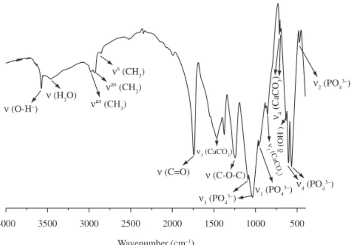
The structures of the composite were analyzed using FT-IR spectroscopy, as shown in Figure 1.
Characteristic structural bands of POVIAC®, CaCO 3 and
HAP-200 were observed in the composite spectrum. The vibration bands at ca. 2975 and 2852 cm-1 can be attributed to the asymmetric
(νas(CH
3)) and symmetric (νs(CH3)) stretching of CH3 respectively,
the band at 2927 cm-1 corresponds to the CH
2 (νas(CH2)) asymmetric
stretching. The band at 1740 cm-1 is due to the CO group stretching
(ν(C=O)) and that at 1245 cm-1 corresponds to the stretching vibration
of the C-O-C group (ν(C-O-C)), confi rming the presenze of the typical side chains of PVAc 30. Phosphate characteristic bands where observed
at: 472 cm-1 ν
2 (PO43-), at 569 and 601 cm-1 ν4 (PO43-), at 960 cm-1 and
at 1088 and 1041 cm-1 ν
1 (PO43-), and ν3 (PO43-). These bands indicate
the classifi cation of the polyhedrons of PO43- in the structure of the
hydroxyapatite31. Besides, at 3570 cm-1 the main ν(OH-) vibration is
observed together with the bands at 3450 due to hydrogen bonded water molecules adsorbed during the preparation process. The band at 633 cm-1 is attributed to the OH- groups (δ)31 and the band at ca.
854 cm–1 may be attributed to the characteristic carbonate out-of-plane
bending (ν
2 mode) vibrations of aragonite. The pair of bands at ca.
713 and 700 cm–1 may be assigned to the in-plane bending modes
(ν4 mode) which are characteristic of the aragonite structure. The band corresponding to the ν1 vibration mode is masked by the doublet corresponding to ν3 (PO43-). No vibration bands characteristic of
Calcite and Vaterite were detected in the FTIR spectrum, further demonstrating that the coral rods are pure aragonite32.
Figure 2 shows the XRD patterns of the POVIAC®/CaCO 3/
HAP-200 composite powder. By comparison with the JCPDS fi les No.71-2396 and No.09-0432, the diffraction peaks can be well identifi ed as due to orthorhombic aragonite with lattice parameters of a0 = 4,961 Å, b0 = 7,970 Å, c0 = 5,739 Å, and space group Pmcn and hexagonal syntetic hydroxyapatite with lattice parameters of a0 = b0 = 9,418 Å, c0 = 6,884 Å, and space group P63/m respectively.
Both in the FT-IR spectra and in the XRD patterns, no signifi cant changes or shifts in the characteristic bands and in the diffraction peaks were observed in the composite biomaterial with respect to the
Figure 1. FT-IR spectra of the POVIAC®/CaCO
starting components (POVIAC®, CaCO
3 and HAP-200), suggesting
that no chemical reactions occurred between the mixed components, which conserve the original properties of the raw materials. The compact structure of the POVIAC®/CaCO
3/HAP-200 composite is
refl ected in the mechanical properties of the obtained composite. This composite present a value of CS = 20 ± 2 MPa, a value in the range observed for trabecular bone (3-20 MPa)33-34. According to recent
reports, the percentage of diminution of mechanical properties by immersion in biological fl uids of similar materials was 25%[16]. Then
we can evaluate that the CS of our formulation after inmersion would be 15 MPa approximately.
3.2. In vitro study
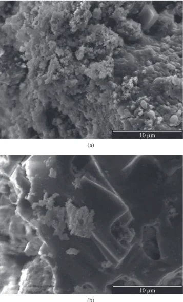
The microscopic observation of the external surfaces showed that the particles of HAP-200 and CaCO3 are agglutinated by the POVIAC®,
and a compact block structure is formed (Figure 3a and 3b). It can also be seen that the composites have a low porosity and the structure of the material is rather dense.
Figures 3c and 3d show that after 30 days in SBF the quantity of pores and the external morphology of the biomaterial composite
Figure 3. Surface morphology of the composite by SEM analysis: before immersion in SBF at different magnifi cations: a) 75×; b) 500×; after soaking in SBF for 30 days at different magnifi cations: c) 75×; and d) 500×.
Figure 2. X-ray diffraction patterns of POVIAC®/CaCO
is signifi cantly different, because of the partial degradation of the calcium carbonate. At the surface of this formulation an erosion phenomenon takes place, causing an increase in the porosity of the biomaterial. This increment could be very advantageous, since it would facilitate the growth of new bone tissue into the implanted material, favoring the process of bone integration.
Moreover, after 30 days of SBF immersion, it can be seen how on the surface of the whole formulation cavities (Figure 3d) were opened, limited by calcium phosphate and carbonate particles. EDX analysis inside one of the above cavities proved that their surface is rich in polymer phase (Figure 4a), as indicated by the low calcium and phosphorus content and the high carbon and oxygen content. Instead, the areas near these caves are rich in the inorganic phase, as shown by the signifi cant increase in phosphorus and calcium content (Figure 4b).
The inorganic phase rich in calcium and phosphorus can be seen at higher magnifi cation (10000×) in Figure 5a. In this case it can be seen that, after 30 days of SBF immersion, the surface of the composite biomaterial is completely covered by a continuous layer. This layer is formed by the deposition of a new, calcium phosphate phase,
characterized by arrangements of nanometric crystallites, typical of the formation of nanodispersed hydroxyapatite. Furthermore, in the region rich in POVIAC®, agglomerates of nanometric hydroxyapatite
crystals fi rmly attached to the polymer (Figure 5b) can be seen. This result is very important because so far there are no reports in the literature demonstrating the deposition of an apatite layer on polyvinyl acetate. It has been recently reported that the incorporation of vinyl acetate in PMMA containing composites increases the probability of apatite layer formation in acrylic bone cements17.
The samples described in this work do not contain PMMA and the formation mechanism of those layers in the two cases is different. In PMMA-PVAc composites the presence of PVAc just increases the hydrophilic character of the acrylic bone cement, while the POVIAC®/
CaCO3/HAP formulation is designed from the very beginning as a hydrophilic composite.
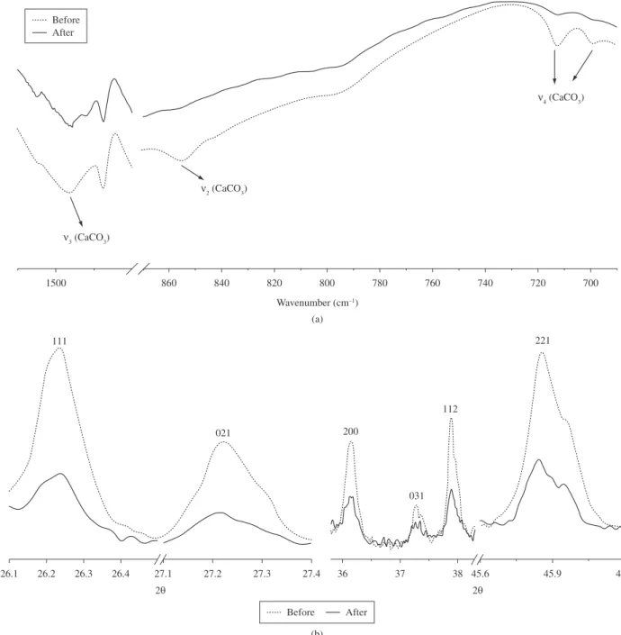
The erosion phenomenon is due to the degradation of calcium carbonate particles present in the biomaterial composite as shown in Figure 6.
The analysis of the composites after immersion in SBF by FT-IR spectroscopy and X-ray diffraction, shows how the intensity of the
Figure 5. Surface morphology of POVIAC®/CaCO
3/HAP-200 composite
after immersion in SBF at higher magnifi cation: a) near the cavity; and b) inside the cavity.
calcium carbonate characteristic structural bands is considerably reduced (Figure 6a), and the intensity of the typical CaCO3 refl exions in the XRD patterns is lowered in a similar way (Figure 6b).
4. Conclusions
A new organic-inorganic composite, that can be used as bone substitute in places where the requested compressive strength is similar to that reported for trabecular bone, was obtained. Physical-chemical characterization showed that there are no chemical interactions between the inorganic fi ller and the polymer matrix. Besides, the biomaterial compound has a dense morphological structure with a low porosity, which increases when in contact with SBF for a period of 30 days. It was observed that the biomaterial was
able to favor the formation and the deposition over the inorganic fi ller and the polymeric matrix of a new calcium phosphate phase, with the typical features of nanometric hydroxyapatite.
Acknowledgements
We thank FONDAZIONE ISI for the fi nancial support for this scientifi c project.
References
1. Roy DM and Linnehan SK. Hydroxyapatite formed from coral skeletal
carbonate by hydrothermal exchange. Nature. 1974; 247(438):220-222.
2. Ripamonti U, Crooks J, Khoali L and Roden L. The induction of bone formation by coral-derived calcium carbonate/hydroxyapatite constructs.
Biomaterials. 2009; 30(7):1428-1439.
Figure 6. Physicochemical characterizations of POVIAC®/CaCO
3/HAP-200 composite, before and after soaking in SBF for 30 days: a) FT-IR analyses; and
3. Piskin E. Biomaterials in different forms for tissue engineering: an
overview. Material Science Forum. 1997; 250:1-14.
4. Zhang Y and Zhang M. Synthesis and characterization of macroporous chitosan/calcium phosphate composite scaffolds for tissue engineering.
Journal of Biomedical Materials Research Part A. 2001; 55(3):304-312.
5. Maquet V and Jerome R. Design of macroporous biodegradable polymer
scaffolds for cell transplantation. Material Science Forum. 1997;
250:15-42.
6. Agrawal CM, Athanasiou KA and Heckman JD. Biodegradable PLA-PGA
polymers for tissue engineering in orthopadics. Material Science Forum.
1997; 250:115-129.
7. Schugens C, Maquet V, Grandfils C, Jerome R and Teyssie P. Biodegradable and macroporous polylactide implants for cell transplantation: I. Preparation of macroporous polylactide supports by
solid–liquid phase separation. Polymer. 1996; 37(6):1027-1038.
8. Schugens C, Maquet V, Grandfils C, Jerome R and Teyssie P. Polylactide macroporous biodegradable implants for cell transplantation. II. Preparation of polylactide foams by liquid-liquid phase separation.
Journal of Biomedical Materials Research Part A. 1996; 30(4):449-461.
9. Agrawal CM and Ray RB. Biodegradable polymeric scaffolds for
musculoskeletal tissue engineering. Journal of Biomedical Materials
Research Part A. 2001; 55(2):141-150.
10. Allard R, Bejui J and Huc A. Substitute bony matrix products promoting
osteogenesis. United State Patent No. US5071436. 1991. Available from:
http://www.boliven.com/patent/US5071436 Access in: 12/2009. 11. Sivakumar M and Panduranga Rao K. Preparation, characterization and
in vitro release of gentamicin from coralline hydroxyapatite–gelatin
composite microspheres. Biomaterials. 2002; 23(15):3175-3181.
12. Gonzalez R and Suzarte A. Composite biomaterial for bone implants.
United State Patent No. 0053951. 2007. Available from: http://www. freepatentsonline.com/20070053951.html. Access in: 12/2009. 13. Schliephake H, Neukam FW, Hutmacher D and Becker J. Enhancement
of bone in-growth into a porous HA-matrix using a resorbable polylactice
membrane. Journal of Oral and Maxillofacial Surgery. 1994; 52(1):57-63.
14. Verheyen CCPM, de Wijn JR, Van Blitterswijk CA, de Groot K and Rozing PM. Hydroxyapatite/poly(l-lactide) composites: an animal study
on push-out strengths and interface histology. Journal of Biomedical
Materials Research Part A. 1993; 27(4):433-444.
15. Durucan C and Brown PW. Biodegradable hydroxyapatite-polymer
composites. Advanced Engineering Materials. 2001; 3(5):227-231.
16. Castro H and Ledea O. Determinación de la bioactividad y la resistencia
a la compresión de bloques de POLIAPATITA®. Química Nova. 2010;
33(4):891-894.
17. Brizuela N, Lopez M and González R. Cementos óseos acrílicos modificados con hidroxiapatita/acetato de vinilo. Caracterización
mecánica, termoanálitica y bioactividad in vitro. Polímeros. 2010;
20(2):98-106.
18. López M, Fuentes G, González R, González J, Peón E and Toledo C. PMMA/Ca2+ Bone cements. Part I. Physico chemical and
thermoanalytical characterization. Latin American Applied Research.
2008; 38(3):227-234.
19. Arias D and González R. Biomaterial de implante óseo compuesto
de HAP-polivinilacetato. Revista CENIC Ciencias Químicas. 2004;
35(2):101-103.
20. Wikesjo UM, Lim WH, Razi SS, Sigurdsson TJ, Lee MB, Tatakis DN et al. Periodontal repair in dogs: a bioabsorbable calcium carbonate coral implant enhances space provision for alveolar bone regeneration in
conjunction with guided tissue regeneration. Journal of Periodontology.
2003; 74(7):957-964.
21. Paul W and Sharma CP. Ceramic drug delivery: a perspective. Journal
of Biomaterials Applications. 2003; 17(4):253-264.
22. Green D, Walsh D, Yang X, Mann S and Oreffo ROC. Stimulation of human bone marrow stromal cells using growth factor encapsulated
calcium carbonate porous microspheres. Journal of Materials Chemistry.
2004; 14:2206-2213.
23. Green DW, Bolland BJRF, Kanczler JM, Lanham SA, Walsh D, Mann S et al. Augmentation of skeletal tissue formation in impaction bone
grafting using vaterite microsphere biocomposites. Biomaterials. 2009;
30(10):1918-1927.
24. Aragón J, Brizuela N and González R. Perfil de liberación controlada de
ASA en biomateriales compuestos de hidroxiapatita-POVIAC. Revista
CENIC Ciencias Químicas. 2006; 37(3):179-181.
25. Aragón J, González R and Fuentes G. Cinética de liberación de cefalexiana
desde un biomaterial compuesto por HAP-200/POVIAC/CaCO3. Anales
de la Real Academia Nacional de Farmacia. 2009; 75(3):345-363.
26. Aragón J, González R, Nayrim B and Oliver L. Estudio cinético de liberación in vitro en un biomaterial compuesto por HAP-200/POVIAC/
CaCO3. Revista Iberoamericana de Polímeros. 2009; 10(2):119-130.
27. Suzarte A, Jordán G, Echevarría M, Iglesias G and Díaz E. Procedures for
obtaining polymers derived from vinyl acetate and their uses. United State Patent No. 0306226. 2009. Available from: http://www.freepatentsonline. com/y2009/0306226.html. Access in: 12/2009.
28. González R, Handal E and Fernández J. Cinética de la reacción de
transformación hidrotérmica del coral a hidroxiapatita. Química Nova.
1993;16(6):513-516.
29. Kokubo T and Takadama H. How useful is SBF inpredicting in vivo bone
bioactivity. Biomaterials. 2006; 27(15):2907-2915.
30. Gómez A. Estudio de Kollidon® SR y su aplicación al diseño de
suspensiones orales opiáceos. [Ph.D. Thesis]. Universidad de Granada;
2007. Available from: http://hera.ugr.es/tesisugr/17241212.pdf. Access in: 02/2010.
31. Fowler BO. Infrared Studies of apatites. I. Vibrational assignments for calcium, strontium, and barium hydroxyapatites utilizing isotopic
substitution. Inorganic Chemistry. 1974; 13(1):194-207.
32. Gen-Tao Z, Qi-Zhi Y, Jie N and Gu J. Formation of aragonite mesocrystals
and implication for biomineralization. American Mineralogist. 2009;
94(2-3):293-302.
33. Comín M, Peris J, Prat J, Dejoz J, Vera P and Hoyos J. Biomecánica de la
fractura ósea y técnicas de reparación. Valencia: Instituto de Biomecánica
de Valencia; 1999.
34. Cowin SC. Bone mechanics handbook. Florida, Boca Raton: CRC Press;