CALCIUM PHOSPHATE BIOMATERIALS FROM MARINE ALGAE. HYDROTHERMAL SYNTHESIS AND CHARACTERISATION
G. Felício-Fernandes e Mauro C. M. Laranjeira*
Departamento de Química - Universidade Federal de Sta. Catarina - 88040-900 - Florianópolis - SC
Recebido em 18/2/99; aceito em 27/1/00
Calcium phosphate compounds such as Hydroxyapatite (HAp) were prepared by hydrothermal syn-thesis with phycogenic CaCO3 as starting material. Material obtained was characterised by usual methods (XRD, FTIR, TG, N2-adsorption, SEM and EDX) in order to study its physical-chemical characteristics. The prepared HAp showed that it may be suitable for use as a biomaterial.
Keywords: hydroxyapatite; calcium phosphate ceramics; algae. ARTIGO
INTRODUCTION
Research on the synthesis of calcium phosphate compounds such as hydroxyapatite (HAp) (Ca10(PO4)6(OH)2) took on great
momentum when it was observed that this substance is present in substantial amounts in the mineralised tissue of the vertebrates – 60-70% of the mineral phase of the human bone1. In order to
propose the biosynthetic processes of the bone, the synthesis of HAp was developed in the laboratory. This calcium phosphate compound was chemically similar to that found in bone and was found to have a great potential for use as an auxiliary in bone regeneration2-6. In recent years bioceramics based on calcium
phosphate salts have received attention as bone substitutes7. These
materials may be resorbable (tricalcium phosphate), bioactive (hydroxyapatite and bioactive glasses), porous for tissue ingrowth (hydroxyapatite-coated metals)8 or composites
(stainless-steel-fibre-reinforced bioglass)9-11.
The human bone is formed basically by an organic phase and other minerals. In the organic phase, the fibres of collagen serve as a matrix for the precipitation of HAp, determining the organisation of the crystals. The collagen gives the bone its elastic resistance. The mineral phase is formed by HAp. The microstructure of the mineral phase of the bone is directly linked to the mechanical requirements of the location in the skeleton12. Two different types of bone tissue are observed:
the compact or cortical and the trabecular or spongy bone tissue4: The trabecular bone forms a homogeneous porous
three-dimensional structure with interconnected pores of characteristic diameters. Both types of bone tissue are anisotropic, a typical characteristic of crystalline bodies. To produce a material with similar microstructure is of great interest in the areas of biomedicine linked to the development of prostheses for bone reconstitution or substitution. This is due to the unique property of bone tissue to regenerate, forming new healthy tissue that grows in the direction of the damaged area, filling in with functional bone. If a physical and
chemically similar material is placed in contact with this healthy bone, then the growth process is accelerated and the material is filled in with new bone and reabsorbed13.
HAp that integrates the mineral phase of the bone is said to be non-stoichiometric and calcium-deficient with a relationship of Ca/P < 1.67. However, some difficulties exist in the study of this HAp that occurs in the bones in the form of small crystals without defined orientation and with a great amount of organic matter and other bound or adsorbed ions at the surface14. Nevertheless, the
substitution of several ions such as CO32- with 4-6% of substitution
is observable12. When the non-stoichiometric HAp is synthesised,
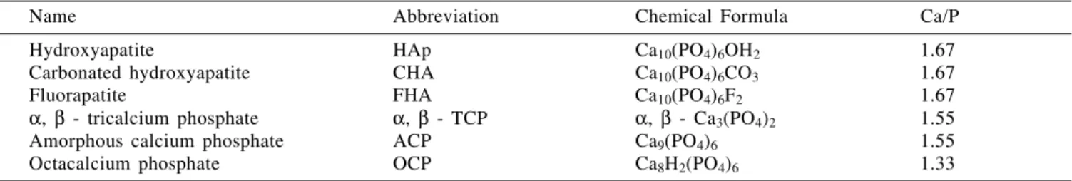
other intermediary products can be formed. An example is octacalcium phosphate (OCP), which forms a mixture of two intercalated layers of HAp/OCP/HAp15-17. Table 1 introduces some
calcium phosphates that can appear as by-products of the synthesis of HAp and whose Ca/P ratios are quite close to one another.
To use a porous material of natural origin as a basic raw ma-terial for the synthesis of calcium phosphates, simulating bone and stimulating its development, is an idea that was put into practice some time ago4,13,20,21. When its aim is to simulate human
bone, e.g., to synthesise HAp with similar microstructure as that found in the bone tissue, hydrothermal synthesis is used, since this method allows the synthesis of a material chemically similar to the bone, presenting good crystalline quality, physiological stability and the maintenance of the morphological characteristics of the initial material, the calcium carbonate of natural origin.
Several processes4,5 are used to produce HAp of wide
application as a temporary substitute for the human bone5,10,11,20,21.
HAp behaves as a temporary substitute by acting as an auxiliary agent in the bone regeneration in such a way that it can be reabsorbed later by the organism22. Thus, the use of HAp in the
areas of orthopaedics20-25, and dentistry26-28 emerges as one of the
most important applications of this material.
Hydrothermal synthesis is characterised by the reaction of aqueous solutions in closed recipients under controlled temperature and/or pressure. The temperature can be elevated above the boiling
Table 1. Intermediate compounds in the formation of HAp18,19.
Name Abbreviation Chemical Formula Ca/P
Hydroxyapatite HAp Ca10(PO4)6OH2 1.67
Carbonated hydroxyapatite CHA Ca10(PO4)6CO3 1.67
Fluorapatite FHA Ca10(PO4)6F2 1.67
α, β - tricalcium phosphate α, β - TCP α, β - Ca3(PO4)2 1.55
Amorphous calcium phosphate ACP Ca9(PO4)6 1.55
point of the water, reaching the pressure of vapour saturation. One specific method of hydrothermal synthesis consists of submitting an aqueous solution containing Ca2+ and PO
43-, to high
temperatures (200oC-500oC). Thus, a calcium phosphate
com-pound is obtained which is able to maintain the morphological structure of the original material (e.g., CaCO3 of corals) and which
possesses physical-chemical characteristics very similar to human bones, is relatively stable in a physiological medium, and also originates a pure crystalline product when it is used as a synthetic reagent17,29-33. The hydrothermal method has been widely5,13 used
for the preparation of materials for prosthetic purposes.
It is believed that the use of natural raw materials for the synthesis of HAp enables it to be accepted better by the organism because of its similar physical-chemical characteris-tics27,28,35. Calcium carbonate for the synthesis of calcium
phosphates similar to bone can be obtained from several natu-ral sources. Only the calcium carbonate originating from marine algae and corals shows characteristic porosity and interconnectivity that makes it like human bone. To use these calcium carbonates as starting material for the synthesis of HAp, they cannot be pulverised. These materials should be used, as taken out, in fragments in the case of algae and in shaped blocks in the case of corals. Thus, the morphological structure of the natural calcium carbonate with macropore and interconnections will be maintained. The way to do this is through hydrothermal synthesis, which allows ionic exchange, without modification of the morphological structure of HAp in order to use it as an auxiliary in bone regeneration4.
The use of corals as the only source of CaCO3 for this
synthesis has the disadvantage of destroying the banks of corals, which form a delicate and important ecosystem for the maintenance of marine life. The use of marine algae appears then as a less aggressive alternative, since the banks of algae can be managed with relative simplicity without destroying the seabed. The exploitation of banks of algae in Brazil is viable, since they are very frequently found on the Brazilian coast, the available stock being of approximately 2 x 10¹¹ tons36. The use
of phycogenic calcium carbonate in the synthesis of HAp has already shown promise in relation to human bone substitution and regeneration27,28.
In recent years, research has been demonstrating that the usefulness of HAp also extends to other areas of great interest. Its use as a support of proteins in column chromatography is already established as a method that unites good performance with high affinity in the separation of several proteins such as
γ-globulin, lysozyme and human albumin, among others37,38.
In general, awareness of the importance of protecting the environment has grown greatly in recent years, and the pro-ducts and processes that aid in the removal of substances harmful to nature are more welcome than hitherto: thus, HAp will have a contribution to make in this field. Several papers 39-43 have demonstrated that this material presents very convenient
qualities for the removal of toxic metals such as Cd, Pb, As, Mn, Al and Ni. The facility of producing ionic exchanges of hydroxyapatite, allied to its application over a great surface area, makes it a material with great potential in the treatment of industrial effluents. The possibility of HAp being used as catalyst for the treatment of toxic gases before they reach the atmosphere is also being researched44,45.
The present work describes the synthesis and characterisation of a calcium phosphate compound such as HAp, which has calcium carbonate from marine algae as starting material.
MATERIAL AND METHODS
Starting material
The biogenic material was obtained from algae of the Rodophycophyta division collected in the coastal area of Santa
Catarina Island. These algae are characterised by a high content of CaCO3 in their vegetative structure. The collected
algae were selected in the laboratory for removal of greater impurities such as mollusk shells or small marine organisms. This material was washed with tap water and than dried in an oven at 80oC for 48 hours. After drying a new selection was
made and the dry weight was measured. The organic matter present was digested through treatment with a dilute aqueous solution (10%) of sodium hypochlorite. The material was then washed until its pH was close to that of physiological pH. This white material formed by particles of 3mm length on average, was dried in the oven at 80oC for 30 hours and then
it was stocked for later analyses and utilisation. The obtained material was considered as being entirely free of organic matter46. The use of CaCO
3 from the algae was evaluated as
80% of the dry weight, a remarkable value considering the natural origin of the material.
The calcium carbonate from the algae was characterised as calcitic according to X-Ray powder diffractometry and FTIR spectroscopy47. The porous microstructure of the material was
analysed by gas adsorption, showing that the material was mesoporous. Photomicrographs by scanning electron microsco-py also confirmed the presence of macropores and interconnec-tions. The porous structure of this phycogenic calcium carbonate, allied to its surface morphology without edges, makes it a promising material as a precursor for biomedical use.
Hydrothermal synthesis
Two different methodologies were used in the synthesis, both involving hydrothermal reactions:
1. (NH4)2HPO4 was dissolved in distilled water in the desired
proportions. This solution was placed in the pressure vessel on top of the phycogenic CaCO3.
2. (NH4)2HPO4 was dissolved in an NH4F (20ppm) aqueous
solution, and this solution alone was later run onto the phycogenic CaCO3 for the synthesis.
The pH was controlled prior to the reactions in the range of 8.5 - 9.0 with use of NH4OH when necessary.
Next, the solutions were placed in the pressure vessels, which were then closed and put into the oven. The temperature was controlled at 200oC. The time of permanence in the oven
varied between 24 and 48 hours. After the withdrawal of the samples from the vessel, they were measured for pH and washed until all excess of HPO42- had been removed and the
pH was close to that of the physiological pH. The samples as prepared were fully dried in an oven at 80oC for 30 hours. The
prepared material with particles not bigger than 3 mm was stocked for analysis.
From the many proportions of the main reagents that were tested, two are presented in this paper because they showed the closest similarity with non-stoichiometric HAp, like bone. The reaction conditions for the hydrothermal synthesis are presented in Table 2.
X-ray diffraction analysis (XRD)
Samples of the synthesised material were pulverised and examined in a Phillips diffractometer model APD15 and Seiffert model 300TT, the XRD patterns being recorded in the range of 5o ≤ 2θ ≤ 50o using CuKα radiation (λ=1.54
nm), with 0.05o 2θ step size and step scanning 1 s per 2θ
Fourier transform-infrared spectrometry (FTIR)
After crushing, the samples were prepared in KBr discs and analysed in Mattson Galaxy 2030 and Nicolet DXB Fourier transform IR spectrometers in the range of 4000cm-1 and
400cm-1. The obtained spectra were compared with that of the
commercial material from Aldrich, used as reference, and with the literature19-20.
Thermogravimetry (TG)
Thermogravimetric analysis in air up to 1000oC was
performed at a heating rate of 10oCmin-1 using a Linseis L81
thermobalance operated by the Lynseis Acquisition Program. For this analysis, the samples were not crushed. A platinum crucible was used as reference.
Scanning Electron Microscopy (SEM)
Fragments of the synthesised material were analysed employing scanning electron microscopy (SEM - ZEISS DSM962). For SEM analyses, the fragments were not crushed so that the material was very similar to the starting raw mate-rial for the synthesis.
Gas adsorption analysis (BET)
For a verification of the morphology of the microstructure of the obtained material, gas adsorption analysis was made, using N2 at boiling point (Micrometrics ASAP 2110 and
Quantachrome Autosorb MGS). The synthesised material was not crushed so results could be obtained from the material without morphological alteration.
Chemical analysis
The phosphorus content was analysed colorimetrically. The complexation titration method was used for the calcium determination. In order to verify the presence of other elements contaminating the material, energy-dispersive electron probe X-ray analysis (EDX-PHILIPS) was used. Some trace elements, which might have been connected with the biocompatibility, were detected by spectrochemical analysis.
RESULTS AND DISCUSSION
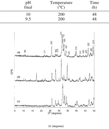
The XRD recorded patterns of the samples are indexed based on the ICDD Card N.9-432, in the recorded diffractogram of the commercial material and in the available literature17,19-50, 20. They
are showed in Figure 1a-c.
It was assumed that the unit cell of all probes was hexago-nal, and the dimensions a and c for all samples were calculated using a least squares refinement program. The lattice parame-ters from ICDD Card N.9-432 used as reference to the refinements, commercial HAp and synthesised samples 1 (without the presence of NH4F) and 2 (in the presence of NH4F)
are given in Table 3.
The peaks on the pattern from sample 2 (Figure 1c), where NH4F was used, seem sharp and well resolved, and can all be
attributed to the hexagonal crystal form of HAp in good
agreement with the patterns on the ICDD Card N.9-432 and the recorded diffractogram from commercial HAp (Figure 1a). The calculated lattice parameters tend to be different from that in the reference (ICDD Card N.9-432). It was shown that the increasing of CO32- content in the HAp lattice can cause a decrease of a
and increase of c51,52, as observed in sample 2. This can be
attributed to a substitution of PO43- ions for CO32- 19,20, typical
for the so called type B-HAp. For sample 1 (Figure 1b), the occurrence of atypical XRD peaks was verified. They are attributed to other phases that can be usually formed in the system (e.g. octacalcium phosphate or tertiary calcium phosphate (TCP)17. Although the lattice parameters were close to that of
the HAp, the peaks were not so well resolved as in sample 2. Figure 2a-c depicts the FTIR spectra of the analysed samples. Spectra of synthesised samples are shown in Figure 2b and Figure 2c. A spectrum of commercial HAp is also shown in Figure 2a.
All the spectra shown give rise to the characteristic absorption bands of HAp53-63. The bands at 565, 602, 1045 and a shoulder at
960 cm-1 can be attributed to the PO
43- ions. Spectra of sample 2
(Figure 2c) show an additional band at 870 cm-1 attributed to
HPO42-. The bands between 1400 and 1550 cm-1 arise from
vibration of CO32- ions. Vibrations of OH- are detected in the
spectrum used as reference (Figure 2a) by the bands located at 635 and 3572 cm-1. A band of low intensity occurred in almost all
the samples at 2300 cm-1, and it is attributed to the CO
2 from the
air19,20. The absence of the bands derived from OH- at 635cm-1 in
Table 2. Reaction conditions for the hydrothermal synthesis
Sample Reagents pH pH Temperature Time
N. (mass / mass) initial final (oC) (h)
1 1CaCO3 : 1(NH4)2HPO4 : 4 H2O 8.5 10 200 48
2 1CaCO3 : 1(NH4)2HPO4 : 4 SFa 9.5 9.5 200 48
a NH
4F (20 ppm) aqueous solution
Figure 1. XRD patterns of the material (a) commercial, (b) synthesised without the presence of NH4F and (c) in presence of NH4F.
Table 3. Reference and calculated lattice parameters.
Sample a (nm) c (nm)
ICDDN. 9-432 0.9418 0.6884
Commercial HAp 0.9432 0.6893
1 0.9357 0.6827
2 0.9377 0.6890
the spectra of the synthesised samples (Figures 2b and 2c) can be explained by the substitution of the groups OH- for CO
32- in
the lattice of HAp, which is characteristic of carbonated type A-HAp49. The band at 3572cm-1 is usually masked in the spectra
by a wide band of H2O between 3500 and 3700cm-1 48. The
extraction of this water included in the lattice is only possible with treatment of the samples at 650oC, bringing the risk of
deterioration of the samples for liberation of the CO32- ions
together with the water, as discussed later. But the absence of the band located at 635cm-1 in the two spectra is an indication
that this type of substitution really occurs.
comparing the three diagrams. For commercial HAp (Figure 3b), called stoichiometric HAp, the weight loss is gradual and smooth. The total percentage of weight loss was 5.12%. For non-stoichiometric HAp synthesised without NH4F (Figure 3a),
a sudden weight loss between 880oC and 931oC occurs. This
weight loss corresponds to 0.45% of the mass in an interval of temperature of only 50oC and is due to the phase change
non-stoichiometric HAp →α-TCP that is determined by the gradual loss of OH- and probably CO
2 from non-stoichiometric HAp
and possible decomposition of the carbonates linked to the chain66-69. This same curve already occurs at 650oC for HAp
synthesised in presence of NH4F (Figure 3c), rising to 782oC.
The relative loss in weight (2.08%) is substantially larger than in the previous case. Also, the process occurs in a temperature range which is twice as that of the previous case. This is attributed to the higher content of carbonate in HAp synthesised without NH4F, reducing the thermal stability of the HAp lattice64.
Figure 3. TG diagrams of the material (a) synthesised without the presence of NH4F, (b) commercial and (c) synthesised in the
presence of NH4F.
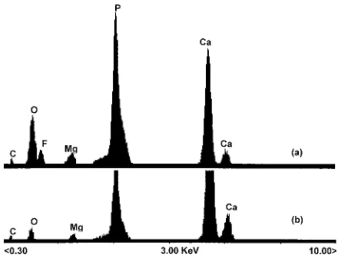
Figure 4 shows the result of the EDX analysis. The presence of a peak of Mg2+ is observed at 1.2keV, and in general the
peaks belong to expected elements - calcium (Kα) at 3.69keV, calcium (Kβ) at 4.01keV, phosphorus at 2.01keV, oxygen at
0.5keV and carbon at 0.2keV. The presence of carbon and oxygen provides further evidence of the ionic substitution in these samples. In non-stoichiometric HAp synthesised in presence of NH4F (Figure 4a), the peaks of C and O are more
intense probably due to substitution of CO32- at the sites of PO4
3-and OH- in the lattice, as discussed before. The presence of
contamination in this material of natural origin is expected, since HAp presents relative facility for ionic exchanges, especially with charged elements and ionic radii similar to Ca2+, as in the
case of Mg2+ and Na+, respectively51. The fact that the EDX
analysis has shown such expressive relative amounts of Mg2+,
indicates the incorporation of the Mg2+ into the formation of the
crystalline lattice of hydroxyapatite, since this ion usually links in small amounts to the hydroxyapatite molecule during its formation56. This ion also occurs in HAp of the human bone.
The photomicrographs obtained by SEM (Figure 5a-b) revealed a porous interconnected structure similar to the phycogenic CaCO3. The maximum distance between two walls
of the pores was an average of 20µm, that is smaller than is generally accepted as the size required for a porous HAp implant. The surface seems to be homogeneous and regular, without ridges (Figure 5a). The interconnected pore structure is similar to the trabecular bone structure4.
Figure 2. FTIR spectra of the material (a) commercial, (b) synthesised without the presence of NH4F and (c) in presence of NH4F.
In sample 2, where NH4F was used (Figure 2c), one can see
the appearance of bands located at 875cm-1 derived from HPO 4
2-ions55. A remarkable increase in the band intensities at 1456 and
1430cm-1 was also observed, besides the appearance of a band
at 864cm-1. These bands are characteristic of carbonated type
B-HAp, in which occurs the substitution of groups PO43- for CO3
2-in the lattice of HAp57-60. These results allied to the absence of
the band at 635cm-1 lead to the conclusion that the synthesised
material is HAp of the type AB. Moreover this agrees with the results of XRD. For this HAp, probably existing together with a fluorapatite phase, due to the high content of F- added, the
appearance of typical bands derived from this ion was expected, but this did not occur. These bands were probably masked by more intense bands of PO43-65.
The TG curves for the synthesised material as well as the commercial HAp from Aldrich are shown in Figure 3a-c. They can be generically described as a gradual weight loss starting with the detachment of adsorbed water on the surface of the sample up to 250oC 64, and continuing with losses of hydroxyls
from the crystalline lattice until the complete conversion to β– TCP, which begins at approximately 650oC. Above 900oC, the
complete decomposition of the material begins. A loss of weight was observed between 200oC and 500oC, probably originating
from the firing of residual organic matter, which is common in view of the biological origin of the material61. These weight losses
might also originate from the detachment of HPO42-, but this
cannot be observed even when submitting the material to temperatures of 450oC and conducting an FTIR analysis in an
attempt to verify the presence or absence of the band at 875cm-1,
typical of this ion. It is accepted49,50 that carbonated HAp with
low cristallinity already loses OH- and CO
32- in considerable
The adsorption isotherms of nitrogen of the synthesised material are of type IV, presenting a very defined and reproducible hysteresis loop of the type H370, typical of
mesoporous materials with slit-like pores (rm≈250 Å). The BET
specific surface area calculated using 5 points, varied between 30-50 m2g-1 in the synthesised samples.
These results, associated to that SEM analysis, indicate the presence of macro and mesopores in these samples. They are si-milar to those found for the phycogenic CaCO3 (BET specific
surface area of 20-30 m2g-1; N
2-adsorption isotherm of type IV,
hysteresis loop of type H3). This indicates that the original physical structure of the material was maintained even after con-version to non-stoichiometric HAp by a parallel way of the synthesis of HAp from corals50. The type of pores determined by
the analysis of N2-adsorption is characteristic of materials formed
by superimposed plates, as takes place with nacre deposition in the mollusk shell71 or the formation of trabecular bone4. The result
is a porous structure with a homogeneous surface (Figures 5a-b). In spite of the fact that the material presents a surface area
and diameter of pores smaller than the material of coralline origin, it may be used as temporary substitute of bone, as demonstrated by Kasperk and collaborators27,28.
The Ca/P ratio rises from 1.5 to 1.8. The Ca/P ratio expected for stoichiometric HAp is approximately 1.67, but in living organisms this value tends to be a little lower. Iijima et al.17
verified that an increase in the concentration of F- during the
synthesis leads to the fastest formation of HAp with poor stoichiometry.
CONCLUSIONS
This study has demonstrated the possibility of non-stoichiometric HAp production from the CaCO3 of marine algae
collected on the Brazilian coast, using hydrothermal synthesis with relatively low expenditure of energy and without alterations in the porous structure of the phycogenic CaCO3.
The fact that synthesised HAp is non-stoichiometric represents an advantage, since human bones themselves are formed of non-stoichiometric HAp.
Results have demonstrated the occurrence of carbonated type AB–HAp, which is the same as that of the human bone.
ACKNOWLEDGMENTS
We thank Prof. Klaus K. Unger of the University of Mainz, Germany, for the facilities available in his laboratory during the studies developed at the University of Mainz, and to DAAD/ CAPES for the concession of the grant to G. Felício-Fernandes.
REFERENCES
1. Constantz, B. R.; Ison, I. C.; Fulmer, M. T.; Posner, R. D.; Smith, S.; Van Wagoner, T. M.; Ross, J.; Goldstein, S. A.; Jupiter; J. B.; Rosenthal, D. I.; Science 1995, 267, 1796.
2. Kawachi, E. Y.; Bertran, C. A.; Kubota, L. T.;
Biomaterials1998, 19, 2329.
3. Neuman, W. F.; Neuman, M. W.; Chem. Rev. 1953, 53, 1.
4. Yaszemsk, M. J.; Payne, R. G.; Hayes, W. C.; langer, R.; Mikos, A. G.; Biomaterials 1996, 17, 175.
5. Liu, H. S.; Chin, T. S.; Lai, L. S.; Chiu, S. Y.; Chung, K. H.; Chang, C. S.; Lui, M. T.; Ceramics International
1997, 23, 19.
6. Williams, D. F.; In: Williams, D. F.; Ed.; Materials science and technology. A comprehensive treatment. Medical and dental materials, V.14, Cap.7, 259, VCH, Weinheim, 1992.
7. Heimke, G; Angew. Chem. 1989, 101, 111.
8. Hench, L. L.; J. Amer. Ceram. Soc. 1991, 74, 1487.
9. Bet, M. R.; Goissis, G.; Plepis, A. M. G.; Quim. Nova
1997, 20, 475.
10. El Deeb, M. E.; Tompach, P. C.; Morstad, A. T.; J. Oral Maxillofac. Surg. 1988, 40, 955.
11. Kohn, D.; in: Williams, D. F.; Ed.; Materials science and technology. A comprehensive treatment. Medical and den-tal materials,.V.14, Cap.2, 70, VCH, Weinheim, 1992.
12. Calvert, P.; in: Brook, R. J.; Ed.; Materials science and technology. A comprehensive treatment. Processing of ceramics; V.17, Cap.12, 51, VCH, Weinheim, 1996.
13. White, E.; Shors, E. C.; Dental Clinics of North America
1986, 30, 49
14. Eanes, E. D.; Termine J. D.; In: Spiro, T. G.; Ed.; Calcium in Biology, Cap. 5, 200, John Wiley & Sons,
New York, 1983.
15. Brown, W. E.; Nature, 1962, 196, 1048.
16. Brown, W. E.; Schoroeder, L. W.; Ferris, J. S.; J. Phys. Chem. 1979, 83, 1385.
17. IIjima, M.; Nelson, D. G. A.; Pan, Y.; Kreinbrink, A. T.; Adachi, M.; Goto, T.; Moriwaki, Y.; Calc. Tissue Int.
1996, 59, 377.
Figure 4. EDX patterns of the material (a) synthesised with NH4F
addition in the reaction and (b) in NH4F absence. Accelerating voltage
of 25keV.
18. Narasaraju, T. S. B.; Phebe, D. E.; J. Mater. Sci. 1996, 31, 1.
19. Nancolas, G. H.; Lore, M.; Perez, L.; Richardson, C.; Zawacki, S. J., The Anatomical Record 1989, 224, 234.
20. Guillemin, G.; Patat, J. L.; Fournie, J.; Chetail, M.; J. Biomed. Mater. Res. 1987, 21, 557.
21. Chiroff, R. T.; White, R. A.; White, E. W.; Weber, J. N.; Roy, D. M.; J. Biomed. Mater. Res. 1977, 11, 165.
22. Ross, P. E.; Scient. Amer. 1989, 261, 31.
23. Holmes, R.; Mooney, V.; Bucholz, R.; Tencer, A.; Clin. Ortop. Relat. Researc. 1964, 188, 252.
24. Boulton, M. I.; Gregson, P. J.; Tuke, M.; Baldwin, T.;
Mater. Lett. 1991, 12, 1.
25. Wolke, J. C. G.; Klein, C. P. A. T.; De Blick-Hogervorst, J. M. A.; De Groot, K.; Proceedings of the 1993 National Thermal Spray Conference 1993, Anahein, 619.
26. Kenney, E. B.; Lekovic, V.; Sá Ferreira, J. C.; Han, T.; Dimitrijevic, B.; Carranza Jr., F. A.; J. Periodontol.
1985, 57, 76.
27. Kasperk, C.; Ewers, R.; Z. Zahnärztl Implantol. 1986, 2, 242.
28. Kasperk, C.; Ewers, R.; Simons, B.; Kasperk, R.; Dtsch. Zanärztl. Z. 1988, 43, 116.
29. Eysel, W.; Roy, D. M.; J. Crystal Growth 1973, 20, 245.
30. Eysel, W.; Roy, D. M.; Zeit. Krist. 1975, 141, 11.
31. González, R.; Melo, M.; Reyes, C. A. P.; Rodrigues,A. C.; Quim. Nova 1993, 16, 509.
32. Perloff, A.; Posner, A. S.; Inorg. Synth. 1960, 6, 16.
33. Roy, D. M.; Linnehan, S. K.; Nature 1974, 247, 220.
34. Driessens, F. C. M.; Boltong, M. G.; Bermudez, O.; Planell, J. A.; Ginebra, M. P.; Fernandez, E.; J. Mater. Sci. Mater. Med. 1994, 5, 164.
35. Murata, H.; Ishikawa, K.; Tenshin, S.; Horiuchi, S.; Nakanishi, M.; Asaoka, K.; Kawata, T.; Yamamoto, T. T.; Caries Res. 1996, 30, 465.
36. Oliveira, E. C.; An. Acad. bras. Ci. 1996, 68, 17.
37. Chicz, R. M.; Regnier, F. E.; Anal. Chem. 1989, 61, 1742.
38. Fernandez, V. L.; Reimer, J. A.; Denn, M. M.; J. Am. Chem. Soc. 1992, 114, 9634.
39. Shimabayashi, S.; Tamura, C.; Nakagani, M.; Chim. Pharm. Bull. 1981, 29, 2116.
40. Reichert, J. G.; Binner, J. G. P.; J. Mater. Sci. Mater. Med. 1996, 31, 1231.
41. Ma, Q. Y.; Traina, S.; Logan, T. J.; Environ. Sci. Technol.
1993, 27, 1803.
42. Ma, Q. Y.; Traina, S.; Logan, T. J.; Ryan J. A.; Environ. Sci. Technol. 1994, 28, 1219.
43. Xu, Y.; Schwartz, F. W.; Traina, S. J.; Environ. Sci. Technol. 1994, 28, 1472.
44. Lee, K. Y.; Houalla, M.; Hercules, D. M.; Hall, W. K.; J. Catal. 1994, 145, 223.
45. Matsumura, Y.; Moffat, J. B.; J. Catal. 1994, 148, 323.
46. Roy, D. M.; 1975, US Patent N.3.929.971
47. Felício-Fernandes, G.; Laranjeira, M. C. M.; Unpublished results.
48. Yubao, L.; De Groot, K.; De Wijn, J.; Klein, C. P. A. T.; Meer, S. V. D.; J. Mater. Sci. Mater. Med. 1994, 5, 326.
49. Li, P.; Kangasniemi, I.; De Groot, K.; Kokubo, T.; J. Am. Ceram. Soc. 1994, 77, 1307.
50. Sivakumar, M.; Kumar, T. S. S.; Shanta, K. L.; RAO, K. P.; Biomaterials 1996, 17, 1709.
51. De Maeyer, E. A. P.; Verbeek, R. M. H.; Naessens, D. E.; Inorg. Chem. 1994, 33, 5999.
52. De Maeyer, E. A. P.; Verbeek, R. M. H.; Naessens, D. E.; Inorg. Chem. 1995, 34, 2085.
53. Nakamoto, K.; Infrared and Raman Spectra of Inorganic and Coordination Compounds, John Wiley & Sons, New
York, 4th Edition; 1986 pp. 106, 107, 112, 124, 138, 383.
54. Yoshimura, M.; Suda, H.; J. Mater. Sci. 1994, 29, 3399.
55. Yasukawa, A.; Takase, H.; Kandori, K.; Ishikawa, T.;
Polyhedron 1994, 13, 3079.
56. Yasukawa, A.; Ouchi, S.; Kandori, K.; Ishikawa, T.; J. Mater. Chem. 1996, 6, 1401.
57. Suchanek, W.; Suda, H.; Yashima, M.; Kakihana, M.; Yoshimura, M.; J. Mat. Res. 1995, 10, 521.
58. Nadal, M.; Trombe, J. C.; Bonel, G.; Montel, G. J.; Chim. Phys. 1970, 67, 1161.
59. Roy, D. M.; Eysel, W.; Dinger, D.; Mat. Res. Bull. 1974, 9, 35.
60. De Maeyer, E. A. P.; Verbeek, R. M .H.; Naessens, D. E.; Inorg. Chem. 1993, 32, 5709.
61. Driessens, F. C. M.; Verbeek, R. M. H.; Kriekens, P.; Z. anorg. allg. Chemie 1983, 504, 195.
62. Rehman, I.; Bonfield, W.; J. Mater. Sci. Mater. Med.
1997, 8, 1.
63. Murray, M. G. S.; Wang, J.; Ponton, C. B.; Marquis, P. M.; J. Mater. Sci. 1995, 30, 3061.
64. Kandori, K.; Yasukawa, A.; Ichikawa, T.; Chem. Mat.
1995, 7, 26.
65. Tucker, B. E.; Cottell, C.; Mauyeung; R. C. Y.; Spector, M.; Nancollas, G. H.; Biomaterials 1996, 17, 631.
66. Correia, R. N.; Magalhães, M. C. F. ; Marques, P. A. A. P.; Senos, A. M. R.; J. Mater. Sci. Mater. Med. 1996, 7, 501
67. Yshikawa, T.; Tanaka, H.; Yasukawa, A.; Kandori, K. J. Mater. Chem. 1995, 5, 1963.
68. Bett, J. A. S.; Christner, L. G.; Hall, W. K.; J. Am. Chem. Soc. 1967, 89, 5535.
69. Monma H.; Kamiya, T.; J. Mater. Sci. 1987, 22, 4247.
70. Gregg, S. J.; Sing, K. S. W.; Adsorption, Surface Area and Porosity, Academic Press, London, 2nd. Ed., 1982,
pp. 130, 131.