Craniofacial skeletal pattern: is it really correlated with
the degree of adenoid obstruction?
Murilo Fernando Neuppmann Feres1, Tomas Salomão Muniz2, Saulo Henrique de Andrade2, Maurilo de Mello Lemos3, Shirley Shizue Nagata Pignatari4
Objective: The aim of this study was to compare the cephalometric pattern of children with and without adenoid obstruc-tion. Methods: The sample comprised 100 children aged between four and 14 years old, both males and females, subjected to cephalometric examination for sagittal and vertical skeletal analysis. The sample also underwent nasofiberendoscopic examina-tion intended to objectively assess the degree of adenoid obstrucexamina-tion. Results: The individuals presented tendencies towards vertical craniofacial growth, convex profile and mandibular retrusion. However, there were no differences between obstructive and non-obstructive patients concerning all cephalometric variables. Correlations between skeletal parameters and the percent-age of adenoid obstruction were either low or not significant. Conclusions: Results suggest that specific craniofacial patterns, such as Class II and hyperdivergency, might not be associated with adenoid hypertrophy.
Keywords:Mouth breathing. Diagnosis. Angle Class II malocclusion.
How to cite this article: Feres MFN, Muniz TS, Andrade SH, Lemos MM, Pignatari SSN. Craniofacial skeletal pattern: is it really correlated with the degree of adenoid obstruction? Dental Press J Orthod. 2015 July-Aug;20(4):68-75. DOI: http://dx.doi.org/10.1590/2176-9451.20.4.068-075.oar
Submitted: September 09, 2014 - Revised and accepted: January 19, 2015 » The authors report no commercial, proprietary or financial interest in the prod-ucts or companies described in this article.
Contact address: Murilo Fernando Neuppmann Feres
Av. São Francisco de Assis, 218, Jardim São José, Bragança Paulista/SP - Brazil CEP: 12916-900 - E-mail: [email protected]
1 Associate professor, Universidade São Francisco, Department of Orthodontics,
Bragança Paulista, São Paulo, Brazil.
2 Private practice, Bragança Paulista, São Paulo, Brazil.
3 Full professor, Universidade Guarulhos, Department of Orthodontics,
Guarulhos, São Paulo, Brazil.
4 Adjunct professor of Otolaryngology, Universidade Federal de São Paulo,
Department of Otolaryngology, Head and Neck Surgery, São Paulo, São Paulo, Brazil.
DOI: http://dx.doi.org/10.1590/2176-9451.20.4.068-075.oar
Objetivo: a presente pesquisa teve como objetivo comparar o padrão cefalométrico de crianças com e sem obstrução adenoidiana. Métodos: a amostra consistiu de 100 crianças, com idades entre 4 e 14 anos, de ambos os sexos, subme-tidas a exames cefalométricos para a avaliação de variáveis cefalométricas horizontais e verticais. A amostra também foi submetida à nasofibroendoscopia, por meio da qual o grau de obstrução adenoidiana foi objetivamente aferido. Resul-tados: os pacientes avaliados demonstraram tendência ao crescimento vertical acentuado, ao perfil convexo e à retrusão mandibular. No entanto, não houve diferenças entre pacientes portadores e não portadores de obstrução, em relação a todas as variáveis cefalométricas. As correlações estabelecidas entre os parâmetros esqueléticos e os percentuais de hiper-trofia foram baixas ou não significativas. Conclusões: os resultados sugerem que padrões faciais específicos, tais como Classe II e hiperdivergência, parecem não estar associados à hipertrofia adenoideana.
INTRODUCTION
Studies on the relationship between respiratory pattern and the development of craniofacial characteristics have been published for a considerable period of time.1-8 The persistence of the interest in this topic might be partially explained by the high prevalence of mouth breathing,9 even among orthodontic patients.10
The presence of this habit is correlated with a number of muscular11 and dento-craniofacial altera-tions, including maxillary constriction, posterior cross-bite, retrusion and clockwise rotation of the mandible, Class II skeletal pattern and excessive vertical growth.1-8
One of the main causes of mouth breathing is adenoid hypertrophy,12 In addition, several studies have demon-strated a signiicant correlation between long face mor-phology and anatomical reduction in the nasopharyngeal airway.1,7,8,13,14,16-19 Class II malocclusion or mandibular retrognathia have also been frequently related to smaller dimensions of the nasopharynx.13,20-23 Therefore, some of these studies7,14,16,18,24 suggest that dimensional reduction of the nasopharynx, due to hyperdivergent craniofacial pattern or mandibular retrusion, might predispose pa-tients to an obstructive breathing status derived from ad-enoid hypertrophy. However, such inferences might be considered mere assumptions rather than scientiic evi-dence, since most of these studies have relied upon inac-curate methods, such as rhinomanometry1,13,21 or lateral cephalometric radiographs,7,8,14,16,17,18,19,20,22 to assess pa-tients’ respiratory pattern. On the other hand, nasoibe-rendoscopic examination has been considered as the gold standard method for adenoid evaluation.23
The primary objective of this study was to describe the craniofacial morphology of patients with com-plaints of nasopharyngeal obstruction. In addition, comparative analysis of cephalometric skeletal features was conducted on patients with and without adenoid hypertrophy, as assessed by nasoiberendoscopic exami-nation. Finally, this study also aimed to investigate the correlations established between skeletal characteristics and the percentage of adenoid obstruction.
MATERIAL AND METHODS
This research was a descriptive-analytical, cross-sectional study approved by Universidade Federal de São Paulo Institutional Review Board (protocol #0181/08).
Between February 2009 and June 2010, 170 chil-dren who attended or were referred to a public
pedi-atric otolaryngology outpatient clinic, were invited to take part in the study, from which 43 refused to participate. The convenience sample thus consisted of 127 individuals, both males and females, aged be-tween four to 14 years old. In order to be eligible to the study, the children should have presented complaints of nasopharyngeal obstruction and/or mouth breathing. At this point, no objective information regarding the degree of adenoid hypertrophy was available.
Children with syndromes or craniofacial malformation, as well as those which had been pre-viously subjected to orthodontic treatment, were not included in the sample.
All eligible participants, along with their parents or legal guardians, were properly informed about the study objectives and procedures, as well as the examinations that would be performed. Those who agreed to par-ticipate formalized their intent by signing an informed consent form previously prepared according to Univer-sidade Federal de São Paulo Institutional Review Board. Initially, the children selected underwent lateral ceph-alometric radiographic examination performed by a radi-ology specialist who used the same device for all of them (Instrumentarium Ortopantomographic OP100; Gen-eral Electric Healthcare, Tuusula, Finland). The focus-ilm distance was 140 cm, while X-ray exposure settings were 70 kV and 12 mA for 0.40 to 0.64 s. During record taking, patients were instructed to breathe exclusively through the nose, keep their mouth closed and their teeth in occlusion. We used 20 x 25 cm ilms (Kodak, Roches-ter, NY) and processed them according to a standardized protocol. Radiographs were identiied by codes, so as to prevent patient identiication.
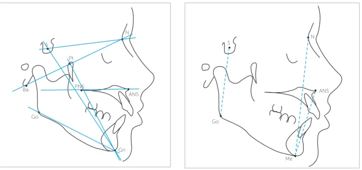
Lateral cephalometric radiographs were manually traced by two independent blind examiners, and sub-sequent measurements (Table 1, Figs 1, 2, and 3) were performed on Ultraphan acetate sheets, with the aid of a light box, a protractor and a digital caliper (model 799A-8/200; Starrett , Itu, Brazil), with 0.01 mm precision.
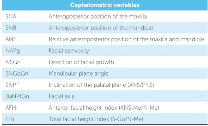
Table 1 - Cephalometric variables evaluated.
Cephalometric variables
SNA Anteroposterior position of the maxilla
SNB Anteroposterior position of the mandible
ANB Relative anteroposterior position of the maxilla and mandible
NAPg Facial convexity
NSGn Direction of facial growth
SNGoGn Mandibular plane angle
SNPP Inclination of the palatal plane (ANS/PNS)
BaNPtGn Facial axis
AFHi Anterior facial height index (ANS-Me/N-Me)
FHi Total facial height index (S-Go/N-Me)
primary video was edited to prevent patient identiica-tion. Edited video clips were then forwarded to another experienced and independent otolaryngologist who had not been involved with subjects’ enrollment, vide-onasopharyngoscopic examination performance, record taking or editing.
In order to evaluate the edited video clips, the examiner used an assessment method originally de-signed to quantify the degree of obstruction caused by the adenoid tissue: measured choanal obstruction (MCO) which has previously proven to be satisfacto-rily reproducible.25 The evaluator was instructed to choose the frame that provided the best view of the
adenoid in relation to the choana, obtained from the most distal portion of the inferior turbinate. In these frames, the patient had to be breathing exclusively through the nose, with no evidence of soft palate el-evation. The selected frame was then converted into a JPEG file and the MCO was calculated by means of Image J,26 an image processing software. MCO rep-resented the percentage of the choanal area occupied by adenoid tissue (Fig 4). When images from both nostrils were available, the mean between right and left side evaluations was calculated to minimize po-tential variations, as proposed by Feres et al.25
Statistical analysis
At irst, radiographic parameters reliability was de-termined ater intra and inter-reproducibility analysis calculated by intraclass correlation coeicient (ICC).
Then, radiographic variables were described (means, standard deviations, minimum and maximum values) for all subjects. Subsequently, the sample was divided into two groups, according to the degree of obstruction caused by adenoid hypertrophy. According to previous parameters,27 patients with MCO ≥ 66.7% were consid-ered to have pathological adenoid hypertrophy, herein denominated the “positive” group; while patients with MCO < 66.7% were not considered to present patho-logical adenoid hypertrophy (“negative” group). Both groups were compared regarding all cephalometric vari-ables, according to Mann-Withney test.
Finally, Pearson correlation analysis between cepha-lometric measurements and MCO was determined for the whole sample. The “strength” of the correlation was characterized according to Vieira28 as: “irrelevant” (0.00 < r ≤ 0.25), “weak” (0.25 < r ≤ 0.50), “moderate” (0.50 < r ≤ 0.75), or “strong” (0.75 < r ≤ 1.00).
Signiicance level for statistical tests was set at 5% (α ≤ 0.05). All analyzes were performed using SPSS 10.0 for Windows.
RESULTS
From the initial sample comprising 127 patients who met the eligibility criteria and agreed to participate in the study, seven were excluded due to inadequate exams and 20 presented inconsistent repeated radiographic measurements. For the 100 remaining patients, all of them had radiographic parameters with satisfactory re-producibility (Table 2).
Figure 1 - Horizontal cephalometric variables (SNA, SNB, ANB, NAPg). N
S
A
B
Figure 2 - Vertical cephalometric variables (NSGn, SNGoGn, SNPP, BaNPtGn).
Figure 4 - Final frame selection (B) derived from the video clip (A), and MCO calculation (C): MCO = (Ad/Cho) x 100.
Figure 3 - Cephalometric variables related to the facial indices AFHi and FHi.
The inal sample comprised 48 female children (48.0%) and 52 male children (52.0%), with a mean age of 9.0 years old (standard deviation = 2.4). The de-scriptive analysis of all cephalometric variables is pre-sented in Table 3. According to universally accepted parameters, the sample of this study showed skeletal patterns with a slight tendency towards excessive verti-cal growth, a convex proile and mandibular retrusion. However, comparison between the cephalometric
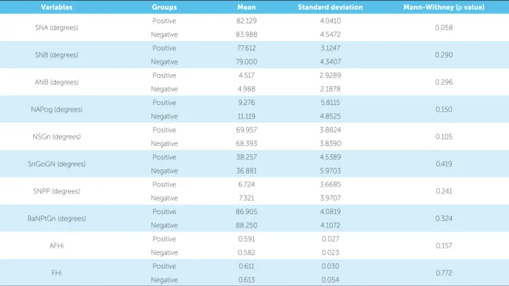
variables of the positive (n = 58, 58.0%) and negative groups (n = 42, 42.0%) showed no statistically signii-cant diferences (Table 4).
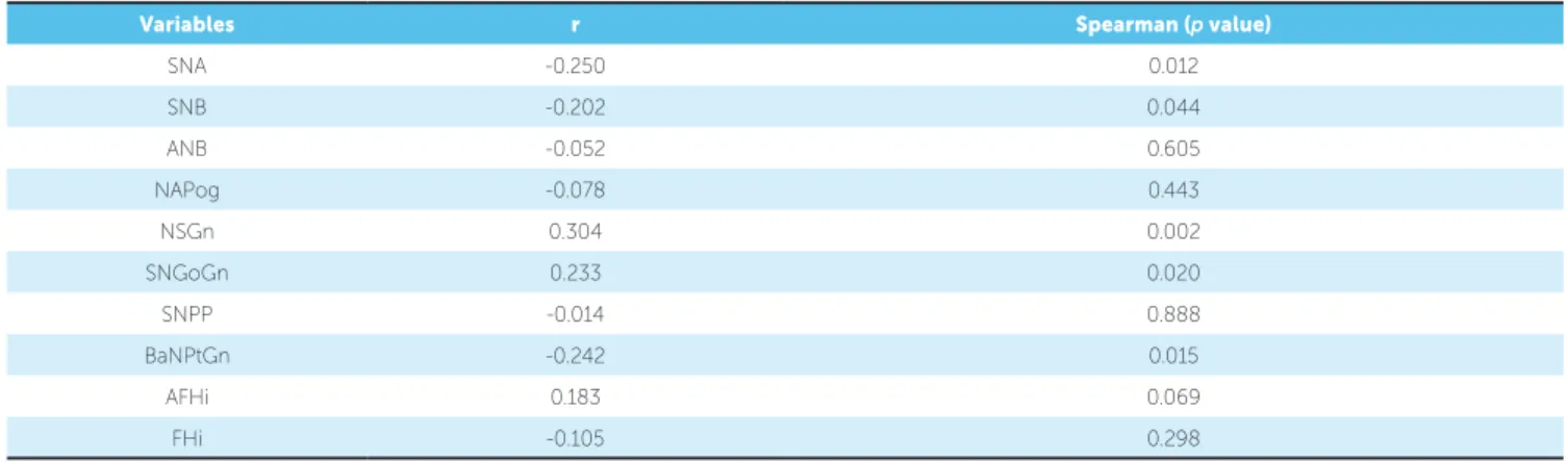
Furthermore, amongst all radiographic variables evaluated herein, SNA, SNB, NSGn, SNGoGn and BaNPtGn were signiicantly correlated with MCO. Al-though statistically signiicant, the magnitude of these correlations was considered either irrelevant (SNA, SNB, SNGoGn, BaNPtGn) or weak (NSGn) (Table 4).
N
S
ANS PNS
Pt
S
N
ANS
Me Go
Go Ba
Gn
Table 2 - Interclass correlation coefficient (ICC) of skeletal parameters (intra and inter-examiner analysis).
Variables Intraexaminer p value Inter-examiner p value
SNA (degrees) 0.942 < 0.001 0.950 < 0.001
SNB (degrees) 0.966 < 0.001 0.970 < 0.001
ANB (degrees) 0.928 < 0.001 0.913 < 0.001
NAPog (degrees) 0.940 < 0.001 0.926 < 0.001
NSGn (degrees) 0.961 < 0.001 0.945 < 0.001
SnGoGN (degrees) 0.911 < 0.001 0.908 < 0.001
SNPP (degrees) 0.923 < 0.001 0.910 < 0.001
BaNPtGn (degrees) 0.988 < 0.001 0.975 < 0.001
AFHi 0.890 < 0.001 0.877 < 0.001
FHi 0.869 < 0.001 0.868 < 0.001
Table 3 - Descriptive analysis of radiographic variables.
Variables Mean Standard deviation Minimum Maximum
SNA (degrees) 82.910 4.3376 70.0 97.5
SNB (degrees) 78.195 3.7281 70.0 93.0
ANB (degrees) 4.715 2.6412 -6.0 11.5
NAPog (degrees) 10.050 5.4802 -8.0 24.0
NSGn (degrees) 69.300 3.9222 56.0 79.0
SnGoGN (degrees) 37.679 5.2047 19.0 48.0
SNPP (degrees) 6.975 3.7902 -4.0 16.0
BaNPtGn (degrees) 87.470 4.1261 78.0 101.0
AFHi 0.588 0.026 0.528 0.705
FHi 0.612 0.041 0.517 0.784
Table 4 - Comparative analysis between positive (MCO ≥ 66.7%) and negative groups (MCO < 66.7%) in relation to the radiographic variables.
Variables Groups Mean Standard deviation Mann-Withney (p value)
SNA (degrees) Positive 82.129 4.0410 0.058
Negative 83.988 4.5472
SNB (degrees) Positive 77.612 3.1247 0.290
Negative 79.000 4.3407
ANB (degrees) Positive 4.517 2.9289 0.296
Negative 4.988 2.1878
NAPog (degrees) Positive 9.276 5.8115 0.150
Negative 11.119 4.8525
NSGn (degrees) Positive 69.957 3.8824 0.105
Negative 68.393 3.8390
SnGoGN (degrees) Positive 38.257 4.5389 0.419
Negative 36.881 5.9703
SNPP (degrees) Positive 6.724 3.6685 0.241
Negative 7.321 3.9707
BaNPtGn (degrees) Positive 86.905 4.0819 0.324
Negative 88.250 4.1072
AFHi Positive 0.591 0.027 0.157
Negative 0.582 0.023
FHi Positive 0.611 0.030 0.772
Table 5 - Correlation coefficient (r) between radiographic variables and MCO.
Variables r Spearman (p value)
SNA -0.250 0.012
SNB -0.202 0.044
ANB -0.052 0.605
NAPog -0.078 0.443
NSGn 0.304 0.002
SNGoGn 0.233 0.020
SNPP -0.014 0.888
BaNPtGn -0.242 0.015
AFHi 0.183 0.069
FHi -0.105 0.298
DISCUSSION
The association between specific skeletal patterns and the presence of obstructive adenoid is a topic which has been debated for years,1,7,8,13-19 although controversy still remains. One of the reasons that might contribute for this debate to persist is related to the varied sorts of assessment methods used to evaluate the level of adenoid obstruction.
This study has demonstrated that children with respiratory complaints might present skeletal fea-tures associated with hyperdivergency and retrogna-thia. However, despite currently accepted hypoth-eses according to which dolichofacial or Class II pa-tients are more anatomically susceptible to present adenoid obstruction, evidence presented herein sug-gests that children are likely to experience it regard-less of their skeletal characteristics.
According to most studies, the size of the naso-pharyngeal airway is significantly correlated with ex-cessively vertical cephalometric features. Research-ers have suggested that this dimensional reduction of the nasopharynx might be attributed to skeletal characteristics which are inherent to hyperdivergent patients, such as maxillomandibular retrusion.7,14,16,18,23 Nevertheless, Santos-Pinto et al16 refuted this hy-pothesis when they demonstrated that individu-als with varying nasopharyngeal dimensions did not significantly differ in relation to the anteroposterior position of the maxilla and the mandible. The data obtained in our study support their findings,16 since the anteroposterior position of the maxilla and the
mandible showed no relevant correlation with the de-gree of adenoid obstruction, as determined by flexible nasofiberendoscopic examination. Moreover, accord-ing to our results, the subjects who were considered to be positive presented similar maxillomandibular sagittal position as those considered to be negative for adenoid obstruction.
That finding might explain why no significant differences were found in relation to ANB when positive and negative groups were compared. In ad-dition, ANB revealed no relevant correlation with the degree of adenoid obstruction. These findings corroborate the results of Freitas et al,17 according to which sagittal malocclusions are not correlated with nasopharyngeal airway depth.
Further evidence provided by this study contra-dicts what other studies7,20,23,25 claim. Some of these researches,7,23 after lateral cephalometric analysis, reported that Class II patients had significantly small-er airway areas. In their latest study on tomographic measurements, Claudino et al28 were unable to de-tect a significant association between nasopharyngeal dimensions and the sagittal skeletal pattern in adoles-cents. The authors demonstrated that more obvious influence of the skeletal pattern could be observed in relatively lower portions of the pharynx, such as the oropharynx, rather than at the nasopharyngeal level.28
correlations were observed between the percentage of adenoid obstruction and any of the skeletal variables in-vestigated. It is our opinion that most of the studies that have been carried out to date7,8,13,14,16-22,24 have actually failed to infer that patients with speciic skeletal pat-terns (dolichofacial and/or Class II) signiicantly present higher frequencies of pathological adenoid obstruc-tion. Considering the data obtained herein, it no longer seems reasonable to assume that a reduction in the na-sopharyngeal airway is directly related to an actual clini-cal obstruction. Thüer et al13 have already reported that there is no signiicant correlation between nasal airlow parameters, derived from rhinomanometry, and the na-sopharyngeal space observed in lateral cephalometric radiograph. The absence of a signiicant correlation be-tween respiratory capacity and anatomical traits of doli-chocephaly has been also reported by Solow et al21 who sought to correlate skeletal morphological patterns with data obtained from rhinomanometry examination.
In addition, although imaging techniques may indeed indicate nasopharyngeal anatomical reduc-tion, these might not be able to promote significant influence on patient’s clinical respiratory conditions, nor necessarily predispose one to effectively develop obstruction. Unlike many other researches, in this study, a direct and visual nasopharyngeal evaluation method was used, which, according to relevant lit-erature,23 is considered to be the gold standard for adenoid evaluation.
However, this study presents signiicant limitations, with the most important one being associated with single cross-sectional evaluation of adenoid hypertrophy. As previously reported,31 the adenoid lymphoid tissue might be susceptible to sudden dimensional changes as
a consequence of allergic sensitization. Therefore, the authors suggest that future studies should address this limitation by performing serial adenoid evaluations, so as to minimize potential variations. In addition, new research is still required to investigate the inluence of other morphological parameters, such as those related to the cranial base,30 on the dimensional reduction of the nasopharynx and the potential establishment of an obstructive respiratory process, since this study was lim-ited to assess only maxillary or mandibular parameters.
CONCLUSION
The sample studied herein showed skeletal pat-terns with a discrete tendency towards excessive vertical growth, a convex profile and mandibular re-trusion. However, no statistically significant differ-ences were found between patients with or without adenoid hypertrophy. The correlations established between the characteristics of craniofacial morphol-ogy and the percentage of choanal obstruction were weak or not significant.
Acknowledgements
This research was financially supported by Fundação de Amparo à Pesquisa do Estado de São Paulo (FAPESP) under the protocol #08/53538-0.
Author contributions
1. Their efect on mode of breathing and nasal airlow and their relationship to characteristics of the facial skeleton and the dentition. A biometric, rhino-manometric and cephalometro-radiographic study on children with and without adenoids. Acta Otolaryngol Suppl. 1970;265:1-132.
2. Melsen B, Attina L, Santuari M, Attina A. Relationships between swallowing pattern, mode of respiration, and development of malocclusion. Angle Orthod. 1987;57(2):113-20.
3. Löfstrand-Tideström B, Thilander B, Ahlqvist-Rastad J, Jakobsson O, Hultcrantz E. Breathing obstruction in relation to craniofacial and dental arch morphology in 4-year-old children. Eur J Orthod. 1999;21(4):323-32.
4. Sabatoski CV, Maruo H, Camargo ES, Oliveira JHG. Estudo comparativo de dimensões craniofaciais verticais e horizontais entre crianças respiradoras bucais e nasais. J Bras Ortodon Ortop Facial. 2002;7(39):246-57.
5. Lopatiene K, Babarskas A. Malocclusion and upper airway obstruction. Medicina (Kaunas). 2002;38(3):277-83.
6. Lessa FC, Enoki C, Feres MF, Valera FC, Lima WT, Matsumoto MA, Breathing mode inluence in craniofacial development. Braz J Otorhinolaryngol. 2005;71:156-60.
7. Wysocki J, Krasny M, Skarzyński PH. Patency of nasopharynx and a cephalometric image in the children with orthodontic problems. Int J Pediatr Otorhinolaryngol. 2009;73:1803-9.
8. Ucar FI, Uysal T Orofacial airway dimensions in subjects with Class I malocclusion and diferent growth patterns. Angle Orthod. 2011;81(3):460-8. 9. de Menezes VA, Leal RB, Pessoa RS, Pontes RM. Prevalence and factors related
to mouth breathing in school children at the Santo Amaro project-Recife, 2005. Braz J Otorhinolaryngol. 2006;72(3):394-9.
10. di Francesco RC, Bregola EGP, Pereira LS, Lima RS. A obstrução nasal e o diagnóstico ortodôntico. Rev Dent Press Ortod Ortop Facial. 2006;11(1):107-13. 11. Valera FC, Travitzki LV, Mattar SE, Matsumoto MA, Elias AM, Anselmo-Lima WT.
Muscular, functional and orthodontic changes in pre school children with enlarged adenoids and tonsils. Int J Pediatr Otorhinolaryngol. 2003;67(7):761-70. 12. Farid M, Metwalli N. Computed tomographic evaluation of mouth breathers
among paediatric patients. Dentomaxillofac Radiol. 2010;39(1):1-10. 13. Thüer U, Kuster R, Ingervall B. A comparison between anamnestic,
rhinomanometric and radiological methods of diagnosing mouth-breathing. Eur J Orthod. 1989;11:161-8.
14. Joseph AA, Elbaum J, Cisneros GJ, Eisig SB. A cephalometric comparative study of the soft tissue airway dimensions in persons with hyperdivergent and normodivergent facial patterns. J Oral Maxillofac Surg. 1998;56(2):135-9; 15. Akcam MO, Toygar TU, Wada T. Longitudinal investigation of soft palate and
nasopharyngeal airway relations in diferent rotation types. Angle Orthod. 2002;72(6):521-6.
16. Santos-Pinto A, Paulin RF, Melo ACM, Martins LP. A inluência da redução do espaço nasofaringeano na morfologia facial de pré-adolescentes. Rev Dental Press Ortod Ortop Facial. 2004;9(3):19-26.
REFERENCES
17. Freitas MR, Alcazar NM, Janson G, Freitas KM, Henriques JF. Upper and lower pharyngeal airways in subjects with Class I and Class II malocclusions and diferent growth patterns. Am J Orthod Dentofacial Orthop. 2006;130(6):742-5. 18. Feres MFN, Enoki C, Anselmo-Lima WT, Matsumoto MAN. Dimensões
nasofaringeanas e faciais em diferentes padrões morfológicos. Dental Press J Orthod. 2010;15(3):52-61.
19. Macari AT, Bitar MA, Ghafari JG. New insights on age-related association between nasopharyngeal airway clearance and facial morphology. Orthod Orthod Craniofac Res. 2012;15(3):188-97.
20. Mergen DC, Jacobs RM. The size of nasopharynx associated with normal occlusion and Class II malocclusion. Angle Orthod. 1970;40(4):342-6. 21. Solow B, Siersbaek-Nielsen S, Greve E. Airway adequacy, head posture, and
craniofacial morphology. Am J Orthod. 1984;86(3):214-23.
22. Krasny M, Wysocki J, Zadurska M, Skarżyński PH. Relative nasopharyngeal patency index as possible objective indication for adenoidectomy in children with orthodontic problems. Int J Pediatr Otorhinolaryngol. 2011;75(2):250-5. 23. Mlynarek A, Tewik MA, Hagr A, Manoukian JJ, Schloss MD, Tewik TL, et al. Lateral neck radiography versus direct video rhinoscopy in assessing adenoid size. J Otolaryngol. 2004;33(6):360-5.
24. Alves M Jr, Franzotti ES, Baratieri C, Nunes LK, Nojima LI, Ruellas AC. Evaluation of pharyngeal airway space amongst diferent skeletal patterns. Int J Oral Maxillofac Surg. 2012;41(7):814-9.
25. Feres MF, Hermann JS, Sallum AC, Pignatari SS. Endoscopic evaluation of adenoids: reproducibility analysis of current methods. Clin Exp Otorhinolaryngol. 2013;6(1):36-40.
26. ImageJ. US National Institutes of Health. 1997 [Access in: 2014 Jan 09]. Available from: http://imagej.nih.gov/ij.
27. Chien CY, Chen AM, Hwang CF, Su CY. The clinical signiicance of adenoid-choanae area ratio in children with adenoid hypertrophy. Int J Pediatr Otorhinolaryngol. 2005;69(2):235-9.
28. Vieira S. Introdução à Bioestatística. Rio de Janeiro: Elsevier; 2008. 29. Claudino LV, Mattos CT, Ruellas AC, Sant’ Anna EF. Pharyngeal airway
characterization in adolescents related to facial skeletal pattern: a preliminary study. Am J Orthod Dentofacial Orthop. 2013;143(6):799-809.
30. Martin O, Muelas L, Viñas MJ. Comparative study of nasopharyngeal soft-tissue characteristics in patients with Class III malocclusion. Am J Orthod Dentofacial Orthop. 2011;139(2):242-51.