Clinical and radiographic evaluation of maxillary central
inci-sors exposure in patients undergoing maxillary advancement
Guilherme dos Santos Trento1, Felipe Bueno Rosettti Bernabé2, Delson João da Costa3, Nelson Luis Barbosa Rebellato3, Leandro Eduardo Klüppel3, Rafaela Scariot4
1 Resident in Oral and Maxillofacial Surgery, Universidade Federal do Paraná
(UFPR), Curitiba, Paraná, Brazil.
2 Specialist in Oral and Maxillofacial Surgery, Universidade Federal do Paraná
(UFPR), Curitiba, Paraná, Brazil.
3 Professor, Universidade Federal do Paraná (UFPR), Residency program in Oral
and Maxillofacial Surgery, Curitiba, Paraná, Brazil.
4 Professor, Universidade Positivo, Undergraduate program in Dentistry,
Curitiba, Paraná, Brazil.
» The authors report no commercial, proprietary or financial interest in the products or companies described in this article.
How to cite this article: Trento GS, Bernabé FBR, Costa DJ, Rebellato NLB, Klüppel LE, Scariot R. Clinical and radiographic evaluation of maxillary central incisors exposure in patients undergoing maxillary advancement. Dental Press J Orthod. 2015 Nov-Dec;20(6):52-9.
DOI: http://dx.doi.org/10.1590/2177-6709.20.6.052-059.oar
Submitted: December 14, 2014 - Revised and accepted: August 04, 2015
Contact address: Guilherme dos Santos Trento
Av. Prof. Lothario Meissner, 632 - Jardim Botânico - Curitiba - PR - Brazil CEP: 80210-170 – E-mail: [email protected]
Introduction: Patients with dentofacial deformities may undergo orthodontic or orthodontic-surgical treatment. Both modalities can affect esthetics. Objective: This study aims to evaluate clinical and radiographic changes in expo-sure of maxillary central incisors occurring after orthognathic surgery for maxillary advancement. Methods: A total of 17 patients who underwent orthognathic surgery for maxillary advancement between September, 2010 and July, 2011 were selected. Exposure of maxillary central incisors was evaluated clinically and by lateral cephalograms. Measurements were taken one week before and three months after surgery. Data were paired in terms of sex, age, nasolabial angle, height and thickness of the upper lip, the amount of maxillary advancement, clinical exposure and inclination of maxillary central incisor by statistical tests (CI 95%). Results: After maxillary advancement, incisor clinical exposure had increased even with relaxed lips and under forced smile. Moreover, there was a mean increase of 23.33% revealed by lateral cephalo-grams. There was an inverse correlation between upper lip thickness and incisors postsurgical exposure revealed by ra-diographic images (p = 0.002). Conclusions: Significant changes in the exposure of maxillary central incisors occur after maxillary advancement, under the influence of some factors, especially lip thickness.
Keywords:Orthognathic surgery. Maxilla. Incisor. Esthetics.
DOI: http://dx.doi.org/10.1590/2177-6709.20.6.052-059.oar
Introdução: pacientes portadores de deformidades dentofaciais podem submeter-se a tratamento ortodôntico ou ortodôntico--cirúrgico. Ambos podem modificar a estética do paciente. Objetivo: esse estudo tem por objetivo avaliar, clinicamente e radiograficamente, as mudanças na exposição dos incisivos centrais superiores em pacientes submetidos à cirurgia ortognática de avanço de maxila. Métodos: foram selecionados 17 pacientes submetidos à cirurgia ortognática de avanço maxilar no perí-odo de setembro de 2010 a julho de 2011. A exposição dos incisivos centrais superiores foi avaliada clinicamente e por meio de radiografias cefalométricas em norma lateral. Essas medidas foram tomadas uma semana antes e três meses depois da cirurgia. Os dados foram, por meio de testes estatísticos (CI 95%), correlacionados por sexo, idade, ângulo nasolabial, altura e espessura do lábio superior, quantidade de avanço maxilar, exposição clínica e inclinação dos incisivos centrais superiores. Resultados:
após o avanço maxilar, houve um aumento da exposição clínica dos incisivos tanto com o lábio superior relaxado quanto sob sorriso forçado. Além disso, obteve-se um aumento médio de 23,33% na exposição dos incisivos nas radiografias cefalométricas em norma lateral. Houve correlação inversa entre a espessura do lábio superior e a exposição pós-cirúrgica dos incisivos nas imagens radiográficas (p = 0,002). Conclusão: mudanças significativas na exposição dos incisivos centrais superiores ocorrem após o avanço maxilar, sob influências de certos fatores, especialmente a espessura do lábio superior.
INTRODUCTION
Severe malocclusion requires combined treatment of surgery and Orthodontics. Less severe dentofacial de-formities can be treated only by orthodontic treatment.1
Changes in the facial skeleton produced by this treat-ment modality afect not only the bones of the facial skeleton, but also the relationship between hard and sot tissues of the face.2 The most widely used technique for
repositioning the maxilla is Le Fort I osteotomy which can be used for correction of vertical, anteroposterior and transverse problems that involve the maxilla by means of osteotomies across the anterior and lateral walls of this structure.3 In cases of Class III malocclusion, maxillary
advancement aims to correct the bite, improve facial es-thetics and harmonize the facial proile. Therefore, it is important for the clinician to be able to predict sot tis-sue changes resulting from alterations of hard tistis-sues.4
Sot tissue changes resulting from maxillary advance-ment via Le Fort I osteotomy have been reported to be between 33% and 100%.5,6 Studies that describe the
in-luence of sot tissue surgical corrections are limited.7-10
Nevertheless, some studies have shown that changes in the sot tissues of the lips are inluenced by the magni-tude and direction of the jaw segment during surgery,11
and mainly by tone and lip thickness.5,12,13,14 In the case
of impaction, and with posterior or anterior movement of the maxilla, it was found that the nasolabial angle is increased despite a wide variation in tissue responses.9,15
Bundgaar, Melsen, and Terp7 hypothesized that angular
changes may be related to muscle function on the site of osteotomy, and assessment of patient’s muscle pattern could be important for predictive tracing of hard and sot tissues. Stella et al,16 in order to assess the predictability
of changes in the sot tissue of the upper lip as a result of maxillary advancement by the Le Fort I technique, se-lected 20 adult patients with a follow-up of six months. Patients were subdivided into two groups based on lip thickness: Group 1 (lips between 10 and 17-mm thick) and Group 2 (greater than 17-mm thick). Most patients showed a reduction in thickness of the upper lip, and presented no increase in thickness. The reduction of lip thickness was greater than 25% in most patients. It was further stated that clinically relevant correlations can-not be made between the change of sot tissues and bone advancement; however, when the reference is the thickness of the upper lip, there is a better relation-ship between these two variables.15,16 In a retrospective
cephalometric study, Van Butsele et al17 evaluated sot
and hard tissue ratios in relation to maxillary advance-ment. The authors concluded that, for each millimeter of maxillary advancement, the upper lip moved upward in almost 30% the amount of advancement, in addition to having an elongation of 1.7 mm. Del Santo et al18
evaluated 19 patients undergoing Le Fort I osteotomy in order to study changes in the lips. The authors con-cluded that signiicant horizontal changes occur in the upper lip when the maxilla is moved signiicantly an-teroposteriorly at a ratio of 0.6 : 1. This is because the vertical changes of the upper lip only occur when there is a signiicant change in the anteroposterior position of the maxillary basal bone. Given the above statement, the objective of this study was to evaluate the clinical and radiographic changes, with exposure of maxillary central incisors, occurring ater maxillary advancement.
MATERIAL AND METHODS
Sample selection: A total of 17 patients were selected to undergo orthognathic surgery for maxillary advance-ment in the Departadvance-ment of Oral and Maxillofacial Sur-gery, Universidade Federal do Paraná, in the period of September, 2010 to July, 2011. All patients (aged 18 or older) included in the study had Class III maloc-clusion and underwent maxillary advancement alone or combined with mandibular surgery, with previous orthodontic decompensation. Those who did not get V-Y closure on the upper lip, did not present central incisors and did not attend postoperative control were excluded from the sample. This research was approved by the Ethical Research Committee on Human Be-ings at the Human Health Department under number CEP/ D: 921.046.10.05 and CAAE: 0033.0.091.000-10. All patients signed an informed consent form.
Clinical analysis: All clinical measurements were performed with patients seated and with their head in natural position. Clinical analysis of maxillary central incisors exposure with a relaxed lip and under forced smile was performed one week before surgery and three months ater surgery. Clinical measurements were taken with the aid of a digital caliper (VonderTM).
These measurements consisted of the distance be-tween the lowest upper lip point and the incisal edge of maxillary incisors.
surgical procedure. All radiographs were performed by the equipment Orthophos model 90 KV/12 mA (Sie-mensTM, Germany) located at the Department of Dental
Radiology. All radiographs were taken with the lips at rest and in natural head position. Presurgical and postsur-gical cephalograms were traced and analyzed at three dif-ferent time intervals in order to perform intra-examiner calibration, and through the intraclass correlation coef-icient (pre ICC = 0.984 and post ICC = 0.993), which allowed radiographic interpretation to be conducted by the same examiner. Ater identifying the cephalometric landmarks of interest to the study, we assessed expo-sure of maxillary central incisors in pre and postsurgi-cal cephalograms. Three planes were traced from the cephalometric landmarks: Frankfort horizontal plane passing through the porion (higher point in the contour of the ear canal) and orbitale (lowest point of the lower edge of the contour of the orbital cavity); a plane passing through the incisal edge of maxillary incisor; and a plane passing through the stomion superius. These last two planes were traced parallel to Frankfort horizontal plane. Thus, ater the three planes had been outlined, a mea-surement was made using Rickets’ rule from the incisal edge of the maxillary central incisor to the stomion su-perius (distance between the plane passing through the incisal edge and the plane passing through the stomion
superius) (Fig 1). Moreover, the nasolabial angle in the presurgical radiographs was traced and measured (angle formed by a line tangent to the columella through the subnasale landmark and by a line tangent to the up-per lip passing through the labial suup-perius) (Fig 2). The height (line joining subnasale and stomion superius) and the width of the upper lip in the presurgical radiographs were also traced and measured (Figs 3 and 4). Finally, inclination of maxillary central incisors before and ater surgery were measured by the angle formed by the long axis of the maxillary central incisor to the sella-nasion line (plane passing through nasion and sella landmarks) (Fig 5). Because surgeries were performed by diferent surgeons, measurements were also taken on pre- and postoperative radiographs to ensure that there were no vertical movements of the maxillary segment. Thus, the Frankfort horizontal plane and a line perpendicular to this plane through the incisal edge were traced. Mea-surement was performed by the distance from the incisal edge to the Frankfort horizontal plane (Fig 6).
Statistical analysis: Results were submitted to de-scriptive and statistical analysis. Statistical evaluation was performed by frequency analysis and speciic statis-tical tests using the Statisstatis-tical Package for Social Scien-cesTM (version 15.0; SPSS Inc., Chicago, IL, USA) with
a 95% conidence interval.
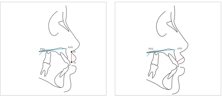
Figure 1 - Radiographic analysis of exposure of maxillary central incisor. Plane 1: Frankfort horizontal plane (FH). Plane 2: Plane passing through sto-mion superius parallel to FH. Plane 3: Plane passing through the edge of maxillary central incisor parallel to FH. Po = Porion. Or = Orbitale.
Figure 2 - Measure of nasolabial angle (angle formed by a line tangent to the columella through the subnasale and by a line tangent to the upper lip passing through the labial superius).
Po
Plane 2
Or
Plane 3
Figure 3 - Measure of upper lip height (plane passing through subnasale and stomion superius). PNS = Posterior nasal spine. ANS = Anterior nasal spine. Sb = Subnasale. Ss = Stomion superiorius.
Figure 5 - Inclination of the upper central incisor to the Sella-Nasion plane. Plane 1: Plane passing through sella and nasion. Plane 2: Plane passing through the long axis of the upper central incisor. S = Sella. Na = Nasion.
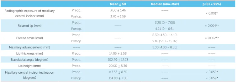
Figure 4 - Measure of upper lip thickness. PNS = Posterior nasal spine. ANS = Anterior nasal spine.
Figure 6 - Measure from the incisal edge to the Frankfort horizontal plane. Plane 1: Frankfort horizontal plane (FH). Plane 2: Plane perpendicular to FH through the incisal edge of the upper central incisor. Po = Porion. Or = Orbitale.
RESULTS
The sample consisted of 17 patients (14 females and 3 males). Sex was not correlated with increased clinical and radiographic exposure of maxillary central incisors ater maxillary advancement (p = 0.423). Patients had a mean age of 23 years in the sample (18-41). Age was
not correlated with increased clinical and radiographic exposure of maxillary central incisors ater orthogna-thic surgery (p = 0.650). Table 1 shows all values found in the clinical and radiographic exposure of maxillary central incisors both pretreatment and post-treatment. Mean clinical exposure of maxillary central incisors
PNS ANS
Sb
Ss
PNS ANS
Plane 2
Plane 2 Or
Plane 1 S
Angle
Na
with relaxed lip was 3.20 mm (0 – 7 mm) at the preop-erative stage and 4.21 mm (0 – 6.60 mm) at the postop-erative stage. Thus, there was a mean increase of 31% ater maxillary advancement. Mean clinical exposure of maxillary central incisors under forced smile was 8.30 mm (4.50 – 14.10 mm) at the preoperative stage and 9.16 mm (5.10 – 15.02 mm) at the postoperative stage. There was a statistical association between pre and post-operative measurements (Wilcoxon test / p = 0.001 - CI 95%). Mean exposure of maxillary central incisors in lateral cephalograms (presurgical) was 3.00 ± 1.46 mm, while postsurgical mean was 3.70 ± 1.59 mm. Thus, there was a mean increase in the exposure of central incisors of 0.70 mm which corresponds to 23.33%. The variables of exposure were also correlated with these same teeth on preoperative and postoperative ra-diographs (paired Student’s t-test / p < 0.001 – CI 95%). The mean amount of maxillary advancement was 5.11 mm. There was no statistical association between increased radiographic exposure of maxillary central incisors and the amount of maxillary advancement (p = 0.951). Mean lip thickness was 14.05 ± 2.58 mm. There was a statistically signiicant correlation be-tween increased exposure of maxillary central inci-sors and lip thickness ater maxillary advancement in
lateral cephalograms (p = 0.002 / r = 0.696 – CI 99%). In this study, the nasolabial angle had a mean value of 101.70 ± 13.30°. This angle is not related to increased exposure of maxillary central incisors in any measures revealed by radiographic images (p = 0.398 – Spearman Correlation Coeicient – CI 95%). Mean lip height was 20.00 ± 2.29 mm. There was no statistically signii-cant correlation between lip height and increased radio-graphic exposure of these teeth ater surgery (Pearson’s Correlation Coeicient – p = 0.357). Mean maxillary incisors inclination was 113.35 ± 8.39° preoperatively. This variable was not correlated with increased radio-graphic exposure of incisors (p = 0.533). Postoperative inclination of maxillary incisors ranged as from an aver-age of 114.88 ± 7.50°. There was no association between postoperative inclination and radiographic exposure of incisors ater surgery (p = 0.814). It was not possible to associate inclination of maxillary central incisors pre-operatively with postpre-operatively by means of paired Student’s t-test (p = 0.059), which had an average in-crease of one degree between these two surgical times. There was no statistical association between increased radiographic exposure of maxillary central incisors and the diference in inclination before and ater surgery (p = 0.259). All results can be seen in Table 1.
Mean ± SD Median (Min-Max) p (CI = 95%)
Radiographic exposure of maxillary central incisor (mm)
Preop. 3.00 ± 1.46 ———
< 0.001* Postop. 3.70 ± 1.59
Relaxed lip (mm) Preop. ——— 3.20 (0 – 7.00) = 0.004**
Postop. 4.21 (0 – 6.61)
Forced smile (mm) Preop. ——— 8.30 (4.50 - 14.10) = 0.002**
Postop. 9.16 (5.10 – 15.02)
Maxillary advancement (mm) ——— ——— 5.00 (4.00 – 8.00) ———
Lip thickness (mm) Preop. 14.05 ± 2.58 ——— ———
Nasolabial angle (degrees) Preop. 102.29 ± 12.73 ——— ———
Lip height (mm) Preop. 20.00 ± 5.36 ——— ———
Maxillary central incisor inclination (degrees)
Preop. 113.35 ± 8.39 ——— = 0.059*
Postop. 114.88 ± 7.50 ——— = 0.059*
Table 1 - Values of clinical and radiographic exposure of maxillary central incisors, pre and postoperatively; maxillary advancement; upper lip thickness preop-eratively; nasolabial angle preoppreop-eratively; preoperatively height of the upper lip, and inclination of maxillary central incisor in pre and postoperative periods.
DISCUSSION
It is essential to be able to predict postoperative hard tissue and facial proile changes resulting from orthog-nathic surgery in order to achieve functional and esthet-ic success of the procedure.13,19,20,21 The literature
sug-gests that the etiology of sot tissue changes is postsurgi-cal edema, increased support of bone tissue and the el-evation of periosteum and muscles near the nose with-out correct repositioning.13 Facial changes in patients
undergoing orthognathic surgery performed in the up-per jaw are multifactorial.15,19 If there was a pattern to
predict the amount of exposure of maxillary central in-cisors ater orthodontic-surgical combined treatment, there would be an ideal preoperative predictability. However, it is diicult to have a standard or a universal method to measure the exposure of teeth with relaxed lips and at smiling because of the number of variables that may be associated with it, such as the degree of muscle activity, individual diversity factors and age.22
In addition, there are diferences in the studies regarding the selection of the sample, namely: the inclusion of pa-tients with birth defects or syndromes in the same sam-ple of patients with facial deformity; the use of diferent radiological equipment to perform presurgical and post-surgical radiographs; the diiculty maintaining the cor-rect position of the head and patients’ lips at the time of radiograph; the exclusion or non-exclusion of segmen-tal surgeries in the sample; only one or multiple motion vectors in the maxilla in this sample; using the same technique of incision and suture in the sample; in addi-tion to osteoplasty (e.g., recontour of the anterior nasal spine) and follow-up time.13,18,19 Other prominent
fac-tors are the complexity of anatomical structures in this region of the face, the technical diiculty of correctly visualizing the outline on the radiograph, the absence of a speciic and unique methodology as the means of per-formance, and comparison of tracings. Due to this di-versity of factors that can alter the results, there is a lim-itation in comparing studies.18 Although women tend
to display greater maxillary incisor exposure at rest and movement than men,23 the study found no relationship
of sex with increased exposure of maxillary central inci-sors ater maxillary advancement. It is worth noting that the research sample had a small number of men (n = 3), which interfered in the analysis of results. Lee, Bailey, and Proit8 claimed that the physiological variation of
age and loss of muscle tone may explain the diference
between movement of sot and hard tissues. Younger adults with lack of dentoalveolar support do not show facial concavity, which is usually associated with an old-er age group. Tonicity and thickness of sot tissues are considered to be responsible for this diference.19
Flexi-bility of sot tissues, especially the lips, is directly inlu-enced by tone and thickness.12 In this study, age was not
related to increased exposure of maxillary central inci-sors ater surgery, since the vast majority of patients were young adults (20 to 30 years old). In most patients who underwent maxillary advancement alone or com-bined with another procedure in the mandible, the re-sults indicated an increase in radiographic exposure of maxillary central incisors ater orthognathic surgery (23.33%). There are two factors described in the litera-ture that may inluence this condition: sot tissue chang-es ater orthognathic surgery and changchang-es in bone and tooth structure itself. Thus, with regard to a potential change of sot tissues, we consider in this study some factors that may contribute to this increase in exposure of maxillary central incisors ater maxillary advance-ment as far as the nasolabial angle, height, and thickness of the lips. With regard to a potential change of hard tissue, the amount of maxillary advancement, the incli-nation of maxillary central incisors before and ater sur-gery, and the diference in inclination of these same structures before and ater surgery were considered. Ac-cording to the results of this study, only sot tissue changes inluenced the increased exposure of incisors ater maxillary advancement. Sot tissue changes ater maxillary advancement may include changes in the po-sitioning of the apex of the nose and nasolabial angle.15
The literature shows an increase in the nasolabial angle of 1.20° with anterior repositioning of the maxilla and a mean value of 0.65° for every 1 mm of advancement, although there is a wide variation in tissue response, some patients show an increase,9,15,19 while others show
a reduction12 in the postsurgical period. Bundgaar,
Melsen, and Terp7 hypothesized that an angular change
may be related to muscle function on the site of the os-teotomy.7 Therefore, this change in the nasolabial angle
measure-ment of a muscular component is unattainable in prac-tice. Radiograph measurements depend on the posi-tioning of the lips during the radiographic procedure which introduces the same signiicant variation.8,10,20
Considering natural lip thickness, the literature shows that lips with thickness greater than 17 mm have a smaller efect in relation to the movement of maxillary advancement; however, the opposite occurs with thin-ner lips in comparison to those that had excellent cor-relation.24 Thinner lips tend to expose more of incisors
ater maxillary advancement, which can be explained by the fact that these lips follow maxillary advancement to a greater degree compared to thicker lips.5,13,14 It is also
known that a thick lip can absorb the upper jaw amount of advancement by distension,18 in addition to having a
irmer grip on the base of the nose, which prevents ver-tical and horizontal movements of the upper lip in re-sponse to maxillary movement.24 In our study, we did
not ind any relation between the preoperative height of the upper lip and increased radiographic exposure of maxillary central incisors ater maxillary advancement. Although we found, in the literature, that shorter lips tend to have greater vertical movement ater surgery,25
small changes were observed in the vertical alteration of the lip with insigniicant statistical correlations ater maxillary advancement.9,21 Additionally, recent
cepha-lometric investigations have found that movement of hard and sot tissues ater orthognathic operations were strongly correlated horizontally but not vertically, and the position of the lips could not be predicted accurate-ly.12 Regarding the inluence of hard tissues that
con-cern the increased exposure of maxillary central incisors ater maxillary advancement, we evaluated the amount of maxillary advancement. This anterior movement is accompanied by a vertical and horizontal movement that inluences the exposure of incisors.17,20 In our study,
the amount of maxillary advancement was not statisti-cally signiicant, since the vast majority of advances were of the same size, between 4 mm (47%) and 5 mm (23%). Another factor that could inluence the inal ex-posure of maxillary incisors could be an increased incli-nation of the incisor ater orthognathic surgery for max-illary advancement, since orthodontic movement of anterior teeth can inluence and result in a change of
upper lip position, thus inluencing the exposure of maxillary central incisors.19 It is worth noting that the
op-tion to measure the inclinaop-tion of maxillary central inci-sors, before and ater surgery, was based on the fact that the second measurement was performed three months ater the procedure, which could lead to a biased result if there was a change in the inclination of incisors by orth-odontic movement, thus inluencing the exposure of maxillary incisors ater surgery. In our study, the difer-ence between inclination (pre- and postsurgical period) of these teeth was not signiicant. There is no relationship between the inclination of maxillary incisors by preop-eratively increasing their exposure ater surgery. There was also no relationship between the diference in incli-nation before and ater surgery of maxillary incisors and increased exposure of these teeth ater surgery.
CONCLUSIONS
1) Signiicant clinical change in the exposure of maxillary central incisors occurs ater maxillary ad-vancement, with a mean increase of 31% with a relaxed upper lip and 10.36% under forced smile.
2) Signiicant radiographic change in the exposure of maxillary central incisors occurs ater maxillary ad-vancement, with a mean increase of 23.33% in lateral cephalograms.
3) Increased exposure of maxillary central incisors ater maxillary advancement is mainly inluenced by upper lip thickness. Thin lips tend to expose more of incisors ater maxillary advancement, while thicker lips expose less due to their greater adherence to the base of the nose and by presenting more consistency.
4) There is a need for further studies relating to the change in exposure of maxillary central incisors ater maxillary advancement.
Author contributions
1. Araújo A. Cirurgia Ortognática. 1ª ed. São Paulo: Ed. Santos; 1999. 2. Chew MT, Sandham A, Wong HB. Evaluation of the linearity of soft- to
hard-tissue movement after orthognathic surgery. Am J Orthod Dentofacial Orthop. 2008 Nov;134(5):665-70.
3. Bell WH. Le Fort I osteotomy for correction of maxillary deformities. J Oral Surg. 1975 Jun;33(6):412-26.
4. Marşan G, Cura N, Emekli U. Soft and hard tissue changes after bimaxillary surgery in Turkish female Class III patients. J Craniomaxillofac Surg. 2009 Jan;37(1):8-17.
5. Lines PA, Steinhauser EW. Diagnosis and treatment planning in surgical orthodontic therapy. Am J Orthod. 1974 Oct;66(4):378-97.
6. Carlotti AE Jr, Aschafenburg PH, Schendel SA. Facial changes associated with surgical advancement of the lip and maxilla. J Oral Maxillofac Surg. 1986 Aug;44(8):593-6.
7. Bundgaard M, Melsen B, Terp S. Changes during and following total maxillary osteotomy (Le Fort I procedure): a cephalometric study. Eur J Orthod. 1986;8(1):21-9.
8. Lee DY, Bailey LJ, Proit WR. Soft tissue changes after superior repositioning of the maxilla with Le Fort I osteotomy: 5-year follow-up. Int J Adult Orthodon Orthognath Surg. 1996;11(4):301-11.
9. Mansour S, Burstone C, Legan H. An evaluation of soft-tissue changes resulting from Le Fort I maxillary surgery. Am J Orthod. 1983 Jul;84(1):37-47. 10. Rosen HM. Lip-nasal aesthetics following Le Fort I osteotomy. Plast Reconstr
Surg. 1988 Feb;81(2):171-82.
11. Kretschmer WB, Zoder W, Baciut G, Bacuit M, Wangerin K. Accuracy of maxillary positioning in bimaxillary surgery. Br J Oral Maxillofac Surg. 2009 Sep;47(6):446-9.
12. Altug-Atac AT, Bolatoglu H, Memikoglu UT. Facial soft tissue proile following bimaxillary orthognathic surgery. Angle Orthod. 2008 Jan;78(1):50-7. 13. Bell WH, Proit WR, White RP. Surgical correction of dentofacial deformities.
Philadelphia: Ed. W. B. Saunders; 1980. p. 2171-209.
REFERENCES
14. Freihofer HP Jr. The lip proile after correction of retromaxillism in cleft and non-cleft patients. J Maxillofac Surg. 1976 May 1;4(3):136-41.
15. Radney LJ, Jacobs JD. Soft-tissue changes associated with surgical total maxillary intrusion. Am J Orthod. 1981 Aug;80(2):191-212.
16. Stella JP, Streater MR, Epker BN, Sinn DP. Predictability of upper lip soft tissue changes with maxillary advancement. J Oral Maxillofac Surg. 1989 Jul;47(7):697-703.
17. Van Butsele BL, Mommaerts MY, Abeloos JS, De Clercq CA, Neyt LF. Creating lip seal by maxillo-facial osteotomies. A retrospective cephalometric study. J Craniomaxillofac Surg. 1995 Jun;23(3):165-74.
18. Del Santo LM, Souza RP, Del Santo Jr M, Marcantonio E. Alteração no peril dos lábios de pacientes submetidos a avanços maxilares em cirurgia ortognática do tipo Le Fort I. Rev Dental Press Ortod Ortop Facial. 2004;9(3):49-63.
19. Louis PJ, Austin RB, Waite PD, Mathews CS. Soft tissue changes of the upper lip associated with maxillary advancement in obstructive sleep apnea patients. J Oral Maxillofac Surg. 2001 Feb;59(2):151-6.
20. Bell WH, Dann III JJ. Correction of dentofacial deformities by surgery in the anterior part of the jaws. Am J Orthod. 1973;64(2):162-87.
21. Bell WH, Scheideman GB. Correction of vertical maxillary deiciency: stability and soft tissue changes. J Oral Surg. 1981 Sep;39(9):666-70.
22. Fudalej P. Long-term changes of the upper lip position relative to the incisal edge. Am J Orthod Dentofacial Orthop. 2008 Feb;133(2):204-9; quiz 328.e1. 23. Mobarak KA, Krogstad O, Espeland L, Lyberg T. Factors inluencing the
predictability of soft tissue proile changes following mandibular setback surgery. Angle Orthod. 2001;71(3):216-27.
24. Epker BN, Stella JP, Fish LC. Dentofacial deformities: integrated orthodontic and surgical correction. St Louis: Mosby; 1995.