www.jbp.org.br
ISSN 1806-3713
Volume 43, Number 4 July | August 2017
HIGHLIGHT
V
olume 43, Number 4
July | A
ugus
t 20
17
Recommendations for
the pharmacological
treatment of COPD
Mechanical ventilation
weaning
Chest CT findings in
patients with
www.jbp.org.br
ISSN 1806-3713
Volume 43, Número 3 maio | junho 2017
DESTAQUE
Diretrizes Br asileiras para Fibrose Cís
tica Impacto da asma no
Brasil Distúrbio do sono
ISSN 1806-3713
Published once every two months J Bras Pneumol. v.43, number 4, p. 247-326 July/August 2017
Publicação Indexada em:
Latindex, LILACS, Scielo Brazil, Scopus, Index Copernicus, ISI Web of Knowledge, MEDLINE e PubMed Central (PMC)
Disponível eletronicamente nas versões português e inglês:
www.jornaldepneumologia.com.br e www.scielo.br/jbpneu
Associação Brasileira de Editores Científicos
I N T E R N A T I O N A L
EDITOR-IN-CHIEF
Rogerio Souza - Universidade de São Paulo, São Paulo - SP
EXECUTIVE EDITORS
Bruno Guedes Baldi - Universidade de São Paulo, São Paulo - SP
Caio Júlio Cesar dos Santos Fernandes - Universidade de São Paulo - São Paulo - SP Carlos Roberto Ribeiro de Carvalho - Universidade de São Paulo, São Paulo - SP Carlos Viana Poyares Jardim - Universidade de São Paulo, São Paulo - SP ASSOCIATE EDITORS
Afrânio Lineu Kritski - Universidade Federal do Rio de Janeiro, Rio de Janeiro, RJ Andre Luis Pereira de Albuquerque - Universidade de São Paulo - São Paulo - SP Bruno Hochhegger - Universidade Federal do Rio Grande do Sul - Porto Alegre – RS Edson Marchiori - Universidade Federal Fluminense, Niterói - RJ
Fernanda Carvalho de Queiroz Mello - Universidade Federal do Rio de Janeiro - Rio de Janeiro - RJ Frederico Leon Arrabal Fernandes - Universidade de São Paulo - São Paulo - SP
Giovanni Battista Migliori - Director WHO Collaborating Centre for TB and Lung Diseases, Fondazione S. Maugeri, Care and Research Institute, Tradate - Italy
Giovanni Sotgiu - University of Sassari, Sassari - Italy Irma de Godoy - Universidade Estadual Paulista, Botucatu - SP
Marcelo Alcântara Holanda - Universidade Federal do Ceará - Fortaleza - CE Pedro Caruso - Universidade de São Paulo - São Paulo - SP
Pedro Rodrigues Genta - Universidade de São Paulo - São Paulo - SP
Renato Tetelbom Stein - Pontifícia Universidade Católica do Rio Grande do Sul, Porto Alegre - RS Ricardo de Amorim Corrêa - Universidade Federal de Minas Gerais - Belo Horizonte - MG Ricardo Mingarini Terra - Universidade de São Paulo - São Paulo - SP
Simone Dal Corso - Universidade Nove de Julho - São Paulo – SP Tomás Pulido - Instituto Nacional de Cardiología Ignacio Chávez - México Ubiratan de Paula Santos - Universidade de São Paulo, São Paulo - SP Veronica Amado - Universidade de Brasília, Brasília - DF
EDITORIAL COUNCIL
Alberto Cukier - Universidade de São Paulo, São Paulo – SP Álvaro A. Cruz - Universidade Federal da Bahia, Salvador, BA Ana C. Krieger - Weill Cornell Medical College - New York – USA
Ana Luiza Godoy Fernandes - Universidade Federal de São Paulo, São Paulo - SP Antonio Segorbe Luis - Universidade de Coimbra, Coimbra - Portugal Ascedio Jose Rodrigues - Universidade de São Paulo - São Paulo - SP Brent Winston - University of Calgary, Calgary - Canada
Carlos Alberto de Assis Viegas - Universidade de Brasília, Brasília - DF
Carlos Alberto de Castro Pereira - Universidade Federal de São Paulo, São Paulo - SP Carlos M. Luna - Hospital de Clinicas, Universidad de Buenos Aires, Buenos Aires - Argentina Carmen Silvia Valente Barbas - Universidade de São Paulo, São Paulo - SP
Celso Ricardo Fernandes de Carvalho - Universidade de São Paulo, São Paulo - SP Dany Jasinowodolinski - Universidade de São Paulo, São Paulo - SP
Denis Martinez - Universidade Federal do Rio Grande do Sul, Porto Alegre - RS Douglas Bradley - University of Toronto, Toronto, ON - Canadá
Emílio Pizzichini - Universidade Federal de Santa Catarina, Florianópolis - SC Fábio Biscegli Jatene - Universidade de São Paulo, São Paulo - SP
Frank McCormack - University of Cincinnati School of Medicine, Cincinnati, OH - USA Geraldo Lorenzi Filho - Universidade de São Paulo, São Paulo - SP
Gilberto de Castro Junior - Universidade de São Paulo, São Paulo - SP
Gustavo Javier Rodrigo - Hospital Central de las Fuerzas Armadas, Montevidéu – Uruguay Ilma Aparecida Paschoal - Universidade de Campinas, Campinas - SP
C. Isabela Silva Müller - Vancouver General Hospital, Vancouver, BC - Canadá J. Randall Curtis - University of Washington, Seattle, Wa - USA
John J. Godleski - Harvard Medical School, Boston, MA - USA José Alberto Neder - Queen’s University - Ontario, Canada
José Antonio Baddini Martinez - Universidade de São Paulo, Ribeirão Preto - SP José Dirceu Ribeiro - Universidade de Campinas, Campinas - SP
José Miguel Chatkin - Pontifícia Universidade Católica do Rio Grande do Sul, Porto Alegre - RS José Roberto de Brito Jardim - Universidade Federal de São Paulo, São Paulo - SP José Roberto Lapa e Silva - Universidade Federal do Rio de Janeiro, Rio de Janeiro - RJ Kevin Leslie - Mayo Clinic College of Medicine, Rochester, MN - USA
Luiz Eduardo Nery - Universidade Federal de São Paulo, São Paulo - SP Marc Miravitlles - University Hospital Vall d’Hebron - Barcelona, Catalonia, Spain Marisa Dolhnikoff - Universidade de São Paulo, São Paulo - SP
Marli Maria Knorst - Universidade Federal do Rio Grande do Sul, Porto Alegre - RS Mauro Musa Zamboni - Instituto Nacional do Câncer, Rio de Janeiro - RJ Nestor Muller - Vancouver General Hospital, Vancouver, BC - Canadá Noé Zamel - University of Toronto, Toronto, ON - Canadá
Oliver Augusto Nascimento - Universidade Federal de São Paulo - São Paulo - SP Paul Noble - Duke University, Durham, NC - USA
Paulo Francisco Guerreiro Cardoso - Universidade de São Paulo, São Paulo - SP Paulo Manuel Pêgo Fernandes - Universidade de São Paulo, São Paulo - SP Peter J. Barnes - National Heart and Lung Institute, Imperial College, London - UK Renato Sotto Mayor - Hospital Santa Maria, Lisboa - Portugal
Richard W. Light - Vanderbili University, Nashville, TN, USA Rik Gosselink - University Hospitals Leuven - Bélgica Robert Skomro - University of Saskatoon, Saskatoon - Canadá Rubin Tuder - University of Colorado, Denver, CO - USA
Sérgio Saldanha Menna Barreto - Universidade Federal do Rio Grande do Sul, Porto Alegre - RS Sonia Buist - Oregon Health & Science University, Portland, OR - USA
Talmadge King Jr. - University of California, San Francisco, CA - USA
ISSN 1806-3713
E
x
p
e
d
ie
n
te
BRAZILIAN THORACIC SOCIETY
Ofice: SCS Quadra 01, Bloco K, Asa Sul, salas 203/204. Edifício Denasa, CEP 70398-900, Brasília, DF, Brazil. Tel. +55 61 3245-1030/+55 0800 616218. Website: www.sbpt.org.br. E-mail: [email protected]
The Brazilian Journal of Pulmonology (ISSN 1806-3713) is published once every two months by the Brazilian Thoracic Society (BTS). The statements and opinions contained in the editorials and articles in this Journal are solely those of the authors thereof and not of the Journal’s Editor-in-Chief, peer reviewers, the BTS, its oficers, regents, members, or employees. Permission is granted to reproduce any igure, table, or other material published in the Journal provided that the source for any of these is credited.
BTS Board of Directors (2017-2018 biennium):
President: Fernando Luiz Cavalcanti Lundgren - PE Secretary-General: Benedito Francisco Cabral Júnior - DF CFO: Simone Chaves Fagondes - RS
Scientiic Director: Ana Luisa Godoy Fernandes - SP
Director, Communications: Fernanda Miranda de Oliveira - GO Director, Education and Professional Practice:: Irma de Godoy - SP Director, Professional Advocacy: Marcelo Gervilla Gregório - SP President, BTS Congress 2018: Marcelo Fouad Rabahi - GO President Elect (2019/2020 biennium): José Miguel Chatkin - RS
Editor-in-Chief of the Brazilian Journal of Pulmonology: Rogério de Souza - SP
AUDIT COMMITTEE:
Active Members: Ronaldo Rangel Travassos Júnior - PB, Eduardo Felipe Barbosa Silva - DF, Filadélia Passos Travassos Martins - CE
Alternates: Leandro Genehr Fitscher - RS, Ciléa Aparecida Victória Martins - ES, Eduardo Pamplona Bethlem - RJ
COORDINATORS, BTS DEPARTMENTS:
Thoracic Surgery – Darcy Ribeiro Pinto Filho -RS Sleep–disordered Breathing – Pedro Rodrigues Genta -SP Respiratory Endoscopy – Mauro Musa Zamboni -RJ Pulmonary Function – Silvia Carla Sousa Rodrigues -SP Imaging – Pablo Rydz Pinheiro Santana -SP
Lung Diseases – Vera Luiza Capelozzi -SP
Pediatric Pulmonology – Marina Buarque de Almeida -SP
COORDINATORS, BTS SCIENTIFIC COMMITTEES:
Asthma – Maria Alenita de Oliveira - SP Lung Cancer – Gustavo Faibischew Prado - SP Pulmonary Circulation – Marcelo Basso Gazzana -SP Advanced Lung Disease – Paulo Henrique Ramos Feitosa -DF Interstitial Diseases – José Antônio Baddini Martinez -SP
Environmental and Occupational Respiratory Diseases – Carlos Nunes Tietboehl-Filho -RS COPD – Frederico Leon Arrabal Fernandes -SP
Epidemiology – Juliana Carvalho Ferreira - SP Cystic Fibrosis – Rodrigo Abensur Athanazio – SP
Respiratory Infections and Mycoses – Mônica Corso Pereira - SP Pleura – Roberta Karla Barbosa de Sales -SP
Smoking – Maria da Penha Uchoa Sales - CE Intensive Care – Eduardo Leite Vieira Costa -SP Tuberculosis – Denise Rossato Silva -RS
ADMINISTRATIVE SECRETARIAT OF THE BRAZILIAN JOURNAL OF PULMONOLOGY Address: SCS Quadra 01, Bloco K, Asa Sul, salas 203/204. Edifício Denasa, CEP 70398-900, Brasília, DF, Brazil. Tel. +55 61 3245-1030/+55 0800 616218.
Assistant Managing Editor: Luana Maria Bernardes Campos. E-mail: [email protected]
Circulation: 4.000 copies
Distribution: Free to members of the BTS and libraries Printed on acid-free paper
ISSN 1806-3713
Published once every two months J Bras Pneumol. v.43, number 4, p. 247-326 July/August 2017
C
o
n
te
n
ts
EDITORIAL
247 - JBP and bibliometric indices Rogério Souza
249 - The dificult task of searching for tools that help predict mechanical ventilator weaning success
Bruno do Valle Pinheiro
CONTINUING EDUCATION: IMAGING
251 - Thickening of the tracheal wall
Edson Marchiori, Bruno Hochhegger, Gláucia Zanetti
CONTINUING EDUCATION: SCIENTIFIC METHODOLOGY
252 - Prognostic studies for health care decision making Cecilia Maria Patino Juliana Carvalho Ferreira
ORIGINAL ARTICLE
253 - Usefulness of radiological signs of pulmonary congestion in predicting failed spontaneous breathing trials
Ana Carolina Peçanha Antonio, Cassiano Teixeira, Priscylla Souza Castro, Ana Paula Zanardo, Marcelo Basso Gazzana, Marli Knorst
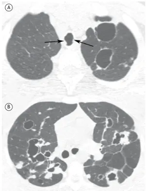
259 - Laryngotracheobronchial papillomatosis: chest CT indings Helena Ribeiro Fortes, Felipe Mussi von Ranke, Dante Luiz Escuissato,
Cesar Augusto Araujo Neto, Gláucia Zanetti, Bruno Hochhegger, Klaus Loureiro Irion, Carolina Althoff Souza, Edson Marchiori
264 - Translation and cultural adaptation of a speciic instrument for measuring asthma control and asthma status: the Asthma Control and Communication Instrument
Michelle Gonçalves de Souza Tavares, Carolina Finardi Brümmer,
Gabriela Valente Nicolau, José Tavares de Melo Jr, Nazaré Otilia Nazário,
Leila John Marques Steidle, Cecília Maria Patino, Marcia Margaret Menezes Pizzichini, Emílio Pizzichini
270 - High-resolution computed tomography indings of pulmonary tuberculosis in lung transplant recipients
Irai Luis Giacomelli, Roberto Schuhmacher Neto, Carlos Schuller Nin, Priscilla de Souza Cassano, Marisa Pereira, José da Silva Moreira, Douglas Zaione Nascimento, Bruno Hochhegger
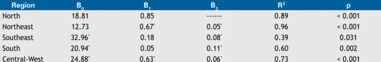
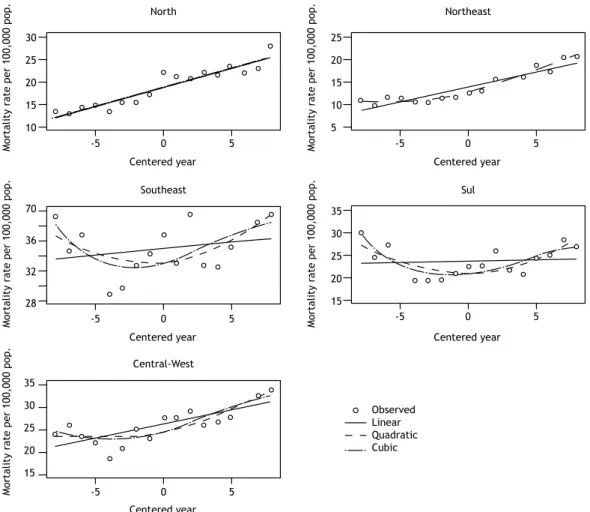
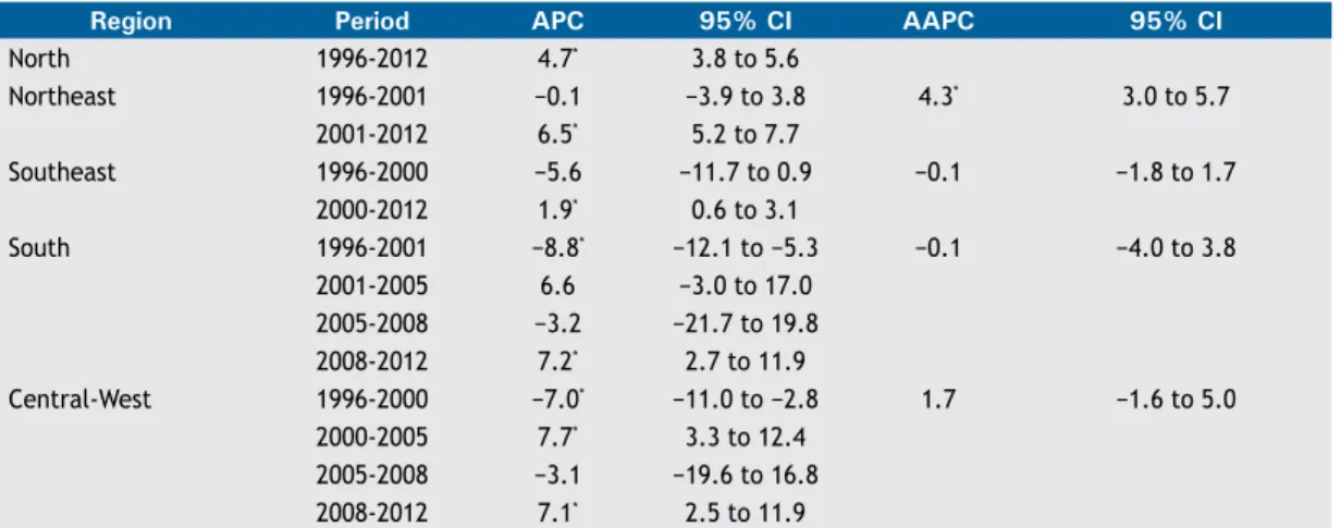
274 - Pneumonia mortality trends in all Brazilian geographical regions between 1996 and 2012
Rosemeire de Olanda Ferraz, Jane Kelly Oliveira-Friestino, Priscila Maria Stolses Bergamo Francisco
280 - Can the six-minute walk distance predict the occurrence of acute exacerbations of COPD in patients in Brazil?
Fernanda Kazmierski Morakami, Andrea Akemi Morita, Gianna Waldrich Bisca, Josiane Marques Felcar, Marcos Ribeiro, Karina Couto Furlanetto,
ISSN 1806-3713
Published once every two months J Bras Pneumol. v.43, number 4, p. 247-326 July/August 2017
C
o
n
te
n
ts
285 - Sleep quality in medical students: a comparison across the various phases of the medical course
Camila de Castro Corrêa, Felipe Kazan de Oliveira, Diego Scherlon Pizzamiglio, Erika Veruska Paiva Ortolan, Silke Anna Theresa Weber
SPECIAL ARTICLE
290 - Recommendations for the pharmacological treatment of COPD: questions and answers
Frederico Leon Arrabal Fernandes, Alberto Cukier, Aquiles Assunção Camelier, Carlos Cezar Fritscher, Cláudia Henrique da Costa, Eanes Delgado Barros Pereira, Irma Godoy, José Eduardo Delini Cançado, José Gustavo Romaldini, Jose Miguel Chatkin, José Roberto Jardim, Marcelo Fouad Rabahi, Maria Cecília Nieves Maiorano de Nucci, Maria da Penha Uchoa Sales, Maria Vera Cruz de Oliveira Castellano, Miguel Abidon Aidé, Paulo José Zimermann Teixeira, Renato Maciel, Ricardo de Amorim Corrêa,
Roberto Stirbulov, Rodrigo Abensur Athanazio, Rodrigo Russo, Suzana Tanni Minamoto, Fernando Luiz Cavalcanti Lundgren
META-ANALYSIS
302 - Long-acting muscarinic antagonists vs. long-acting β2 agonists in COPD exacerbations: a systematic review and meta-analysis
Israel Silva Maia, Mariângela Pimentel Pincelli, Victor Figueiredo Leite, João Amadera, Anna Maria Buehler
REVIEW ARTICLE
313 - Chest CT indings in patients with dysphagia and aspiration: a systematic review Betina Scheeren, Erissandra Gomes, Giordano Alves, Edson Marchiori, Bruno Hochhegger
IMAGING IN PULMONARY MEDICINE
319 - Multislice CT in the diagnosis of bronchopleural istula Bruno Hochhegger, Gláucia Zanetti, Edson Marchiori
CASE REPORT
320 - Pulmonary foreign body granulomatosis in a chronic user of powder cocaine Shruti Khurana, Ankit Chhoda, Sandeep Sahay, Priyanka Pathania
LETTER TO THE EDITOR
322 - The reversed halo sign: also think about chronic eosinophilic pneumonia Gaetano Rea, Giorgia Dalpiaz, Alessandro Vatrella, Stefania Damiani, Edson Marchiori
324 - Pneumothorax: between the beach and the stratosphere Eduardo Kaiser Ururahy Nunes Fonseca, Adham do Amaral e Castro, Yoshino Tamaki Sameshima
http://dx.doi.org/10.1590/S1806-37562017000400002
JBP and bibliometric indices
Rogério Souza1,21. Disciplina de Pneumologia, Instituto do Coração, Hospital das Clínicas, Faculdade de Medicina, Universidade de São Paulo, São Paulo (SP) Brasil. 2. Editor-Chefe do JBP - Jornal Brasileiro de Pneumologia, Brasília (DF) Brasil.
The process of publishing a scientiic journal has complexities that go well beyond the choice of manuscripts, although this process alone has intrinsic peculiarities. Initially, it is necessary to consider the context surrounding the journal.(1) The JBP is the leading journal in the ield
of respiratory medicine in Latin America, a fact that has recently been conirmed with the release of the 2016 bibliometric indices. We achieved an impact factor of 1.496, according to the Thomson Reuters index, and, according to the Scopus database, which uses the same methodology, we achieved an index of 1.609. These are the highest values ever achieved by our Journal and place us in the second quartile of the respiratory medicine journals. In addition, if we observe other indicators, we can infer that the trend is toward growth. For instance, international collaboration has grown consistently in recent years, increasing from 8.5% in 2013 to 16.9% in 2016, which demonstrates the improved representativeness of the JBP.
It is important to emphasize the concept that the indices used for the evaluation of the various scientiic publications are not the sole determinant of the relevance of such publications and, sometimes, even create additional complicating factors.(2) We need to maintain our commitment to increasing our visibility without losing focus on the formative character that our Journal has, particularly in Brazil. However, the metric by which the national publications are evaluated in the Brazilian graduate system does not take that into account, giving importance only to the impact factor and making large research groups less interested in the national publications. This is a problem that needs to be addressed directly if we want to further increase the editorial relevance of the JBP.
Over the past two years, we have been able to balance all that. The proile of the most often cited articles includes review of topics that are most prevalent(3,4) and original articles addressing prevalent topics or rarer conditions. (5,6) However, it should also be considered that the JBP is the oficial organ of the Brazilian Thoracic Association, and, therefore, all related ields should be covered, regardless of the citation potential of each one of them, since it is well known that smaller or still incipient ields are less likely to be cited over the time period used in the bibliometric indices. All of these aspects should be considered together in analyzing the relevance of the JBP in the respiratory medicine setting.
For such discussions to become increasingly present in the JBP, the participation of the associate editors in
editorial decisions has been most relevant. They are the ones mainly responsible for the growth of the Journal and the consolidation of our indices. For this to be even more long-lasting, the position of Vice Editor of the JBP was created. It is the Vice Editor’s responsibility to participate in the most signiicant editorial decisions, together with the Editor-in-Chief, for a period of two years, after which he will take on the editorial leadership for the customary period of four years. The creation of this new position was aimed at enabling smoother transitions, allowing changes in editorial policies in a context known to all parties involved. The Vice Editor selection process was disseminated through our media and will be completed in July of 2017, and the results should be known by the time the September/October issue of the JBP comes out.
While on one hand the decentralization of editorial policies is underway, several barriers have yet to be overcome. As a result of the increased visibility of the JBP, there has been a signiicant increase in the number of submissions. While such an increase is desirable, because it relects our representativeness, it carries with it an even greater demand for reviewers. We have had the unequivocal collaboration of a large number of colleagues, who, almost anonymously, have contributed signiicantly with their critical and analytical thinking and their insight. There is a need for greater recognition to be given to these colleagues, to whom the entire editorial board expresses its eternal gratitude. The Brazilian Thoracic Association has studied alternatives for achieving this objective. This is not a characteristic of ours alone; the major international journals are discussing how to give better recognition to their reviewers and, at the same time, attract more people to this position, a position that is key to the routine of any journal known for excellence, such as ours is. Critical analysis of scientiic studies needs to be made part of the daily life of pulmonologists in training. In the long term, the result of this process will be better education of researchers and faculty. An increased critical mass of reviewers and potential editors will be a very beneicial secondary effect of this process.
All in all, we have much to celebrate from the growth of the JBP, but we still have numerous challenges ahead, both known and unknown. To overcome all of them, the participation of the JBP’s readership is essential. Therefore, here is an invitation: give your opinions, ideas, criticisms, and suggestions! This will allow the JBP to relect the concerns of those for whom it is intended.
J Bras Pneumol. 2017;43(4):247-248
JBP and bibliometric indices
REFERENCES
1. Souza R. 2015--another step along the road in a 40-year journey. J Bras Pneumol. 2015;41(1):1-2. https://doi.org/10.1590/S1806-37132015000100001
2. Souza R. Consolidating in the present, with an eye to the future. J Bras Pneumol. 2016;42(6):399-400. https://doi.org/10.1590/s1806-37562016000600002
3. Torres-Sánchez I, Rodriguez-Alzueta E, Cabrera-Martos I, López-Torres I, Moreno-Ramírez MP, Valenza MC. Cognitive impairment in COPD: a systematic review. J Bras Pneumol. 2015;41(2):182-90. https://doi.org/10.1590/S1806-37132015000004424
4. Caruso P, Albuquerque AL, Santana PV, Cardenas LZ, Ferreira JG, Prina E, et al. Diagnostic methods to assess inspiratory and
expiratory muscle strength. J Bras Pneumol. 2015;41(2):110-23. https://doi.org/10.1590/S1806-37132015000004474
5. Freitas CS, Baldi BG, Araújo MS, Heiden GI, Kairalla RA, Carvalho CR. Use of sirolimus in the treatment of lymphangioleiomyomatosis: favorable responses in patients with different extrapulmonary manifestations. J Bras Pneumol. 2015;41(3):275-80. https://doi. org/10.1590/S1806-37132015000004553
6. Stelmach R, Fernandes FL, Carvalho-Pinto RM, Athanazio RA, Rached SZ, Prado GF, et al. Comparison between objective measures of smoking and self-reported smoking status in patients with asthma or COPD: are our patients telling us the truth? J Bras Pneumol. 2015;41(2):124-32. https://doi.org/10.1590/S1806-37132015000004526
http://dx.doi.org/10.1590/S1806-37562017000400001
The dificult task of searching for tools that
help predict mechanical ventilator weaning
success
Bruno do Valle Pinheiro1
1. Serviço de Pneumologia e Terapia Intensiva, Hospital Universitário, Universidade Federal de Juiz de Fora, Juiz de Fora (MG) Brasil. Mechanical ventilation (MV) is one of the most common
supportive measures employed in ICU and is fundamental in maintaining life under certain conditions.(1)However,
MV can be associated with serious complications, such as pneumonia, tracheobronchitis, critical illness neuropathy and myopathy, delirium, barotrauma, and MV-induced lung injury, making its interruption desirable once the patient is able to breathe spontaneously in a safe manner and without need of a tracheal cannula.(2)This process of disconnecting the patient from the ventilator is designated weaning.
Weaning remains one of the great challenges of MV, especially because it is impossible to predict, with the desired accuracy, whether extubation will be successful or whether reintubation will be necessary. The rates of patients who undergo unscheduled extubation and are successfully weaned range from 25% to 75%; these data show that exaggerated conservatism may delay MV weaning in some cases. However, the mean rates of reintubation after elective extubation remain between 10% and 12%, regardless of the indices used to in order to predict weaning success.(4) Nevertheless, these
are mean values and certainly vary depending on the complexity of MV weaning: it can be simple — patients extubated after a irst spontaneous breathing trial (SBT) — dificult — patients who fail on the irst SBT and require up to three SBTs or up to 7 days after the irst weaning trial — or prolonged — patients requiring more than three SBTs or more than 7 days after the irst weaning trial.
Among the causes of weaning failure, especially in dificult and prolonged cases, it is worth noting cardiac dysfunction, associated or not with luid overload. When the patient spontaneously breathe without the help of positive pressure of the ventilatory support, either during a T-piece trial or after extubation, the negative intrathoracic pressure during inhalation promotes an increase in venous return, with a consequent increase in the preload of the right and left ventricles, as well as a decrease in the left ventricular ejection pressure gradient, causing an increase in left ventricular afterload. At the same time, right after spontaneous breathing is initiated, there may be an increase in adrenergic tone, with increased levels of catecholamines and increases in left ventricular preloads and afterloads. These changes altogether increase oxygen consumption by the myocardium and may even generate ischemia in patients with previous coronary disease. Another possible consequence is the inability of the heart to deal with the increases in preload and afterload, resulting in increased illing pressures and pulmonary congestion. Pulmonary
congestion increases the work of breathing and may be responsible for MV weaning failure.(6)
Given the importance of cardiac dysfunction and hypervolemia in weaning failure, it is expected that the identiication of these conditions can be useful in the evaluation of these patients. In this sense, Antonio et al.,(7) in the current issue of the JBP, evaluated whether the presence of signs of pulmonary congestion on chest X-rays correlated with SBT failure. To that end, the authors evaluated patients older than 18 years of age undergoing MV for more than 24 h, depending on their clinical or surgical conditions. The patients were evaluated daily and were considered eligible for weaning if the cause of their respiratory failure improved and if they had good level of consciousness, adequate gas exchange, absence of respiratory acidosis, hemodynamic stability, and a rapid shallow breathing rate ≤ 105 breaths/min/L. In such cases, a T-tube was placed for 30-120 min, and the following signs of failure were observed: RR > 30 breaths/ min; arterial oxyhemoglobin saturation < 90%; use of accessory muscles; HR > 140 bpm; systolic blood pressure < 90 mmHg or < 20% of basal levels; and altered level of consciousness. The presence of any of these indings indicated SBT failure, whereas the absence of all of the signs meant SBT success, and extubation was carried out. A radiologist, blinded to the SBT result, evaluated the chest X-ray performed within 24 h prior to the trial and used a radiological score, described by Shochat et al.,(8) in order to assess pulmonary congestion.
The authors evaluated 170 patients, the majority of whom had simple weaning — 78.3% were extubated on irst attempt, and the duration of MV before weaning was 4 days (interquartile range [IQR]: 2-4 days) among those who had SBT success and 6 days (IQR: 4-11 days) among those who had SBT failure. The radiological score was not able to discriminate SBT results; the scores were similar between the patients showing SBT success or SBT failure (median = 3 days; IQR: 2-4 days) in both groups. ROC curve analysis revealed no cut-off point that accurately discriminated between SBT success and SBT failure. The results led the authors to conclude correctly that there is no indication to perform chest X-rays in order to evaluate pulmonary congestion as an additional tool to recommend the use of SBT in patients who meet the commonly accepted criteria to start the trial.
In some respects, this negative result could already be expected. The vast majority of patients had simple weaning; therefore, failure rates were low. Failure can occur due to various causes, cardiovascular failure being only one of them. In addition, among the patients studied in that cohort, less than half had systolic or diastolic J Bras Pneumol. 2017;43(4):249-250
The dificult task of searching for tools that help predict mechanical ventilator weaning success
dysfunction, which would be risk factors for weaning failure due to cardiovascular disease or hypervolemia. However, this negative result does not rule out the possibility that the evaluation of cardiac dysfunction or hypervolemia may be useful as a predictor of weaning success. In this sense, at least two points deserve to be discussed. The irst point is whether such an assessment is necessary before SBT is performed in each and every patient. Performing additional evaluations in patients with a low probability of failure can be only a delaying factor in extubation, increasing the chances of MV complications. Therefore, it would be interesting to study a population at a greater risk of failure, even with an increased risk of cardiovascular failure or hypervolemia. Some criteria that could deine this population would be the classiication of weaning as dificult or prolonged, history of heart disease, or other risk factors, such as advanced age. The second
point is whether a chest X-ray is the ideal tool for this sort of investigation or whether other options have higher yields. Among these options, some studies have demonstrated the usefulness of echocardiography and B-type natriuretic peptide quantiication in identifying patients who fail SBT due to heart disease.(9,10)
In summary, another study showed the ineffectiveness of an isolated parameter in predicting SBT success or SBT failure; in this case, a radiological score for pulmonary congestion. Although the parameter itself might be inadequate, it should be considered that the result might have been due to the population studied, which consisted of patients who had simple weaning, with a low probability of weaning failure. Increasing the number of predictors is not only unnecessary but can lead to delayed extubation, as the authors have properly discussed.
REFERENCES
1. Esteban A, Ferguson ND, Meade MO, Frutos-Vivar F, Apezteguia C, Brochard L, et al. Evolution of mechanical ventilation in response to clinical research. Am J Respir Crit Care Med. 2008;177(2):170-7. https://doi.org/10.1164/rccm.200706-893OC
2. Boles JM, Bion J, Connors A, Herridge M, Marsh B, Melot C, et al. Weaning from mechanical ventilation. Eur Respir J. 2007;29(5):1033-56. https://doi.org/10.1183/09031936.00010206
3. Kiekkas P, Aretha D, Panteli E, Baltopoulos GI, Filos KS. Unplanned extubation in critically ill adults: clinical review. Nursing Crit Care. 2012;18(3):123-34. https://doi.org/10.1111/j.1478-5153.2012.00542.x
4. Peñuelas O, Thille AW, Esteban A. Discontinuation of ventilatory support: new solutions to old dilemmas. Curr Opin Crit Care. 2015;21(1):74-81. https://doi.org/10.1097/MCC.0000000000000169 5. Perren A, Brochard L. Managing the apparent and hidden dificulties
of weaning from mechanical ventilation. Intensive Care Med. 2013;39(11):1885-95. https://doi.org/10.1007/s00134-013-3014-9 6. Teboul JL. Weaning-induced cardiac dysfunction: where are
we today? Intensive Care Med. 2014;40(8):1069-79. https://doi.
org/10.1007/s00134-014-3334-4
7. Antonio ACP, Teixeira C, Castro PS, Zanardo AP, Gazzana MB, Knorst M. Radiological signs of pulmonary congestion do not predict failed spontaneous breathing trial. J Bras Pneumol. 2017;43(4):253-58. 8. Shochat M, Shotan A, Trachtengerts V, Blondheim DS, Kazatsker
M, Gurovich V, et al. A novel radiological score to assess lung luid content during evolving acute heart failure in the course of acute myocardial infarction. Acute Card Care. 2011;13(2):81-6. https://doi. org/10.3109/17482941.2011.567279
9. Lamia B, Maizel J, Ochagavia A, Chemla D, Osman D, Richard C, et al. Echocardiographic diagnosis of pulmonary artery occlusion pressure elevation during weaning from mechanical ventilation. Crit Care Med. 2009;37(5):1696-701. https://doi.org/10.1097/ CCM.0b013e31819f13d0
10. Mekontso Dessap A, Roche-Campo F, Kouatchet A, Tomicic V, Beduneau G, Sonneville R, et al. Natriuretic peptide-driven luid management during ventilator weaning: a randomized controlled trial. Am J Respir Crit Care Med. 2012;186(12):1256-63. https://doi. org/10.1164/rccm.201205-0939OC
http://dx.doi.org/10.1590/S1806-37562017000000074
Thickening of the tracheal wall
Edson Marchiori1, Bruno Hochhegger2, Gláucia Zanetti11. Universidade Federal do Rio de Janeiro, Rio de Janeiro (RJ) Brasil.
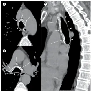
2. Universidade Federal de Ciências da Saúde de Porto Alegre, Porto Alegre (RS) Brasil. A 63-year-old female patient presented with a 1-year history of recurrent polyarthritis and a 3-month history of dry cough. Her laboratory test results were normal. Physical examination revealed deformity in both ears, with inlammatory signs. A chest CT showed diffuse thickening of the walls of the trachea and main bronchi (Figure 1). Her lung parenchyma was normal.
Diffuse thickening of the tracheal wall has a large number of possible etiologies—amyloidosis; relapsing polychondritis (RP); tracheopathia osteochondroplastica (TPO); infections, such as tuberculosis, paracoccidioidomycosis, and rhinoscleroma; granulomatosis with polyangiitis;
sarcoidosis; lymphomas; etc. Some imaging features can be useful in narrowing the differential diagnosis, such as the presence of calciications, and deine whether the entire tracheal circumference is affected or whether the lesion spares the posterior membranous wall, affecting only the cartilaginous portion. In the case presented here, two CT features are of note: the wall thickening has calciications in its entire longitudinal extension; and the posterior membranous portion of the tracheal wall is spared.
Tracheal wall calciications can be observed in healthy patients, being related to senility. However, calciications associated with wall thickening can be seen in amyloidosis, TPO, and RP. In amyloidosis, involvement is circumferential, also affecting the posterior membranous wall. Therefore, in the case presented here, the differential diagnosis is restricted to two diseases: TPO and RP.
TPO is a disease of unknown etiology, characterized by the formation of small, usually calciied submucosal nodules protruding into the tracheal lumen. The disease restricts itself to the tracheobronchial tree. It may be asymptomatic, or it may result in cough, dyspnea, wheezing, or, occasionally, hemoptysis.
RP is characterized by recurrent, potentially severe episodes of inflammation of cartilaginous tissues, including the cartilage of the ear, nose, peripheral joints, and tracheobronchial tree. Airway symptoms include progressive dyspnea, cough, stridor, and hoarseness, which are due to destruction and ibrosis of the cartilaginous rings of the larynx and trachea, leading to luminal collapse, and also to airway narrowing caused by inlammation and cicatricial ibrosis.
Our patient had, in addition to characteristic tracheobronchial changes on chest CT, a seronegative arthropathy and auricular chondritis. On the basis of these elements, a diagnosis of RP was made. In most cases, biopsy is not necessary for diagnostic conirmation. Figure 1. Axial CT slices (in A and B) and coronal CT
reconstruction (in C) showing diffuse thickening of the anterior
wall of the trachea and main bronchi, with calciications
(arrows). Note that the posterior wall (arrowheads) is spared.
A
B
C
RECOMMENDED READING
1. Marchiori E, Pozes AS, Souza Junior AS, Escuissato DL, Irion KL, Araújo Neto Cd, et al. Diffuse abnormalities of the trachea: computed tomography indings. J Bras Pneumol. 2008;34(1):47-54. https://doi.org/10.1590/S1806-37132008000100009
J Bras Pneumol. 2017;43(4):251-251
ISSN 1806-3713 © 2017 Sociedade Brasileira de Pneumologia e Tisiologia
http://dx.doi.org/10.1590/S1806-37562017000000241
Prognostic studies for health care decision
making
Cecilia Maria Patino1,2 Juliana Carvalho Ferreira1,3
1. Department of Preventive Medicine, Keck School of Medicine, University of Southern California, Los Angeles (CA) USA.
2. Methods in Epidemiologic, Clinical and Operations Research (MECOR) program, American Thoracic Society/Asociación Latinoamericana del Tórax, Montevideo, Uruguay. 3. Divisão de Pneumologia do Instituto do Coração (InCor) do Hospital das Clínicas, Faculdade de Medicina da Universidade de São Paulo, São Paulo (SP) Brasil.
PRACTICAL SCENARIO
A multicenter retrospective cohort study was conducted to develop and validate a prognostic model to predict 1-year mortality among adult patients receiving at least 14 uninterrupted days of mechanical ventilation. Likely prognostic variables were chosen, a priori, based on published literature and clinical judgment (10 variables). During the development phase of the study, the prognostic variables were included in a logistic regression model to evaluate how well each variable predicted 1-year mortality by calculating discrimination (ability to correctly classify patients into those who did and did not die) using ROC curves and area under the curve (AUC). The authors found that 5 of the 10 variables maximized the prognostic capability of the model for 1-year mortality (age, platelet count, vasopressor use, hemodialysis, no trauma diagnosis) showing very good discrimination (AUC = 0.80; 95% CI: 0.76-0.83). For the validation phase, the authors used the β-coeficient values estimated for each variable in the development cohort logistic regression model to predict 1-year mortality in a new cohort of patients and showed that discrimination was also very good (AUC = 0.78; 95% CI: 0.72-0.83), thus showing that the 5-variable model was valid. Then the authors created a clinical prediction rule, a point system used to easily calculate the probability of 1-year mortality for each patient, based on the strength of association of each variable (β-coeficient) with mortality in the development model. All β-coeficients were assigned 1 point except for the category “age ≥ 65 years,” which was assigned 2 points. Lastly, the authors validated this point system by showing that, as the number of points increased, the probability of 1-year mortality increased.(1)
WHY PROGNOSTIC STUDIES ARE USEFUL
The overall goal of prognostic research for clinical settings is to help clinicians, patients, and families make informed health-care decisions based on information available on each patient in the present to predict outcomes in the future. In our example, identifying patients at high risk of dying within 1 year justiies clinicians’ recommendation for closer outpatient-monitoring after discharge. Additionally, it helps patients and family think
about appropriate end-of-life decisions for those at very high risk of dying, as well as to identify individualized interventions to prevent future hospitalizations due to respiratory failure.
HOW TO DEVELOP A CLINICAL PREDICTION RULE
The process involves designing a retrospective or prospective cohort study that measures prognostic variables among participants at study baseline (entry), that follows them during a pre-speciied time and that assesses whether they develop the outcome or not. Using data from a subset of the participants, called the development cohort, a logistic regression model with the outcome (in our example, 1-year mortality) as the dependent variable and plausible predictive variables as independent variables is built and the AUC is calculated (Figure 1). In a second step, the mathematical equation (β-coeficients) from the development model is tested in another subgroup of similar patients, called the validation cohort. The clinical prediction rule is built by assigning points to each predictive variable based on their strength of association with the outcome.(2)
Figure 1. The ROC curve is used to quantify model discrimination by plotting the true positive rate (sensitivity)
against the false positive rate (1 − speciicity) for different
possible cut-off values of a prognostic model. The greater the area under the curve (AUC), the better the model discriminates
the subjects with the outcome from those without it. This igure was created with ictitious data.
Se
n
si
ti
vi
ty
1.00
0.75
0.50
0.25
0.00
AUC = 0.8392
1 − specificity
0.00 0.25 0.50 0.75 1.00
REFERENCES
1. Hough CL, Caldwell ES, Cox CE, Douglas IS, Kahn JM, White DB, et al. Development and validation of a mortality prediction model for patients receiving 14 days of mechanical ventilation. Crit Care Med. 2015;43(11):2339-45. https://doi.org/10.1097/CCM.0000000000001205
2. Steyerberg EW, Moons KG, van der Windt DA, Hayden JA, Perel P, Schroter S, et al. Prognosis Research Strategy (PROGRESS) 3: prognostic model research. PLoS Med. 2013;10(2):e1001381. https:// doi.org/10.1371/journal.pmed.1001381
J Bras Pneumol. 2017;43(4):252-252
252
http://dx.doi.org/10.1590/S1806-37562016000000360
ABSTRACT
Objective: Inspiratory fall in intrathoracic pressure during a spontaneous breathing trial (SBT) may precipitate cardiac dysfunction and acute pulmonary edema. We aimed to determine the relationship between radiological signs of pulmonary congestion prior to an SBT and weaning outcomes. Methods: This was a post hoc analysis of a prospective cohort study involving patients in an adult medical-surgical ICU. All enrolled individuals met the eligibility criteria for liberation from mechanical ventilation. Tracheostomized
subjects were excluded. The primary endpoint was SBT failure, deined as the inability to
tolerate a T-piece trial for 30-120 min. An attending radiologist applied a radiological score on interpretation of digital chest X-rays performed before the SBT. Results: A total of 170 T-piece trials were carried out; SBT failure occurred in 28 trials (16.4%), and 133 subjects
(78.3%) were extubated at irst attempt. Radiological scores were similar between
SBT-failure and SBT-success groups (median [interquartile range] = 3 4] points vs. 3 [2-4] points; p = 0.15), which, according to the score criteria, represented interstitial lung congestion. The analysis of ROC curves demonstrated poor accuracy (area under the
curve = 0.58) of chest x-rays indings of congestion prior to the SBT for discriminating between SBT failure and SBT success. No correlation was found between luid balance
in the 48 h preceding the SBT and radiological score results (ρ = −0.13). Conclusions:
Radiological indings of pulmonary congestion should not delay SBT indication, given
that they did not predict weaning failure in the medical-surgical critically ill population.
(ClinicalTrials.gov identiier: NCT02022839 [http://www.clinicaltrials.gov/])
Keywords: Radiography; Pulmonary edema; Ventilator weaning.
Usefulness of radiological signs of
pulmonary congestion in predicting failed
spontaneous breathing trials
Ana Carolina Peçanha Antonio1,2, Cassiano Teixeira2, Priscylla Souza Castro2,3, Ana Paula Zanardo2, Marcelo Basso Gazzana2, Marli Knorst4
Correspondence to:
Ana Carolina Peçanha Antonio. Rua Ari Marinho, 11, apto. 210, CEP 90520-300, Porto Alegre, RS, Brasil. Tel.: 55 51 3359-8000. E-mail: [email protected]
Financial support: None.
INTRODUCTION
Weaning from mechanical ventilation (MV) is a gradual process that involves withdrawing the patient from the ventilator and removing the endotracheal tube. The weaning process can account for as much as 42% of the total duration of MV.(1-3) MV is associated with signiicant complications that are time-dependent in nature, prolonged intubation resulting in an increased incidence of complications, such as ventilator-associated pneumonia and increased mortality.(4,5) However, impetuous attempts
at weaning from MV are also hazardous. A failed attempt at extubation is associated with an 8-fold increase in the odds ratio for nosocomial pneumonia and a 6-to-12-fold increase in mortality risk.(6) Therefore, the clinical challenge is to balance aggressiveness against safety.
Large clinical trials conducted in the 1990s showed that clinicians frequently miss opportunities to wean patients from MV. The fact that most patients (75%) are extubated on the same day on which the weaning process is initiated suggests that the process can be initiated earlier, misconceptions and “slow” weaning procedures accounting for delayed weaning.(7,8)
Weaning-induced cardiac dysfunction is recognized as an important cause of weaning failure.(9) During a spontaneous
breathing trial (SBT), an abrupt drop in intrathoracic pressure during inhalation tends to increase the systemic venous return pressure gradient and to decrease the left ventricular (LV) ejection pressure gradient, thus resulting in increased LV illing pressure. A marked increase in the work of breathing can result in increased cardiac work and myocardial oxygen demand.(9,10)
Chest X-rays (CXRs) are commonly used in order to detect pulmonary edema. Radiographic signs that suggest accumulation of luid in the lung interstitium or alveolar space include vascular redistribution, septal lines (Kerley’s A and B lines), interlobular septal thickening, peribronchial cufing, bilateral opacities (“bat wing” pattern), air bronchogram, and pleural effusion. In patients with pulmonary edema due to heart failure, the heart is commonly enlarged.(11,12) Some experts recommend that CXRs be routinely taken before an SBT in order to conirm “disease reversal”, discard luid overload, and deine eligibility.(2,13-15) However, these criteria have been neither deined nor prospectively evaluated in a randomized controlled trial. In addition, CXR accuracy is signiicantly limited by acquisition techniques and clinical issues that override standardization procedures, especially in the ICU.(11,12)
1. Unidade de Terapia Intensiva Adulto, Hospital de Clínicas de Porto Alegre, Universidade Federal do Rio Grande do Sul, Porto Alegre (RS) Brasil.
2. Hospital Moinhos de Vento, Porto Alegre (RS) Brasil.
3. Unidade de Terapia Intensiva, Hospital Mãe de Deus, Porto Alegre (RS) Brasil.
4. Programa de Pós-Graduação em
Pneumologia, Universidade Federal do Rio Grande do Sul, Porto Alegre (RS) Brasil.
Submitted: 6 December 2016.
Accepted: 18 June 2017.
Study carried out at Hospital Moinhos de Vento, Porto Alegre (RS) Brasil.
J Bras Pneumol. 2017;43(4):253-258
Usefulness of radiological signs of pulmonary congestion in predicting failed spontaneous breathing trials
Shochat et al.(16) developed a radiographic score (RS) to evaluate lung luid content in individuals with acute heart failure following acute myocardial infarction. In that prospective single-center study, the novel RS was found to be able to assess lung edema severity and its changes over time, as well as correlating signiicantly with patient clinical status.
In a recent study, our research group showed loss of lung aeration during the weaning process, as estimated by bedside lung ultrasound; however, the presence of interstitial syndrome before initiation of weaning from MV failed to distinguish between individuals in whom weaning was successful and those in whom weaning failed.(17) We assumed that CXR indings of lung edema also lack predictive power to discriminate between weaning success and weaning failure; therefore, radiological signs of pulmonary congestion should not delay the decision to initiate the weaning process. The objective of the present study was to assess prospectively whether radiological signs of pulmonary congestion prior to an SBT correlated with weaning outcomes in a heterogeneous group of mechanically ventilated patients.
METHODS
Between January of 2011 and March of 2013, nonconsecutive patients over 18 years of age and undergoing invasive MV for at least 24 h were selected from among those treated at a semiclosed medical-surgical ICU in a private hospital, with 24-h intensivists. Patients with a tracheostomy were excluded. The local research ethics committee approved the study and waived the requirement for informed consent. The present study was a post hoc analysis of a prospective cohort study designed to investigate the potential role of lung ultrasound in predicting weaning outcomes.
Patients were assessed daily for eligibility to weaning according to the following parameters: clinical improvement of the underlying condition that led to acute respiratory failure; alertness and ability to communicate; adequate gas exchange, as indicated by a PaO2 of at least 60 mmHg and an FiO2 < 0.40; no signiicant respiratory acidosis (i.e., pH > 7.3); rapid shallow breathing index ≤ 105 breaths/min/L; and vasoactive drugs at low and stable doses (norepinephrine doses < 0.12 µg/kg/min or equivalent dopamine doses).
Attending physicians ordered digital CXRs on a heterogeneous pattern. Confirmation of disease resolution is a typical reason for the prescription of CXRs in our center, notably during MV. Anteroposterior CXR views were performed with the patients in the semi-upright position. For every ready-to-wean subject, a sole attending blinded radiologist was asked to interpret the most recent CXR available, usually obtained in the preceding 1-24 h, in accordance with the RS suggested by Shochat et al.(16) Then, the staff team, blinded to the CXR indings, coordinated ventilator discontinuation by means of a T-piece trial.
Each selected radiological sign of lung congestion was ascribed a predetermined value (Table 1), based
on the sum of the RS sign scores: greater increases in lung luid content represented higher RS scores, relecting luid accumulation. However, one single adjustment had to be made in one of the RS parameters: cardiothoracic ratio ≥ 60% was considered abnormal given that patients were in the semi-upright position during image acquisition.(18) Examples of CXRs indings
of lung edema are presented in Figures 1A and 1B. The primary outcome in this post hoc analysis was SBT failure, deined as the inability to tolerate a T-piece trial for 30-120 min, subjects not being extubated in this case. SBT was interrupted if the subject developed signs of respiratory discomfort (RR > 35 breaths/ min; arterial oxyhemoglobin saturation < 90%; use of accessory respiratory muscles or paradoxical thoracoabdominal ventilation); tachycardia (HR > 140 bpm); hemodynamic instability (systolic blood pressure < 90 mmHg or 20% over basal levels); or change in mental status (drowsiness, coma, or anxiety). There were no secondary endpoints in the present study.
Demographic data, including age, gender, and race, as well as comorbidities, severity of illness at the time of ICU admission, reason for MV initiation, physiological weaning predictors, and luid balance (total inputs minus total outputs) 48 h before the SBT were recorded. The presence of diastolic or systolic LV dysfunction (the latter condition being deined as an ejection fraction < 45%) was documented based on a formal echocardiogram report dated up to six months prior to admission. A diagnosis of COPD was based on history, physical examination, CXR, and previous pulmonary function tests, if available.
Shochat et al.(16) found a mean raise of 4.8 points in the RS of individuals who developed overt acute heart failure during hospitalization, whose mean baseline values were 0.6. Hence, our inal sample of 170 subjects available for the analysis of the primary outcome had a 99% power to detect the same difference between SBT-success and SBT-failure groups, at a two-sided alpha level of 0.05.
The results were expressed as mean ± SD, median (interquartile range [IQR]), and proportions, as appropriate. The normal distribution of the various parameters was investigated by the distribution of data and using the Kolmogorov-Smirnov test. We used the Student’s t-test or the Mann-Whitney U test to compare continuous variables, and the chi-square test or Fisher’s exact test to compare proportions, as appropriate. A ROC curve was generated based on predictive RS results and SBT outcomes. Spearman’s correlation coeficient was determined in order to correlate luid balance and RS results. The analyses were performed with the use of the IBM SPSS Statistics software package, version 20.0 (IBM Corporation, Armonk, NY, USA).
RESULTS
We obtained complete data for 170 weaning procedures. Overall, SBT failure occurred in 28
Antonio ACP, Teixeira C, Castro PS, Zanardo AP, Gazzana MB, Knorst M
(16.4%). Table 2 shows the baseline characteristics of the cohort according to the outcome. Patients who were successfully extubated had been intubated for a shorter duration than had those who were not (median duration of MV: 4 days vs. 6 days; p = 0.003). In our cohort, 133 patients (78.3%) were extubated at irst attempt. Sepsis of any source was the main reason for MV initiation, in approximately 40% of all individuals
in both groups. Approximately 11% of the individuals were intubated owing to congestive heart failure, and the same amount had a pre-existing diagnosis of systolic LV dysfunction.
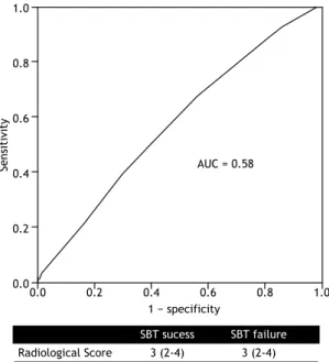
The RS results were similar between SBT-failure and SBT-success groups (median = 3 [IQR: 2-4] vs. 3 [IQR: 2-4]; p = 0.15), which corresponded to interstitial lung congestion. The analysis of the ROC curve showed that the RS prior to a T-piece trial had poor accuracy in discriminating between SBT failure and SBT success (area under the curve = 0.58; p = 0.2; Figure 2). There was no correlation between luid balance 48 h prior to the SBT and RS results (ρ= −0.13; p = 0.1).
DISCUSSION
In a heterogeneous cohort of mechanically ventilated patients who were candidates for a SBT, we found no association between radiological signs of pulmonary congestion indicated by the RS and SBT outcomes. Our study suggests that incorporating radiological estimations of lung edema as a readiness criterion for weaning from MV potentially retards it, as long as interstitial pulmonary congestion was observed on SBTs. To the extent of our knowledge, this is the irst report encompassing such a topic.
The rationale behind delaying an SBT due to radiological signs of pulmonary congestion might be the belief that MV patients could not succeed in a T-piece trial unless they were “dry” again, given that cardiorespiratory interactions under negative pressure promote increases in both LV preload and afterload. However, a myriad of changes in respiratory mechanics and in the cardiovascular system related to weaning failure is not evident until clinical manifestations of distress appear, which promptly demand interruption of the trial or reintubation.(1) Fluid balance cannot predict
SBT outcomes in a slightly larger, mixed medical-surgical
Table 1. Radiological score parameters and values.a
Parameter Value
Redistribution of lung vessels
No 0
Yes 1
Width of the cardiac silhouette > 60%
No 0
Yes 1
Peribronchial cufing
No 0
Yes 1
New pleural effusion
No 0
Unilateral 1
Bilateral 2
Kerley’s A, B, or C lines
None 0
Uncertain 1
Deinite 2
Lung opacity
None 0
Lung opacity 1
Ground-glass opacity 2
“Bat wing” pattern 3
Based on Shochat et al.(16) aSeverity of pulmonary
edema was determined as follows: normal chest X-ray,
0-1 points; interstitial lung congestion, 2-4 points;
and mild, moderate, and severe alveolar edema,
respectively, 5-6 points, 7-8 points, and 9-10 points,
respectively.
Figure 1. In A, a chest X-ray of a 68-year-old female patient shows peribronchial cufing and opacity in a “bat wing”
pattern, revealing edema, compounding a radiological score of 4 points, characterized as interstitial lung congestion.(16)
In B, a chest X-ray of a 57-year-old male patient shows a cardiothoracic ratio > 60%, peribronchial cufing, lung vessel
redistribution, Kerley’s A line, and lung opacity, resulting in a score of 5 points, characterized as mild alveolar edema.(16)
A SEMI-UPRIGHT POSITION B SEMI-UPRIGHT POSITION
PERIBRONCHIAL CUFFING
PERIBRONCHIAL CUFFING
CARDIOTHORACIC RATIO < 0.6 CARDIOTHORACIC RATIO > 0.6
LUNG VESSEL REDISTRIBUTION
KERLEY'S A LINE
LUNG OPACITY "BAT WING" EDEMA
Usefulness of radiological signs of pulmonary congestion in predicting failed spontaneous breathing trials
ICU population either, perhaps being more relevant for COPD patients, as we published previously.(19) An
observational study involving 100 patients immediately before a T-piece trial demonstrated that baseline levels of brain natriuretic peptide—a surrogate marker of congestive heart failure—were moderately elevated exclusively in SBT-failure individuals, who eventually failed owing to cardiac dysfunction.(20) Moreover, overtreatment based on isolated CXR interpretations might be harmful in terms of weaning readiness.(10)
An interobserver agreement study examined the extent to which medical intensivists and a radiologist could agree on whether a CXR revealed diffuse bilateral iniltrates for the diagnosis of ARDS or not and concluded that intensivists without formal consensus training can achieve moderate levels of agreement.(21) Accordingly, in real clinical practice, an expert radiological opinion is not immediately available, and delaying the weaning process based upon poor interpretations of CXRs might be even more questionable. Since the physicians in our study were not aware of the most recent CXR results, we were unable to prove that hypothesis.(24)
A systematic review(22) highlighted the safety of abandoning routine CXRs in favor of a more restrictive approach. Arguments for adopting a restrictive approach included varying interpretations of CXRs depending on clinician and patient factors, low incidence of clinically unsuspected abnormalities, potential harm arising from unnecessary treatment of minor or false-positive indings, costs, radiation exposure and adverse events arising from the repositioning of the patient to obtain CXRs.(23) Likewise, the importance of negative CXR indings for worklow, eficiency, and clinical decision-making might be overestimated. A study collecting the opinions of experienced ICU physicians regarding the appropriateness of performing routine CXRs in various situations encountered in adult ICUs showed there was no consensus regarding the usefulness of obtaining a routine CXR prior to extubation.(24)
It should be pointed out that our study consisted of a relatively small number of patients and absolute number of failure events, with a high prevalence of elderly patients and a low prevalence of systolic LV dysfunction. Nonetheless, our sample had the same
Table 2. Characteristics of the study cohort (N = 170).a
Characteristic Groups p
SBT success (n = 142)
SBT failure (n = 28)
Age, years 76 (66-84) 67 (52-80) 0.15
Female gender 62 (43.7) 13 (46.4) 0.79
APACHE II score 21 ± 6.9 23 ± 7.8 0.16
SOFA score 5 (3-9) 5 (2-10) 0.50
BMI, kg/m2 25 (23-28) 25 (23-29) 0.97
RSBI, f/VT 53 (41-75) 52 (36-71) 0.94
MV duration, days 4 (2-6) 6 (4-11) 0.003
Fluid balance 48 h prior to the SBT, mL 1,219 ± 2,912 1,838 ± 1,896 0.48
Comorbidities
COPD 14 (9.9) 5 (17.9) 0.32
EF < 45% 15 (10.6) 4 (14.3) 0.52
LV diastolic dysfunction 55 (38.7) 8 (28.6) 0.30
Ischemic coronary disease 28 (19.7) 4 (14.3) 0.50
RRT 23 (16.2) 7 (25.0) 0.28
Presence of ascites 3 (2.1) 2 (7.1) 0.19
Reason for MV
Respiratory sepsis 25 (17.6) 6 (21.4) 0.63
Nonrespiratory sepsis 32 (22.5) 5 (17.9) 0.58
CHF 18 (12.7) 1 (3.6) 0.16
Coma 29 (20.4) 4 (14.3) 0.45
Postoperative ARF 7 (4.9) 2 (7.1) 0.63
COPD/asthma 2 (1.4) 2 (7.1) 0.13
Pulmonary embolism 6 (4.2) 1 (3.6) 1.00
ARDS 10 (7.0) 4 (14.3) 0.25
Simple weaning 108 (76.1) 25 (89.3) 0.27
Vasopressor infusion during T-piece trial 27 (19.0) 4 (14.3) 0.55
Vasodilator infusion during T-piece trial 11 (7.7) 2 (7.1) 1.00
SBT: spontaneous breathing trial; APACHE II: Acute Physiology and Chronic Health Evaluation II; SOFA: Sequential Organ Failure Assessment; BMI: body mass index; RSBI: rapid shallow breathing index; f/VT: ratio of RR to tidal volume; MV: mechanical ventilation; EF: ejection fraction; LV: left ventricular; RRT: renal replacement therapy; CHF: congestive heart failure; and ARF: acute respiratory failure. aData are presented as median (interquartile
range), mean ± SD, or n (%).
Antonio ACP, Teixeira C, Castro PS, Zanardo AP, Gazzana MB, Knorst M
expected pre-test probability of SBT failure as the ordinary medical-surgical ICU population. Our original study focused on lung ultrasound assessment of ready-to-wean subjects. Since CXR results were part of a secondary analysis, we did not standardize the moment of CXR acquisition, so not all exams were performed immediately before the SBT but rather within a period of 24 h prior to the trial. At the beginning of our research, the limited number of experts on bedside lung ultrasound, as well as lack of any research funding, forced us to close enrollment on weekends.
Other limitations include the observational design, with all its intrinsic methodological laws, and the absence of high scores of radiological signs of lung edema on the RS, which might imply lesser overall severity in this cohort of patients or relect the pointlessness in requiring CXRs to advance the weaning process. Our
ready-to-wean population showed modest median values on the RS. In the original Shochat et al. paper,(16) an RS of 4 or more represented overt acute heart failure in 95% of the patients reaching this level. Hence, it seems unlikely that an individual presenting with a high RS score should be eligible for an SBT.
We decided on SBT failure as the major outcome since we aimed at predicting the earliest time that a patient might resume spontaneous breathing. Moreover, the exact reason for extubation failure often escapes identiication. Reintubation is usually performed because of an apparently new episode of respiratory distress, which may be related to primary respiratory failure, congestive heart failure, aspiration, ineffective cough causing accumulation of airway mucus, or upper airway obstruction. Other reasons for reintubation include the onset of a new episode of sepsis, surgical complications, acute coronary syndrome, and neurological impairment.(1)
The RS presented by Shochat et al.(16) was chosen by virtue of its comprehensive analysis of dynamic changes, its good correlation with severity of lung edema, the utilization of lung impedance as the gold-standard method, and the sensitivity for the detection of subtle radiological signs of pulmonary congestion. Currently, that RS is the only method available that proposes a quantitative assessment of CXRs in terms of lung luid content. In that cohort,(16) high intraobserver (κ = 0.86; p = 0.0001) and interobserver correlations (κ = 0.82; p = 0.0001) were found regarding RS interpretation. However, its main drawbacks are the lack of assessment in the ICU population—although it included patients admitted to a coronary care unit—its single-center design, and the absence of large-scale validation. Therefore, for the sake of generalizability of our indings, the RS must be further studied.
In conclusion, since the radiological signs of pulmonary congestion demonstrated by the RS were unable to predict SBT failure in this medical-surgical critically ill population, we inferred that speciic CXR reports on signs of pulmonary congestion are insuficient to preclude hemodynamically stable, suficiently oxygenated patients from performing an SBT.
Figure 2. A ROC curve of the ability of the radiological score to predict spontaneous breathing trial failure. The
area under the curve (AUC) is 0.58 (p = 0.2), revealing
poor accuracy.
0.0 0.2 0.4 0.6 0.8 1.0
1 − specificity
1.0
0.8
0.6
0.4
0.2
0.0
Se
ns
it
iv
it
y
AUC = 0.58
SBT sucess SBT failure
Radiological Score 3 (2-4) 3 (2-4)
REFERENCES
1. Tobin MJ. Weaning from Mechanical Ventilation. In: Tobin MJ, editor. Principles and Practice of Mechanical Ventilation. 3rd ed. New York: McGraw-Hill; 2012. p. 1185-220.
2. MacIntyre NR, Cook DJ, Ely EW Jr, Epstein SK, Fink JB, Heffner JE, et al. Evidence-based guidelines for weaning and discontinuing ventilatory support: a collective task force facilitated by the American College of Chest Physicians; the American Association for Respiratory Care; and the American College of Critical Care Medicine. Chest. 2001;120(6 Suppl):375S-95S. https://doi.org/10.1378/chest.120.6_ suppl.375S
3. Boles JM, Bion J, Connors A, Herridge M, Marsh B, Melot C, et al. Weaning from mechanical ventilation. Eur Respir J. 2007;29(5):1033-56. https://doi.org/10.1183/09031936.00010206
4. Cook DJ, Walter SD, Cook RJ, Grifith LE, Guyatt GH, Leasa D, et al. Incidence of and risk factors for ventilator-associated pneumonia in critically ill patients. Ann Intern Med. 1998;129(6):433-40. https://doi. org/10.7326/0003-4819-129-6-199809150-00002
5. Ely EW, Baker AM, Dunagan DP, Burke HL, Smith AC, Kelly PT, et al. Effect on the duration of mechanical ventilation of
identifying patients capable of breathing spontaneously. N Engl J Med. 1996;335(25):1864-9. https://doi.org/10.1056/ NEJM199612193352502
6. Frutos-Vivar F, Esteban A, Apezteguia C, González M, Arabi Y, Restrepo MI, et al. Outcome of reintubated patients after scheduled extubation. J Crit Care. 2011;26(5):502-9. https://doi.org/10.1016/j. jcrc.2010.12.015
7. Esteban A, Frutos F, Tobin MJ, Alía I, Solsona JF, Valverdú I, et al. A comparison of four methods of weaning patients from mechanical ventilation. Spanish Lung Failure Collaborative Group. N Engl J Med. 1995;332(6):345-50. https://doi.org/10.1056/ NEJM199502093320601
8. Brochard L, Rauss A, Benito S, Conti G, Mancebo J, Rekik N, et al. Comparison of three methods of gradual withdrawal from ventilatory support during weaning from mechanical ventilation. Am J Respir Crit Care Med. 1994;150(4):896-903. https://doi.org/10.1164/ ajrccm.150.4.7921460
Usefulness of radiological signs of pulmonary congestion in predicting failed spontaneous breathing trials
org/10.1007/s00134-014-3334-4
10. Teboul JL, Monnet X, Richard C. Weaning failure of cardiac origin: recent advances. Crit Care. 2010;14(2):211. https://doi.org/10.1186/ cc8852
11. Lange NR, Schuster DP. The measurement of lung water. Crit Care. 1999;3(2):R19-R24. https://doi.org/10.1186/cc342
12. Khan AN, Al-Jahdali H, Al-Ghanem S, Gouda A. Reading chest radiographs in the critically ill (Part II): Radiography of lung pathologies common in the ICU patient. Ann Thorac Med. 2009;4(3):149-57. https://doi.org/10.4103/1817-1737.53349
13. Martin KT. Extubation: Guidelines and Procedures [monograph on the Internet]. [cited 2016 Nov 1] Corona, CA: Respiratory Care Educational Consulting Services Inc. and Western Schools. Available from: http://www.rcecs.com/MyCE/PDFDocs/course/V7020.pdf 14. Nickson C. Extubation Assessment in the ICU. Life in the Fastlane
[serial on the Internet]. Mountain View, CA: Creative Commons; c2007-17 [cited 2015 Jul 9]; [about 10 screens]. Available from: http:// lifeinthefastlane.com/ccc/extubation-assessment/2014
15. Macintyre NR. Evidence-based assessments in the ventilator discontinuation process. Respir Care. 2012;57(10):1611-8. https:// doi.org/10.4187/respcare.02055
16. Shochat M, Shotan A, Trachtengerts V, Blondheim DS, Kazatsker M, Gurovich V, et al. A novel radiological score to assess lung luid content during evolving acute heart failure in the course of acute myocardial infarction. Acute Card Care. 2011;13(2):81-6. https://doi. org/10.3109/17482941.2011.567279
17. Antonio AC, Teixeira C, Castro PS, Savi A, Maccari JG, Oliveira RP,
et al. Behavior of Lung Ultrasound Findings during Spontaneous Breathing Trial. Rev Bras Ter Intensiva. Epub 2017.
18. van der Jagt EJ, Smits HJ. Cardiac size in the supine chestilm. Eur J Radiol. 1992;14(3):173-7. https://doi.org/10.1016/0720-048X(92)90080-S
19. Antonio AC, Teixeira C, Castro PS, Savi A, Oliveira RP, Gazzana MB, et al. 48-Hour Fluid Balance Does Not Predict a Successful Spontaneous Breathing Trial. Respir Care. 2015;60(8):1091-6. https:// doi.org/10.4187/respcare.03172
20. Zapata L, Vera P, Roglan A, Gich I, Ordonez-Llanos J, Betbese AJ. B-type natriuretic peptides for prediction and diagnosis of weaning failure from cardiac origin. Intensive Care Med. 2011;37(3):477-85. https://doi.org/10.1007/s00134-010-2101-4
21. Meade MO, Cook RJ, Guyatt GH, Groll R, Kachura JR, Bedard M, et al. Interobserver variation in interpreting chest radiographs for the diagnosis of acute respiratory distress syndrome. Am J Respir Crit Care Med. 2000;161(1):85-90. https://doi.org/10.1164/ ajrccm.161.1.9809003
22. Ganapathy A, Adhikari NK, Spiegelman J, Scales DC. Routine chest x-rays in intensive care units: a systematic review and meta-analysis. Crit Care. 2012;16(2):R68. https://doi.org/10.1186/cc11321 23. Tolsma M, van der Voort PH, van der Meer NJ. Why intensivists want
chest radiographs. Crit Care. 2015;19:100. https://doi.org/10.1186/ s13054-015-0816-x
24. Hejblum G, Ioos V, Vibert JF, Böelle PY, Chalumeau-Lemoine L, Chouaid C, et al. A web-based Delphi study on the indications of chest radiographs for patients in ICUs. Chest. 2008;133(5):1107-12. https://doi.org/10.1378/chest.06-3014