1MD, PhD, Eduardo de Menezes Hospital, FHEMIG, Belo Horizonte MG, Brazil; 2MD, PhD, Department of Infectious Diseases, Federal
University of Minas Gerais, Belo Horizonte, Brazil; 3D e p a rtment of Infectious Diseases, Federal University of Minas Gerais, Belo
Horizonte, Brazil; 4MD, PhD Department of Neurology, University of São Paulo, São Paulo, Brazil.
Received 11 April 2005, received in final form 4 July 2005. Accepted 25 August 2005.
Dr. Paulo Pereira Christo - Av. Prof. Alfredo Balena 189/1708 - 30130-100 Belo Horizonte MG - Brasil. E-mail: [email protected]
HIV-1 RNA LEVELS IN CEREBROSPINAL FLUID
AND PLASMA AND THEIR CORRELATION WITH
OPPORTUNISTIC NEUROLOGICAL DISEASES IN
A BRAZILIAN AIDS REFERENCE HOSPITAL
Paulo P. Christo
1, Dirceu B. Greco
2, Agdemir W. Aleixo
3, Jose A. Livramento
4ABSTRACT - B a c k g ro u n d: Plasma HIV RNA levels reflect systemic viral replication but in CNS it may occur
relatively independent of systemic infection, yet clinical application of CSF HIV-1 RNA levels is less clear. O b j e c t i v e: to compare CSF and plasma HIV-1 RNA levels of patients with diff e rent opportunistic neuro l o g i-cal diseases to those without neurologii-cal disease, as well as to correlate these levels with the outcome of
the disease and use of HAART. M e t h o d: 97 patients who had lumbar puncture for routine work up of
suspected neurological diseases, were divided in 2 groups: without neurological disease (23) and with
neu-rological disease (74). NASBA was used for plasma and CSF HIV RNA. Results: Median CSF viral load was
higher in toxoplasmic encephalitis, cryptococcal meningitis, HIV dementia and neurological diseases with-out a defined etiology when compared to patients withwith-out neurological disease. There was no diff e re n c e between plasma viral load in patients with and without neurological diseases. Median viral load was high-er in plasma and CSF among patients who died when compared to those successfully treated. CSF and
plas-ma viral load were lower in patients with opportunistic diseases on HAART than without HAART. C o n c l u
-sion: CSF viral load was higher in patients with any neurological disease, but this difference was not
pres-ent in plasma viral load, suggesting that neurological disease influences more the CSF than plasma compar-tments. Notwithstanding diff e rent neurological diseases were not possible to be diferentiated by the leve-ls of CSF HIV-1.
KEY WORDS: AIDS, HIV, cere b rospinal fluid, HIV-1 RNA, opportunistic infections, viral load, neuro l o g i c a l disease.
Níveis de RNA do HIV-1 no líquido cefalorraqueano e plasma e sua correlação com doença neu-rológica oportunística em um hospital referência em AIDS
RESUMO - I n t ro d u ç ã o: Os níveis de RNA do HIV-1 no plasma refletem a replicação viral sistêmica e a re p l i c
a-ção no sistema nervoso central pode ocorrer independentemente da infeca-ção sistêmica, mas a utilidade
da medida destes níveis no líquido cefalorraqueano (LCR) permanece indefinida. O b j e t i v o: Comparar os
níveis de RNA do HIV-1 no LCR e plasma de pacientes sem doenças neurológicas e com diferentes doenças
n e u rológicas, bem como correlacionar estes níveis com a sua evolução e o uso de antire t ro v i r a i s . M é t o d o:
Foram avaliados 97 pacientes com suspeita de doença neurológica que realizaram punção lombar e que foram divididos em dois grupos: sem doenças neurológicas (23) e com doenças neurológicas (74). Metodologia
NASBA foi usada para quantificação do RNA do HIV-1. Resultados: A mediana da carga viral do LCR foi
maior em pacientes com neurotoxoplasmose, neurocriptococose, demência pelo HIV e doença neurológi-ca sem etiologia definida quando comparada aos pacientes sem doenças neurológineurológi-cas. Não houve difere n-ça da carga viral do plasma entre os pacientes com e sem doenn-ça neurológica. A mediana da carga viral do plasma e LCR foi maior nos pacientes que faleceram em relação aos tratados com sucesso. A carga viral do LCR e plasma foi menor nos pacientes com doenças oportunísticas que usavam HAART em relação aos
que não a usavam. C o n c l u s ã o: A carga viral no LRC foi maior nos pacientes com qualquer doença
neuro-lógica em relação aos sem doenças neuroneuro-lógicas, mas isto não ocorreu no plasma, sugerindo que doença n e u rológica influencia mais o compartimento do LCR que o do plasma, mas não foi possível diferenciar as doenças neurológicas pelos níveis de RNA do HIV-1 do LCR
O p p o rtunistic neurological diseases and neuro-logical diseases primarily related to HIV-1 are com-mon manifestations announcing the onset of AIDS and also during the course of the infection1. The
i n t roduction of highly active antire t roviral thera-py (HAART) has led to immunological impro v em e n t of patients and a consequent reduction in mor-bidity and mortality and in the incidence of oppor-tunistic diseases2 - 8. However, these diseases,
includ-ing those affectinclud-ing the central nervous system (CNS), continue to occur, especially in underd e v e l-oped countries where the acquisition of antire t ro-viral (ARV) drugs is difficult. In Brazil, despite a pu-blic health system that provides drugs free of char-ge, opportunistic infections, especially highly inca-pacitating neurological diseases, continue to occur due to irregular immunovirological monitoring and late diagnosis of the infection.
The virus enters the CNS during primary HIV in-fection9,10and can be present during all stages of
the disease, with replication in the CNS occurr i n g in a manner relatively independent of systemic in-f e c t i o n1 1 - 1 5. There is evidence of intrathecal re p l i c
a-tion of HI1 in patients with opportunistic and H I V-related neurological diseases, with the prompt d i a g-nosis and treatment of these patients becoming i m p o rt a n t1 6 , 1 7. Plasma HIV-1 RNA levels reflect the
status of systemic viral replication and re p re s e n t the best predictive marker of HIV disease pro g re s-sion, being an important tool in monitoring and in studies regarding aspects of the viral dynamics of HIV infection1 8 - 2 0. In contrast, the clinical
useful-ness of HIV-1 RNA levels in the cere b rospinal flu-id (CSF) of patients with opportunistic neurologi-cal diseases, or the effect of opportunistic diseases on CSF HIV levels in patients under HAART has not been well defined21,22.
The objective of the present study was to com-p a re HIV-1 RNA levels in CSF and com-plasma of com-patients with diff e rent opportunistic neurological diseases to those without neurological disease, as well as to c o rrelate these levels with the evolution of the dis-ease, CD4+ T lymphocyte count, and use of HAART.
METHOD
Patients – We prospectively evaluated 97 HIV- i n f e c t-ed patients who were submittt-ed to lumbar puncture bet-ween May 2001 and May 2002 as part of the work-up for a suspicion of neurological disease at a public AIDS Refe-rence Hospital (Eduardo Menezes Hospital, FHEM I G ) , Belo Horizonte, Brazil. The study was approved by the Re-s e a rch EthicRe-s Committee of the inRe-stitution and written in-f o rmed consent was obtained in-from all part i c i p a n t s .
Data collected from each patient included gender, age, CD4+ lymphocyte count, use and type of ARV ther-a p y, durther-ation of ARV therther-apy, presence ther-and type of neu-rological disease, and clinical outcome.
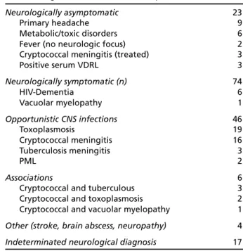
The patients were divided into groups with and with-out neurological diseases. Seventy-four (76.6%) of the 97 patients presented with neurological diseases, including 46 (62%) with opportunistic diseases, 17 (23%) with neuro-logical diseases of undetermined etiology and 7 (9%) with H I V- related neurological diseases, and the remaining 23 patients (23.4%) did not have active opportunistic or HIV-related neurological diseases. In the case of patients with-out neurological diseases the clinical diagnoses were pri-m a ry headache, pri-metabolic/toxic disorders, and fever with a non-neurological focus. Patients submitted to lumbar p u n c t u re to exclude neurosyphilis or to assess clearance of cryptococcal meningitis after correct medication for 8 weeks were also studied. No clinical or laboratory evi-dence of active infection was observed in these patients. The diagnoses are summarized in Table 1.
N e u rological diseases were diagnosed based on the following criteria: criteria of the Working Group of the
American Academy of Neurology Task Forc e2 3for the
diagnosis of HIV-associated dementia (HIV-D) and vacu-olar myelopathy, a suggestive image on a skull comput-ed tomography scan and a clinical and tomographic ima-ge response to specific drug treatment for the diagno-sis of cerebral toxoplasmodiagno-sis, and a positive India ink result, a specific cryptococcal antigen test or positive CSF culture for the diagnosis of meningitis. Tu b e rc u l o u s meningitis was diagnosed based on clinical-neuro l o g i-cal signs of lymphocytic meningitis and the presence of alcohol-acid resistant bacilli or a positive CSF culture .
Table 1. Diagnosis obtained for the 97 patients studied.
Neurologically asymptomatic 23
Primary headache 9
Metabolic/toxic disorders 6
Fever (no neurologic focus) 2
Cryptococcal meningitis (treated) 3
Positive serum VDRL 3
Neurologically symptomatic (n) 74
HIV-Dementia 6
Vacuolar myelopathy 1
Opportunistic CNS infections 46
Toxoplasmosis 19
Cryptococcal meningitis 16
Tuberculosis meningitis 3
PML 2
Associations 6
Cryptococcal and tuberculous 3
S t e reotactic biopsies were obtained for the diagnosis of pro g ressive multifocal leukoencephalopathy and bac-terial abscess. An undetermined diagnosis was conside-red when the patients showed neurological syndro m e s characterized by meningitis, encephalitis or expansive focal brain lesions and when no etiology could be estab-lished after work-up.
Laboratory analysis – CSF and plasma samples were collected within an interval of 48 hours between each
other and stored at - 700C until the time of pro c e s s i n g
within a maximum period of 6 months. HIV-1 RNA was quantified by the nucleic acid sequence based amplifica-tion (NASBA) technique using centrifuged plasma and n o t centrifuged CSF according to manufacturer instru c t i o n s (Nuclisens HIV-1 QT, Organon TeKniKa, Boxtel,
Nether-lands), with a sensitivity of 80 copies/ml (1.90 log1 0c
o-pies/ml). CSF samples containing more than 10 red c e l l s / m l w e re excluded from the analysis. In addition, the num-ber of cells and protein concentration were determ i n e d in CSF samples.
Among the samples collected, HIV-1 RNA was quanti-fied in 90 CSF and 73 plasma samples. Seventy samples of 70 patients had valid quantification in both plasma and CSF.
Statistical analysis – The nonparametric Mann-Whitney test was used to determine the effect of the presence of n e u rological disease and use of ARV therapy on CSF and plasma viral load. Viral load and the characteristics of the d i ff e rent groups of patients with and without neuro l o g-ical diseases were compared by the nonparametric Mann-Whitney test and chi-square test. The correlation between viral load and CD4 count was determined using nonpara-metric Spearm a n ’s rank correlation. p values of < 0.05 w e re considered to indicate statistical significance.
CSF and plasma viral loads were log10 transformed since the HIV-1 RNA levels showed no normal distribu-tion. For patients with undetectable HIV-1 RNA, a log-scale value of 0 was assigned to avoid the problem of
e x p ressing zero logarithmically. All statistical analyses were performed with the SPSS 8.0 program (SPSS Inc.).
RESULTS
Patient characteristics – The mean CD4+ T
lym-phocyte count was 137 cells/mm3 and no diff e re n-ces were observed in mean or median CD4 counts between patients with diff e rent types of neuro-logical diseases and between those underg o i n g A RV therapy or not. Fifty-two (54%) of the 97 pa-tients used ARV drugs, all in the HAART re g i m e n . Seven (13.5%) patients used one drug that cro s s e d the blood-brain barr i e r, 24 (46.2%) used two dru g s , 20 (38.5%) used three drugs, and one patient used four drugs. The mean duration of ARV therapy w a s 10.4 months.
Comparison between the groups with and with-out active neurological diseases showed no signifi-cant difference in gender, CD4 count, use of ARV therapy or mean CSF protein content. In contrast, a significant diff e rence was observed in terms of t h e number of cells in CSF, presence of pleocytosis in C S F, duration of HIV infection, and death during hospi-talization (Table 2).
Association between CSF and plasma viral load
and neurological disease – Median HIV-1 RNA
lev-els in CSF were higher in patients with neurolog-ical diseases than in neurologneurolog-ically asymptomatic subjects, as was the median plasma viral load but the latter was not statistically significant (Table 2). The CSF and plasma HIV-1 RNA means were , re s p e c t i v e l y, 0.84 and 2.39 log1 0copies/mL in pa-tients without neurological disease, while they we-re 2.44 and 3.68 log1 0copies/mL in patients with n e u rological disease. The ratio of mean CSF viral
Table 2. Clinical and laboratory characteristics of the patients with and without neurological disease.
With disease Without disease p*
(N=74) (N=23)
Mean age (years) 36.5 35.8 0.547
Male gender (%) 55 14 0.214
Mean time since diagnosis (months) 5 48 0.014
Pleocytosis in CSF (%) 60.8 4.3 0.000
Median CSF WBC 8.0 2.0 0.000
Median CSF protein (mg/dL) 61.0 43 0.083
HAART use (%) 50 65.2 0.223
Median CD4+ count (cells/mm3) 96.5 89 0.504
Death (%) 36.5 13 0.034
Detectable CSF viral load (%) ** 63.4 26.3 0.004
Detectable plasma viral load (%) ** 82.8 60 0.058
Median CSF viral load (log10) 3.15 <1.9 0.002
Median plasma viral load (log10) 4.35 2.18 0.071
load of patients with and without neurological di-sease was 2.9 and the ratio of mean plasma viral load of patients with and without neurological di-sease was 1.5, showing that CSF viral load was al-most 3 times higher in patients with neuro l o g i c a l disease compared to those without neuro l o g i c a l disease, whereas plasma viral load was 1.5 times higher in patients with neurological disease.
The median of CSF HIV-1 RNA was <1.9 log1 0 copies/mL in patients without neurological disease, 2.99 log1 0in patients with general opportunistic di-sease, 2.62 log1 0in patients with diseases dire c t l y related to HIV, 2.99 log1 0in patients with cere b r a l toxoplasmosis, 3.69 log1 0in patients with cry p t o-coccal meningitis, and 3.96 log1 0in patients with n e u rological disease of undetermined etiology. HIV-1 RNA levels in CSF were higher in all diff e rent gro u-ps of patients with neurological diseases compare d to those without neurological diseases, but no dif-f e rence in median HIV RNA levels in CSF was obser-ved between these groups of diseases (Fig 1).
In plasma, diff e rences in median viral load were only observed between patients with undeterm i-ned neurological diseases (3.96 log1 0c o p i e s / m L ) and those without neurological diseases (<1.9 log1 0 copies/mL) and between patients with undeterm i-ned neurological diseases and those with opport u-nistic diseases (2.99 log10copies/mL).
C o rrelation between CSF viral load and immu
-n i t y– A negative correlation was observed
bet-ween CSF viral load and CD4 cell count (r = -0.307, p = 0.012) (Figure 2) and also between plasma viral load and CD4 T cell count (r = -0.283, p = 0.038). When analyzing patients with and without neuro-logical disease, a correlation between CSF viral load and CD4 count was only observed in patients with neurological disease (r = -0.307, p = 0.03), while CSF viral load was not correlated with the number of CD4 cell in patients without neuro l o g i-cal disease (r = -0.341, p = 0.901).
Association between CSF viral load and outco -m e– A diff e rence in CSF viral load was observ e d between patients who were discharged and those who died, with the median HIV-1 RNA levels in CSF being higher in patients who died (3.26 versus <1.9 l o g1 0; p = 0.027). The same was observed for plas-ma viral load (4.70 versus 3.26 log10; p = 0.033).
Association between CSF viral load and use of A RV therapy. There was a significant effect of the
use of ARV therapy on CSF viral load – Patients
Fig 3. Correlation between cere b rospinal fluid (CSF) viral load and use of ARV therapy.
Fig 2. Correlation between cere b rospinal fluid (CSF) viral load and CD4 count.
who did not use ARV therapy showed a significan-tly higher median CSF viral load than those who did (3.30 versus 0,82 log1 0/ml; p < 0.001). The same was o bs e rved for plasma viral load (4.47 versus 1.87 log10/ml; p < 0.001).
Of the 43 patients with opportunistic neuro l o-gical diseases submitted to HIV quantification in C S F, 26 received ARV therapy (median of <1.9 log1 0) a n d 17 did not (median of 3.82 log1 0). Of the 35 pat i e n t s with opportunistic diseases submitted to HIV-1 q u a n-tification in plasma, 20 used ARV drugs (median of 2.82 log1 0) and 15 did not (median of 4.52 log1 0). M e-dian HIV RNA levels in CSF and plasma were signi-ficantly higher in patients not taking ARV dru g s (p < 0.001) (Fig 3).
DISCUSSION
Association between CSF viral load and neuro logical disease. In the present study, we determ i -ned HIV-1 RNA levels in CSF and plasma of patients without and with diff e rent HIVassociated neuro l o
-gical diseases– A correlation was observed
bet-ween CSF viral load and neurological disease. The median viral load in CSF was higher in patients with neurological disease compared to those with-out neurological disease, and the same was also o b s e rved for plasma viral load, although in this c o m p a rtment the diff e rence was not statistically significant.
The ratio of mean viral load in patients with a n d without neurological disease was higher in CSF than in plasma, i.e., CSF viral load was almost 3 ti-mes higher in patients with neurological disease compared to those without neurological disease, w h e reas plasma viral load was only 1.5 times high-er in patients with neurological disease, demon-strating a greater influence of neurological disea-se on CSF viral load than on plasma load, i.e., neu-rological disease caused a pro p o rtionally gre a t e r i n c rease in CSF than in plasma. This finding may have been due to intrathecal replication or incre a-sed passage of the virus from the plasma to the CSF compartment, since patients with neuro l o g i-cal diseases showed a higher median cell count in CSF than those without neurological disease.
D i ff e rences in median CSF viral load were ob-s e rved between patientob-s with diob-seaob-seob-s primarily re-lated to HIV and those without neurological dise-ases and between patients with opportunistic neu-rological diseases and those without neuro l o g i c a l diseases, but not between groups with HIV- re l a t e d d i s o rders and opportunistic diseases. This cro s s - s e
c-tional analysis did not permit the discrimination between subjects with opportunistic infections and those with HIV- related disorders in terms of CSF viral load or between patients with cerebral toxoplasmo-sis and cryptococcal meningitis. The absence of any significant diff e rence in terms of HIV levels between patients with HIV-D and opportunistic diseases sug-gests that HIV-1 RNA levels in CSF are not a good marker for the diagnosis of this disease2 4 , 2 5.
CSF viral load has been shown to be higher in patients with HIV-D than in those with mild or no n e u rological symptoms2 5 - 2 7. However, higher CSF
HIV loads have been detected in patients with HIV-D, but significant concentrations have also been found in patients without any HIV- related neuro l o-gical disease. On the other hand, low HIV RNA lev-els have been detected in both patients with and without HIV-induced neurological disease2 8. The
as-sociation between HIV RNA levels in CSF and neuro-logical status in HAART- t reated individuals seems to be weaker now than in the pre - H A A RT era2 9.
Viral load in CSF was higher in patients with op-p o rtunistic diseases than in individuals without n e u rological diseases. No significant diff e rence bet-ween these two groups was observed for plasma HIV levels. Certain opportunistic diseases of the CNS a ffect HIV RNA levels in CSF, with elevated concen-trations being observed in patients with lympho-cytic meningitis such as cryptococcal and tuberc u-lous meningitis1 6 , 2 5. These conditions are
characteri-zed by a meningeal inflammatory infiltration and also by a correlation between the number of lym-phocytes in CSF and viral load2 8. High HIV RNA
lev-els in CSF may not be simply due to transport of the v i rus from plasma to CSF, since some studies were unable to show a correlation between CSF viral load and integrity of the blood-brain barr i e r1 6 , 2 5.
The quantity of HIV RNA in the CSF of patients with neurological diseases is more complex compa-red to asymptomatic individuals and other sourc e s might contribute to its presence in CSF. The chro n-ic presence of a lymphocytn-ic inflammatory pro c e s s may re p resent an ideal opportunity for the uncon-trolled replication of HIV-1, particularly at an im-munologically privileged site like the CNS1 6and
in-filtrative lymphocytes may harbor HIV-1, thus re p-resenting an exogenous source that also contribu-tes to CSF viral load. In vitro studies3 0 , 3 1have shown
that cryptococcosis and tuberculosis or their anti-gens can also directly increase viral replication.
who died during hospitalization than in those who i m p roved and were discharged, suggesting that t h e determination of CSF viral load might be a prog-nostic factor in patients with active neuro l o g i c a l disease, especially opportunistic diseases.
C o rrelation between CSF viral load and CD4 T
lymphocyte count– In the present study, a negative
c o rrelation was observed between CSF and plasma viral load and CD4+ T lymphocyte count, with an i n c rease in viral load thus being associated with a reduction in the number of CD4 lymphocytes.
In plasma, the correlation between viral load and the degree of immunity has been well establi-shed, with plasma viral load increasing as the CD4 cell count decre a s e s1 8. With respect to CSF, this
as-sociation is not as well established and there are re p o rts in the literature indicating a corre l a t i o n between CSF viral load and the degree of immuni-ty determined on the basis of CD4 lymphocyte c o u n t2 6, but most studies suggest that no such
cor-relation exists2 4 , 2 7 , 3 2 - 3 6, or even that the CSF viral
load is lower in patients with a CD4 count below 200 cells/mm3,37.
When the patients were stratified into two g roups, with and without neurological disease, a c o rrelation between CD4 count and CSF viral load was only observed in the group of patients with neu-rological disease in which opportunistic diseases pre-dominated. These patients showed a higher CSF viral load and also a higher median number of cells in C S F, possibly as a result of intrathecal viral re p l i c a-tion or passage of the virus from plasma to CSF.
No correlation between CSF viral load and CD4 T lymphocyte count was observed in the group of patients without neurological diseases. This gro u p p resented a median CSF viral load lower than that o b s e rved for patients with neurological diseases and an equally low CD4 count. This finding sug-gests that a control of viral load is achieved with the use of ARV therapy in these patients but in the absence of immune recovery.
The influence of CD4 count on CSF viral load seems to depend on other factors since pre v i o u s studies have re p o rted discordant results re g a rd-ing its correlation, with these re p o rts varyrd-ing in t e rms of the characteristics of the population eval-uated such as use of ARV therapy, number of cells in CSF and the presence of neurological diseases.
Association between CSF viral load and use of
A RV therapy– An association was observed
betwe-en the use of ARV therapy and CSF and plasma vi-ral load. Patients not receiving ARV therapy had a significantly higher viral load than those who did. Comparison of CSF and plasma viral load bet-ween patients with opportunistic diseases who received ARV therapy and those who did not also showed higher HIV RNA levels in those not under-going ARV therapy. This finding demonstrates the i m p o rtance and efficacy of ARV therapy in the con-t rol of HIV RNA levels bocon-th in plasma and CSF, even in patients with a potentially higher viral load in CSF such as those with opportunistic diseases.
C o n t rol of the CSF compartment may re s u l t f rom the control of the plasma compartment, with s u p p ression of the virus at the periphery and a consequent reduction in the amount of virus that enters the CNS, or even from penetration of the d rug through the blood-brain barrier into the CNS and its consequent action on HIV in CSF, since 87% of the patients who received ARV therapy used at least two drugs known to cross the blood-brain b a rr i e r. The most frequently used ARV drugs were those that cross the blood-brain barrier such as l a-mivudine, zidovudine, efavirenz and stavudine3 8 -40.
The CNS serves as a re s e rv o i r, permitting escape cases to be identified more fre q u e n t l y2 1. It has
be-en suggested that drugs crossing the blood-brain b a rrier need to be included in the ARV re g i m e n4 1.
CSF suppression is correlated with predicted CNS antiretroviral drug penetrance42.
Several studies have evaluated the response of HIV in CSF to ARV therapy. Studies conducted be-f o re the advent obe-f HAART did not be-find signibe-ficant d i ff e rences in the detection rate of HIV RNA or mean HIV levels in CSF with the use or duration of A RV therapy2 6. However, since the introduction of
H A A RT several studies have documented a rapid decline in CSF viral load in patients with diff e re n t stages of the disease, including neuro l o g i c a l l y asymptomatic individuals, patients with mild or s e v e re HIV- related symptoms, and patients with o p p o rtunistic cerebral diseases4 3 - 4 7, with the decline
being more important in naive individuals2 1. Most
HIV-infected individuals show a short-term fall in CSF viral load similar to that observed in plasma4 7,
although prolonged therapy may be re q u i red to suppress HIV levels within the CNS36.
HIV levels, suggesting that the presence of neuro-logical diseases has a greater influence on the CSF than on the plasma compartment. It was not pos-sible to differentiate patients with cerebral toxo-plasmosis, cryptococcosis or HIV-D based on HIV- 1 RNA levels in CSF. HIV-1 RNA levels in CSF were lo-wer in patients undergoing ARV therapy, even in those with an opportunistic neurological disease.
REFERENCES
1. B e rger JR, Moskoswitz L, Fischl M, Kelley RE. Neurologic disease as the presenting manifestation of acquired immunodeficiency syndro m e . South Med J 1987 80:683-686.
2. B rodt HR, Kamps BS, Gute P, et al. Changing incidence of A I D S - d e f i n-ing illnesses in the era of antire t roviral combination therapy. A I D S 1997;11:1731-1738.
3. Lambotte O, Kumaran D, Ta rdieu M. HIV-1 persistence, viral re s e r v o i r, and the central nervous system in the HAART era. Brain Pathol 2003; 13:95-103.
4. Albright AV, Samantha SS, González-Scarano F. Pathogenesis of human immunodeficiency virus-induced neurological disease. J Neuro v i ro l 2003;9:222-227.
5. N e u e n b e rg JK, Herndier BG, Bickel M, et al. HIV-1 related neuro p a t h o l-ogy, 1985 to 1999: rising prevalence of HIV encephalopathy in the era of highly active antire t roviral therapy. J Acquir Immune Defic Syndr 2002;31:171-177.
6. Sackor N, Lyles RH, Skolask R, et al. HIV-associated neurologic dis-ease incidence changes: Multicenter AIDS Cohort Study, 1990-1998. Neurology 2001;56:257-260.
7. d’Armino MA, Cinque P, Mocroft A, et al. Changing incidence of cen-tral nervous system disease in the Euro S I D A cohort. Ann Neurol. 2004; 55:320-328.
8. Sackor N. The epidemiology of human immunodeficiency viru s - a s s o c i a-ted neurological disease in the era of highly active antiretroviral ther-apy. J Neurovirol 2002 (Suppl 2):S115-S121.
9. Resnick L, Berger JR, Shapshak P, Tourtellotte WW. Early penetration of the blood-brain barrier by HIV. Neurology 1988;38:9-14.
10. Davis LE, Hjelle BL, Miller VE, et al. Early viral brain invasion in iatro-genic human immunodeficiency virus infection. Neurology 1992; 42:1736-1739.
11. Antinori A, Giancola ML, Grisetti S, et al. Factors influencing viro l o g i-cal response to antire t roviral drugs in cere b rospinal fluid of advanced HIV-1 infected patients. AIDS 2002;16:1867-1876.
12. Tambussi G, Gori A, Capilippi B, et al. Neurological symptoms during primary human immunodeficiency vírus (HIV) infection correlate with high levels of HIV RNA in cere b rospinal fluid. Clin Infect Dis 2000; 30:962-965.
13. Chiodi F, Keys B, Albert J, et al. Human immunodeficiency virus type 1 is present in the cere b rospinal fluid of a majority of infected individu-als. J Clin Microbiol 1992;30:1768-1771.
14. S o n n e r b o rg AB, Ehrnst AC, Bergdahl SK, Pehrson PO, Skoldenberg BR, Strannegard OO. HIV isolation from cere b rospinal fluid in re l a t i o n in immunological deficiency and neurological symptoms. AIDS 1988; 2:89-93.
15. Schacker T, Collier AC, Hughes J, Shea T, Corey L. Clinical and epide-miologic features of primary HIV infection. Ann Intern Med 1996; 125:257-264.
16. Morris L, Silber E, Sonnenberg P, et al. High human immunodeficien-cy virus type 1 RNA load in the cere b rospinal fluid of patients with lymphocytic meningitis. J Infect Dis 1998;177:473-476.
17. Ellis RJ, More DJ, Childers ME, et al. Pro g ression to neurological impair-ment in human immunodeficiency virus infection predicted by elevat-ed cere b rospinal fluid levels of human immunodeficiency virus RNA. Arch Neurol 2002;59:909-912.
18. Piatak M, Saag MS, Yang LC, et al. High levels of HIV-1 in plasma dur-ing all stages of infection determined by competitive PCR. Science 1993;259:1749-1754.
19. P e relson AS, Neumann AU, Markowitz M, Leonard JM, Ho DD. HIV-1 dynamics in vivo: virion clearance rate, infected cell life-span, and viral generation time. Science 1996;271:1582-1586.
20. Mellors JW, Munoz A, Giorgi JV, et al. Plasma viral load and CD4 + lymphocytes as prognostic markers of HIV-1 infection. Ann Intern Med 1997;126:946-954.
21. McArthur JC, Haughey N, Gartner S, et al. Human immunodeficien-cy virus-associated dementia: an evolving disease. J Neurovirol 2003; 9:205-221.
22. Marra CM, Lockhart D, Zunt JR, Perrin M,Coombs RW, Collier A C . Changes in CSF and plasma HIV-1 RNA and cognition after starting potent antiretroviral therapy. Neurology 2003;60:1388-1390. 23. Working Group of America Academy of Neurology AIDS Task Force.
N o m e n c l a t u re and re s e a rch case definitions for neurologic manifesta-tions of human immunodeficiency virus-type 1(HIV-1) infection. Neuro-logy 1991;41:778-785.
24. Bossi P, Dupin N, Coutellier A, et al. The level of human immunode-ficiency virus (HIV) type I RNA in cere b rospinal fluid as a marker of HIV encephalitis. Clin Infect Dis 1998;26:1072-1073.
25. B rew BJ, Pemberton L, Cunningham P, Law MG. Levels of human immunodeficiency virus type 1 RNA in cere b rospinal fluid corre l a t e with AIDS dementia stage. J Infect Dis 1997;175:963-966.
26. McArthur JC, McClernon DR, Cronin MF, et al. Relationship between human immunodeficiency virus-associated dementia and viral in cere-brospinal fluid and brain. Ann Neurol 1997;42:689-698.
27. Ellis RJ, Hsia K, Spector SA, et al. Cere b rospinal fluid human immun-odeficiency virus type 1 RNA levels are elevated in neuro c o g n i t i v e l y i m p a i red individuals with acquired immunodeficiency syndrome. A n n Neurol 1997;42:679-688.
28. Cinque P, Bestetti A, Moreli P, Presti S. Molecular analysis of cere b ro s-pinal fluid: potential for the study of HIV-1 infection of central nerv-ous system. J Neurovirol 2000;6:(Suppl):S95-S102.
29. Sevigny JJ, Albert SM, McDermott MP, et al. Evaluation of HIV RNA and markes of immune activation as predictors of HIV-associated de-mentia. Neurology 2004;63:2084-2090.
30. Pettoello-Mantovani M, Casadevall A, Kollmann TR, Rubinstein A , Goldstein H. Enhancement of HIV-1 infection by the capsular polysac-charide of Cryptococcus neoformans. Lancet 1992;339:21-23. 31. Lederman MM, Georges DL, Kusner DJ, Mudido P, Giam CZ, To o s s i
Z. Mycobacterium tuberculosis and its purified protein derivative activate expression of the human immunodeficiency virus. J A c q u i r Immune Defic Syndr 1994;7:727-733.
32. Conrad AJ, Schmidt P, Syndulko K, et al. Quantifying HIV-1 RNA u s i n g the polymerase chain reaction on cere b rospinal fluid and serum of sero-positive individuals with and without neurological abnormalities. J Acquir Immune Def Syndr Hum Retroviral 1995;10:425-435. 3 3 . Lafeuillade A, Poggi C, Pellegrino P, Corti K, Profizi N, Sayada C. HIV- 1
replication in the plasma and cere b rospinal fluid. Infection 1996;24:367-371. 34. Robertson K, Fiscus S, Kapoor C, et al. CSF, plasma viral load and HIV
associated dementia. J Neurovirol 1998;4:90-94.
35. Polis MA,Suzman DL, Yoder CP, et al. Suppression of cere b ro s p i n a l fluid HIV burden in antire t roviral naive patients on a potent four- d ru g antiretoviral regimen. AIDS 2003;17:1167-1172.
36. S t a n k o ff B, Calvez V, Suarez S, et al. Plasma and cere b rospinal fluid human immunodeficiency virus type-1 (1) RNA levels in HIV-related cognitive. Eur J Neurol 1999;6:669-675.
37. Gisslén M, Fuchs D, Svennerholm B, Hagberg L. Cere b rospinal fluid viral load, intrathecal immunoactivation, and cere b rospinal fluid mono-cytic cell count in HIV-1 infection. J Acquir Immune Defic Syndr 1999; 21:271-276.
38. De Luca A, Ciancio BC, Larussa D, et al. Correlates of independent H I V-1 replication in the CNS and of its control by re t rovirals. Neuro l o g y 2002;59:342-347.
39. Enting RH, Hoetelmans RM, Lange JM, Burger DM, Beijnen JH, Por-tegies P. A n t i re t roviral drugs and the central nervous system. A I D S 1998;12:1941-1955.
40. Wynn HE, Brundage RC, Fletcher CV. Clinical implications of CNS penetration of antiretroviral drugs. CNS drugs 2002;16:595-609. 41. Ellis RJ, Gamst AC, Capparelli E et al. Cere b rospinal fluid HIV RNA
originates from both local CNS and systemic sources. Neurology 2000; 54:927-936.
42. Sackor N, Tarwater PM, Skolask RL, et al. CSF antire t roviral drug pen-etrance and the treatment of HIV-associated psychomotor slowing. Neurology 2001;57;542-544.
43. Gisslén M, Norkrans G, Svennerholm B, Hagberg L. The effect of cere-b rospinal fluid HIV RNA levels after initiation of zidovudine or didano-sine. J Infet Dis 1997;175:434-437.
44. Staprans S, Marlowe N, Glidden D, et al. Time course of cere b ro s p i n a l fluid responses to antire t roviral therapy: evidence for variable compart-mentalization of infection. AIDS 1999;13:1051-1061.
45. Itimovici E, Rabian C, Burg a rd M, Peytavin G, Rouzioux C, Vi a rd JP. Longitudinal comparison of HIV-1 RNA b u rden in plasma and cere b ro s-pinal fluid in two patients starting triple combination antire t ro v i r a l therapy. AIDS 1998;12:535-537.
46. Foudraine NA, Hoetelmans RM, Lange JM, et al. Cere b rospinal fluid H I V-1 RNA and drug concentrations after treatment with lamivudine plus zidovudine or stavudine. Lancet 1998;351:1547-1551.