Arq Neuropsiquiatr 2006;64(2-A):207-210
Cardiomyopathy Clinic, Federal University of Bahia, Salvador BA, Brazil.
Received 17 June 2005, received in final form 29 September 2005. Accepted 12 November 2005.
Dr. Jamary Oliveira-Filho - Rua Prof. Sabino Silva 282 / 701 - 40155-250 Salvador BA - Brasil. E-mail: [email protected]
COGNITIVE DYSFUNCTION IN
CONGESTIVE HEART FAILURE
Transcranial Doppler evidence
of microembolic etiology
Pedro A.P. Jesus, Rodrigo M. Vieira-de-Melo, Francisco J.F.B. Reis,
Leila C. Viana, Amanda Lacerda, Jesangeli S. Dias, Jamary Oliveira-Filho
ABSTRACT - Cognitive symptoms are common in patients with congestive heart failure (CHF) and are usu-ally attributed to low cerebral blood flow. In the present study, we aimed to evaluate global cognitive function (Mini Mental State Exam - MMSE) in relation to both cardiac function (evaluated by echocardio-gram) and cere b rovascular hemodynamics (evaluated by transcranial Doppler - TCD) in CHF patients. In 83 patients studied, no correlation was found between echocardiographic parameters and MMSE scores. In contrast, a significant correlation was found between right middle cerebral art e ry (RMCA) mean flow veloc-ity and MMSE score (r=0.231 p=0.039), as well as between RMCA pulsatilveloc-ity index and MMSE score (rs= –0.292 p=0.015). After excluding patients with a previous history of stroke, only RMCA pulsatility index c o rrelated with MMSE score (rs=–0,314 p=0,007). The relationship between high cere b rovascular re s i s t a n c e and worse cognitive scores suggest that microembolism may be responsible for a significant pro p o rtion of cognitive symptoms in CHF patients.
KEY WORDS: transcranial Doppler, congestive heart failure, cognitive dysfunction cere b rovascular disord e r s .
Disfunção cognitiva na insuficiência cardíaca congestiva: evidência de etiologia micro e m b ó l i-ca ao Doppler transcraniano
RESUMO - Sintomas cognitivos são comuns em pacientes com insuficiência cardíaca congestiva (ICC) e são geralmente atribuídos a um regime de baixo fluxo sanguíneo cerebral. Neste estudo, objetivamos avaliar a função cognitiva global (Mini Exame do Estado Mental - MEEM) em pacientes com ICC e sua relação com o grau de disfunção cardíaca (avaliada pelo ecocardiograma) e a hemodinâmica cerebral (avaliada pelo Doppler transcraniano - DTC). Em 83 pacientes estudados, nenhuma correlação foi encontrada entre a pon-tuação no MEEM e parâmetros ecocardiográficos. Em contraste, uma correlação significativa foi encontra-da entre a velociencontra-dade média na artéria cerebral média direita (ACMD) e a pontuação no MEEM (r=0,231 p=0,039), assim como entre o índice de pulsatilidade na ACMD e a pontuação no MEEM (rs=–0,292 p=0,015). Após excluir pacientes com histórico prévio de acidente vascular encefálico, somente o índice de pulsatili-dade na ACMD manteve uma correlação com a pontuação no MEEM (rs=–0,314 p=0,007). A relação entre maior resistência vascular cerebral e pior desempenho cognitivo sugere que microembolia pode ser re s p o n-sável por uma proporção significativa de sintomas cognitivos em pacientes com ICC.
PALAVRAS-CHAVE: Doppler transcraniano, insuficiência cardíaca congestiva, acidente vascular cerebral.
Congestive heart failure (CHF) is a clinical syndro-me characterized by pro g ressive signs and symptoms of ventricular dysfunction, such as dyspnea and flu-id re t e n t i o n1. Burden of disease is considerable, with
f requent hospital admissions and high mortality rate as ventricular function worsens. Patients with CHF f requently complain of cognitive symptoms such as m e m o ry or attention difficulties, which predict poor
p ro g n o s i s2. Cognitive symptoms may not only
indica-te more advanced cardiac disease, but also impair patient health due to decreased medication compli-ance.
208 Arq Neuropsiquiatr 2006;64(2-A)
able to demonstrate a direct correlation between the d e g ree of ventricular dysfunction and cognitive symp-t o m s3. The other mechanism that could lead to
im-p a i red cognition is microembolism from the heart causing multiple, small strokes. Diff e rentiating the operative mechanism is important, as most patients with CHF will receive therapy directed only towards i m p roving pump function, but not at decre a s i n g thrombus formation at the heart chambers.
In the present study, we aim to evaluate both car-diac function and cere b rovascular hemodynamics in relation to cognitive dysfunction in CHF patients.
METHOD
Consecutive patients from a cardiomyopathy clinic were studied. The Cardiomyopathy Clinic at the Federal University of Bahia is a re f e rence outpatient clinic, admitting patients with clinical suspicion of CHF. All patients are evaluated by a cardiologist and have neurology consultants available on site. Most patients who are re f e rred have card i o m y o p a t h y, since other subspecialty clinics are available at the same hospital, such as valvulopathy and coro n a ry art e ry disease clinics. Patients w ere included if they had a transthoracic e c h o c a rdiogram available within the past year. The study was approved by the local Research Ethics Comitee.
After informed consent, patients underwent a stru c t red evaluation including cardiovascular and cere b ro v a s c u-lar risk factor assessment, demographic data, complete phy-sical and neurologic examination. Cognitive evaluation was p e rf o rmed through the Mini Mental State Exam (MMSE). C o rrection for educational level was perf o rmed using a val-idated scoring system for the Brazilian population, which considers 4 educational levels (illiterate; low - one to four years of education; medium - 5 to 8 years of education; and
high - greater than 8 year s of educat ion)4. “Cutoff” val
-ues for scoring a MMSE as “abnormal” were 13 for illiter-ates, 18 for low and medium levels, and 26 for high level.
Risk factor definitions were as follows: hypert e n s i o n was considered present when blood pre s s u re was above 140 mmHg (systolic) or 90 mmHg (diastolic) on tw o inde-pendent readings or if the patient was taking anti-hyper-tensive medications; diabetes mellitus was defined by a p revious history or by anti-diabetic medication use; smok-ing was only considered if the patients currently smoked. Transthoracic echocardiograms were perf o rmed on diff e r-ent machines and by multiple examiners, with the follow-ing data collected for analysis: ejection fraction, left atri-um diameter, left ventricle systolic and diastolic diameters, evidence of ventricular systolic dysfunction and evidence of ventricular diastolic dysfunction.
A single investigator who was blinded to cl inical and e c h o c a rdiographic data perf o rmed a transcranial Doppler
(TCD) on all patients, in Nicolet Legend equipment. Studies
w e re perf o rmed on the same day of cognitive evaluation, t h rough temporal window insonation of the m ajor bral arteries (terminal internal carotid art e ry, middle cere-bral art e ry - M1 segment, proximal anterior cerecere-bral art e ry,
and posterior cerebral art e ry - segments P1 and P2). For each art e ry, peak systolic (PSV), end diastolic (EDV), and mean velocities (MV) were re c o rded, as well as pulsatility indexes (PI) derived from the formula: PI=(PSV-EDV)/MV.
Data were entered into an electronic database for analy-sis (SPSS, version 12.0). Echocardiographic and TCD contin-uous variables were correlated with MMSE scores. Pearson c o rrelation was used if both variables had normal distribu-tion; Spearman correlation was used otherwise. For cate-gorical echocardiographic data, median MMSE scores where c o m p a red through Mann-Whitney tests. According to edu-cational level, categories of “normal” or “abnormal” MMSE s c o res were compared to other categorical variables by Fi-s h e r’Fi-s exact teFi-st, and to other continuouFi-s variableFi-s thro u g h S t u d e n t ’s t test. A p-value of less than 0.05 was considere d significant.
RESULTS
We studied 83 patients from January to August, 2004. Mean age (±SD) was 55 (±12); 47 (56.6%) pa-tients were male. The major etiology for card i o m y o-pathy was Chagas disease (50.6%), followed by hyper-tension (19.3%) and coro n a ry art e ry disease (13.2%). Mean ejection fraction (±SD) was 39.1% (±9.4). Most patients were of a low educational level (24% were illiterate and 82% had less than 8 years of educat i o n ) . Nine (11%) patients had a previous history of stroke, with NIH Stroke Scale scores ranging from zero to eight.
One patient with severe aphasia was unable to complete the MMSE. The remaining 82 patients had a median MMSE score of 23, ranging from 7 to 30. A significant correlation was found between educatio-nal level and MMSE scores (rs=0.642, p<0.001). After c o rrection for educational level, 17 (21%) patients had abnormal scores on the MMSE.
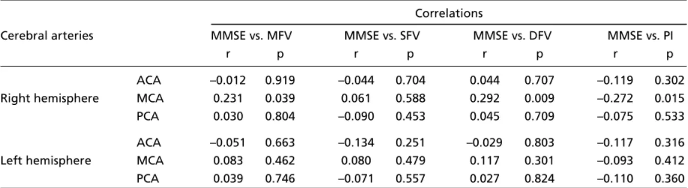
Table 1 shows TCD data in relation to MMSE sco-res. A significant correlation was found between right middle cerebral artery (RMCA) TCD parameters and MMSE scores. There was a direct correlation between MMSE scores and both mean (r=0.231 p=0.039) and diastolic (r=0.292 p=0.009) flow velocities. For pul-satility indexes (PI), there was an inverse corre l a t i o n between RMCA PI and MMSE (rs=-0.292 p=0.015). When excluding patients with a previous history of s t roke, only RMCA PI remained with a significant cor-relation to MMSE score (rs=–0.314 e p=0.007). In con-trast, none of the ecocardiographic parameters cor-related with MMSE scores, including ejection frac-tion, left atrium diameter, systolic and diastolic diam-eter of the left ventricle (data not shown).
Arq Neuropsiquiatr 2006;64(2-A) 209
significant relationship was found between right an-terior cerebral art e ry (RACA) flow velocities and rected MMSE scores. For normal versus abnormal cor-rected MMSE scores, RACA systolic flow velocity was 6 2 . 1±17.8 vs. 49.4±14.5 cm/s (p=0.015), diastolic flow velocity was 25.6±13.0 vs. 17.3±10.7 (p=0.028) and mean flow velocity was 40.0±14.7 vs. 30.3±10.7 (p= 0.023). The results remained unaltered after
exclud-ing patients with a history of stroke. No re l a t i o n s h i p was found between demographic, risk factor or echo-c a rdiographiecho-c variables and echo-correecho-cted MMSE secho-core s .
DISCUSSION
Cognitive symptoms are seen in 37% to 57% of patients with CHF and have been shown to adverse-ly affect pro g n o s i s2 , 5. In general practice, physicians
Table 2. Predictors of cognitive dysfunction expressed by abnormal Mini Mental State Examination (MEEM) scores.
Variables Normal MMSE Abnormal MMSE p
score (n=66) score (n=17)
Clinical
Age, years (mean +/- SD) 54+12 55+13 NS
Male gender, n(%) 37 (56) 10 (59) NS
Hypertension, n(%) 34 (52) 7 (41) NS
Diabetes, n(%) 4 (6) 1 (6) NS
CAD, n(%) 8 (12) 4 (24) NS
Previous stroke, n(%) 6 (9) 3 (18) NS
Current smoking, n(%) 5 (9) 3 (18) NS
Mean (±SD) admission
systolic/diastolic BP, mmHg 123/81±22/14 126/82±31/16 Transcranial doppler
RACA MFV, cm/s 40±15 30±10 0.016
RACA SFV, cm/s 62±18 49±14 0.008
RACA DFV, cm/s 26±13 17±10 0.018
Echocardiogram
Ejection fraction (%) 41±11 38±9 NS
LAD, cm 40±6 41±9 NS
LVSD, cm 51±9 54±7 NS
LVDD, cm 65±9 67±6 NS
Intracardiac thrombus, n(%) 0 (0) 0 (0) NS
BP, blood pressure; CAD, coronary artery disease; DFV, diastolic flow velocity; LAD, left atrium diame-ter; LVDD, left ventricle diastolic diamediame-ter; LVSD, left ventricle systolic diamediame-ter; MFV, mean flow velocity; NS, non-significant; RACA, right anterior cerebral artery; SFV, systolic flow velocity. Table 1. Correlation between transcranial Doppler parameters and Mini Mental State Exame scores.
Correlations
Cerebral arteries MMSE vs. MFV MMSE vs. SFV MMSE vs. DFV MMSE vs. PI
r p r p r p r p
ACA –0.012 0.919 –0.044 0.704 0.044 0.707 –0.119 0.302 Right hemisphere MCA 0.231 0.039 0.061 0.588 0.292 0.009 –0.272 0.015
PCA 0.030 0.804 –0.090 0.453 0.045 0.709 –0.075 0.533
ACA –0.051 0.663 –0.134 0.251 –0.029 0.803 –0.117 0.316 Left hemisphere MCA 0.083 0.462 0.080 0.479 0.117 0.301 –0.093 0.412
PCA 0.039 0.746 –0.071 0.557 0.027 0.824 –0.110 0.360
210 Arq Neuropsiquiatr 2006;64(2-A)
will manage CHF symptoms with medications that i m p rove overall ventricular function. However, no s u c h medications decrease cardioembolic potential, which a re a possible cause of cognitive dysfunction. Pre-vious re p o rts of TCD in patients with CHF have shown low mean flow velocities which improve after car-diac transplantation, but no cognitive data were pre-s e n t e d6 , 7. Others have studied the prevalence of
mi-c roemboli detemi-cted on TCD during mi-cardiami-c surg e ry and correlated these findings to cognitive dysfunc-t i o n8. One study showed a decreased cere b ro v a s c
u-lar reactivity in patients with CHF compared to con-t ro l s9. To our knowledge, no previous study
attempt-ed to correlate the various TCD parameters such as PI and flow velocities in different arterial territories to cognitive changes.
The main finding of our study was a corre l a t i o n between right-hemisphere TCD parameters and cog-nitive dysfunction, in general showing low flow veloc-ities and high pulsatility indexes. Right MCA param-eters showed a significant correlation to uncorre c t-ed MMSE scores, while right ACA parameters showt-ed significant correlation to MMSE scores corrected for educational level. Even in patients without a pre v i-ous history of stroke, right hemisphere parameters remained significant predictors of cognitive impair-ment. The reasons for this finding are speculative, but several studies have documented a greater pro-p o rtion of silent infarcts in the right hemispro-phere1 0 , 1 1,
possibly because right hemisphere symptoms such as anosognosia often remain unnoticed by patients and c a regivers. We hypothesize that microemboli to the right hemisphere may be responsible for concomi-tant cognitive impairment and the changes observ e d in cerebral hemodynamics.
Another intriguing finding was the lack of corre-lation between echocardiographic parameters and cognitive impairment. Most data on CHF show a d i rect relationship between the degree of card i a c dysfunction (measured by the ejection fraction) and cognitive changes1 2 - 1 4. However, at least one pre v
i-ous study did not show a correlation between ejec-tion fracejec-tion and MMSE score3, which suggests that,
in some populations, microemboli may surpass low c e rebral perfusion as the predominant mechanism of cognitive dysfunction. One such unique character-istic of our population was the high prevalence of Chagas disease the etiology of CHF, which is known to be a highly embolic condition15.
T h e re are two limitations to our study. First, echo-c a rdiography was perf o rmed by multiple examiners and equipment, unlike the TCD examinations, which w e re done by a single blinded examiner. Second, sin-ce patients without a history of stroke did not for-mally re q u i re imaging studies, no such data is pre s e n-ted. As such, the TCD signature of low flow veloci-ties and high pulsatility indexes, although suggesti-ve, cannot be definitively attributed to micro e m b o l i with silent brain infarcts.
In conclusion, cognitive changes in patients with CHF were common, unrelated to cardiac systolic dys-function, and significantly related to cere b ro v a s c u-lar parameters suggestive of microembolic etiology. Such findings should be confirmed in studies evalu-ating simultaneously cere b rovascular parameters and imaging of the brain parenquima.
REFERENCES
1. Hunt SA, Baker DW, Chin MH, et al. A C C / A H A guidelines for the evaluation and management of chronic heart failure in the adult: exec-utive summary report of the American College of Card i o l o g y / American Heart Association Task Force on Practice Guidelines. Circulation 2001;104:2996-3007.
2. Zuccala G, Pedone C, Cesari M, et al. The effects of cognitive impair-ment on mortality among hospitalized patients with heart failure. A m J Med 2003;115:97-103.
3. Almeida OP, Tamai S. Congestive heart failure and cognitive function-ing amongst older adults. Arq Neuropsiquiatr 2001;59:324-329. 4. Bertolucci PHF, Brucki SMD, Campacci SR, Juliano Y. O Mini-Exame
do Estado Mental em uma população geral: impacto da escolaridade. Arq Neuropsiquiatr 1994;52:1-7.
5. Trojano L, Antonelli Incalzi R, Acanfora D, Picone C, Mecocci P, Rengo F. Congestive Heart Failure Italian Study Investigators. Cognitive impairment: a key feature of congestive heart failure in the elderly. J Neurol 2003;250:1456-1463.
6. M a s s a ro AR, Almeida DR, Dutra A P, et al. Transcranial Doppler in patients with refractory congestive heart failure before and after heart transplantation. Arq Neuropsiquiatr 2004;62:22.
7. G ruhn N, Larsen FS, Boesgaard S, et al. Cerebral blood flow in patients with chronic heart failure before and after heart transplantation. Stro k e 2001;32:2530-2533.
8. Clark RE, Brillman J, Davis DA, Lovell MR, Price TR, Magovern GJ. M i c roemboli during coronary artery bypass grafting: genesis and eff e c t on outcome. J Thorac Cardiovasc Surg 1995;109:249-257.
9. G e o rgiadis D, Sievert M, Cencetti S, et al. Cere b rovascular reactivity is impaired in patients with cardiac failure. Eur Heart J 2000;21:407-413. 10. EAFT Study Group. Silent brain infarction in nonrheumatic atrial
fib-rillation. Neurology 1996;46:159-165.
11. B rott T, Tomsick T, Feinberg W, et al. Baseline silent cerebral infarc t i o n in the Asymptomatic Carotid A t h e ro s c l e rosis Study. Stroke 1994;25:11 2 2 -1129.
12. Bornstein RA, Starling RC, Myerowitz PD, Haas GJ. Neuro p s y c h o l o g i c a l function in patients with end-stage heart failure before and after car-diac transplantation. Acta Neurol Scand 1995;91:260-265.
13. Zuccala G, Cattel C, Manes-Gravina E, Di Niro MG, Cocchi A, Bernabei R. Left ventricular dysfunction: a clue to cognitive impairment in old-er patients with heart failure. J Neurol Neuro s u rg Psychiatry 1997; 63:509-512.
14. Zuccala G, Onder G, Pedone C, et al. Cognitive dysfunction as a major determinant of disability in patients with heart failure: results from a multicentre survey. J Neurol Neurosurg Psychiatry 2001;70:109-112. 15. Samuel J, Oliveira M, Correa de Araujo RR, Navarro MA, Muccillo G.