Electromyography biofeedback in the treatment of neurogenic
orofacial disorders: systematic review of the literature
Biofeedback
eletromiográfico no tratamento das disfunções
orofaciais neurogênicas: revisão sistemática de literatura
Gabriela Silva de Freitas1, Claudia Tiemi Mituuti1, Ana Maria Furkim1, Angela Ruviaro Busanello-Stella1,
Fabiane Miron Stefani1, Marcela Maria Alves da Silva Arone2, Giédre Berretin-Felix3
ABSTRACT
Purpose: To determine whether the use of electromyographic biofeedback in the therapy of orofacial functions (facial expression, chewing, swallowing, phonation and speech) will result in beneficial effects for individuals with neurological diseases. Research strategy: A keyword search was conducted in the MEDLINE, LILACS and SciELO databases, using the terms “electromyographic biofeedback”, “swallowing”, “speech”, “chewing”, “phonation”, and “facial expression”. The database search and the selection of papers were conducted independently by two researchers. In case of any disagreement, there was a discussion based on the inclusion and exclusion criteria, so that they could reach a common ground. Selection criteria: This work has included experimental studies in humans, in English and Portuguese, which described and discussed the use of electromyographic biofeedback in the treatment of orofacial function diseases resulting from neurological illness. Results: A total of 175 papers were found, wherein only 10 fitted the inclusion criteria. Most works were case studies, followed by case series, case control, and only one randomized controlled trial. Most of studies addressed the therapy with electromyographic biofeedback in the swallowing function, followed by speech function, and only one study addressed the use of electromyographic biofeedback in therapy to improve facial expression. No studies addressing speech therapy using electromyographic biofeedback in patients with neurological diseases in the functions of phonation and chewing were found. Conclusion: The use of electromyographic biofeedback in the therapy for orofacial functions can result in beneficial effects for individuals with neurological diseases in the swallowing, speech, and facial expression functions.
Keywords: Electromyography; Deglutition; Speech; Mastication; Pho-nation; Facial expression
RESUMO
Objetivo: Investigar se o uso do biofeedback eletromiográfico na terapia voltada às funções orofaciais (expressão facial, mastigação, deglutição, fonação e fala) produz efeitos benéficos para os indivíduos com doenças neurológicas. Estratégia de pesquisa: Foi realizada busca nas bases de dados MEDLINE, LILACS e SciELO, por meio dos descritores “electromyographicbiofeedback”, “swallowing”, “speech” “chewing”, “phonation”, e “facial expression”. A busca nas bases de dados e a seleção dos artigos foram realizadas independentemente, por duas pesquisadoras e, nos casos de não concordância, houve discussão fundamentada nos critérios de inclusão e exclusão para que chegassem a um consenso. Critérios de seleção: Foram incluídos estudos experimentais em seres humanos, em inglês e português, que descreveram e discutiram a utilização do biofeedback eletromiográfico no tratamento das alterações das funções orofaciais provenientes de doenças neurológicas. Resultados: Foram encontrados 175 artigos, sendo que somente 10 se adequaram aos critérios de inclusão. A maioria dos trabalhos relacionou-se a estudo de caso, seguido por estudos de série de casos, caso controle e ensaio clínico randomizado. A maior parte dos artigos abordou a aplicação da terapia com biofeedback
eletromiográfico na função da deglutição, seguida da função da fala e apenas um artigo utilizou esta modalidade de tratamento na terapia para melhora da expressão facial. Não foram encontrados estudos que abordassem o tratamento fonoaudiológico utilizando o biofeedback
eletromiográfico em pacientes com doenças neurológicas, nas funções de fonação e mastigação. Conclusão: O uso do biofeedback
eletromiográfico na terapia voltada às funções orofaciais pode produzir efeitos benéficos para os indivíduos com doenças neurológicas, nas funções de deglutição, fala e expressão facial.
Descritores: Eletromiografia; Deglutição; Fala; Mastigação; Fonação; Expressão facial
Work developed in the Undergraduate Course of Special Coordination of Speech Therapy from the Universidade Federal de Santa Catarina – UFSC – Santa Catarina (SC), Brazil, in partnership with the Graduate Course in Speech Therapy, from the College of Bauru from Universidade de São Paulo – USP – Bauru (SP), Brazil. (1) Speech Therapy School, Universidade Federal de Santa Catarina – UFSC – Santa Catarina (SC), Brazil.
(2) Bauru Base Hospital, Bauru (SP), Brazil.
(3) Speech Therapy School, Bauru School of Dentistry, Universidade de São Paulo – USP – Bauru (SP), Brazil. Conflict of interests: No
Authors’ contribution:GSF: data collection and analysis, writing of scientific paper; CTM: data collection and analysis, research advice and writing of scientific paper; AMF, ARBS and FMS: writing of scientific paper; MMASA writing of paper, data collection and analysis; GBF creator of this work, research project advice and correction of this paper.
INTRODUCTION
The main objective of speech therapy applied to irregula-rities of the stomatognathic system is to restore the breathing, chewing, swallowing and speaking abilities, aiming to find the myofunctional balance in individuals with or without anato-mical and / or functional irregularities. This therapy may be applied to prevent, rehabilitate, or activate these functions(1). In
the therapy awareness phase, therapists make individuals aware of their patterns, as opposed to the normal pattern. Eventually, the new pattern learned in therapy should be put into practice on a daily basis, namely the automation phase.
The training of an irregular function occurs in between the steps of awareness and automation of speech therapy. The trai-ning consists of the therapist providing guidance to the patient, facilitating learning strategies, in order to regulate a specific function, bringing it closer to the normal pattern. Several techniques are used as tools for training orofacial functions. The visual tool is the one most commonly described, whereas most studies addressing the use of this strategy include the use of mirrors(2).
Researchers of speech therapy have investigated neural me-chanisms engaged in therapies that include the use of mirrors, as opposed to simple observation of movement, considering body limbs. The literature states that the use of mirrors incre-ases the activation of the primary and superior visual areas, contralateral to the limb observed; it is concluded that the activation of brain lateralization is elicited by inverting the visual feedback (mirror)(3).
Tactile strategies are also described, especially in the trai-ning of speech, with blowing strategies on the back of the hands to work the direction of airflow in the fricative and affricate phonemes(4), as well as the use of proprioceptive tools to
re-gulate the positioning of the tongue in some liquid phonemes. Still regarding the training of speech and vocal production, hearing and visual feedback is referred to as facilitators, with recording strategies to further analyze patients(5) and delayed
feedback for the speech training of patients with dysfluency(6).
Some instruments used for evaluation are also described as supporting methods in the treatment of orofacial functions, namely nasometry and surface electromyography.
Nasometry, which quantifies relative nasal acoustic energy during the production of oral speech, is also used as a resource in therapy for patients with cleft palate and velopharyngeal dysfunction. A nasometer is used to increase visual perception of nasal and oral air flow in the practice of velopharyngeal functioning during speech production. Therapists make speech tasks more difficult, as patients improve their perception of the direction of the oral and nasal air flow(4).
Another technique used as therapeutic methodology, yet rarely described in the field of speech therapy, is the use of electromyographic biofeedback. As a therapeutic strategy, it can be used in the aid of muscle relaxation and coordination,
as well as in the engagement of a larger number of motor units during muscle activity(7).
Some studies have demonstrated clinical efficacy in the treatment of a variety of muscular disorders and, in the field of Speech Therapy, although poorly addressed, the technique is commonly used in neurological cases of paralysis, spasticity, and neurological hyperfunction(7).
The possibility of applying surface electromyography (EMG) to clinical speech therapy brings important contribu-tions. One is related to the fact that it is an objective examination which allows for numerical results that may quantify muscle function. Furthermore, equipment-generated visual images help patients and therapists understand the muscle function to be assessed and treated. Although there is still little standardiza-tion of therapeutic procedures and assessment of the muscles involved in orofacial functions, EMG provides a great range of applications intended to prove the therapeutic efficacy of exercises and to improve the work of speech therapists when dealing with orofacial myofunctional disorders.
OBJECTIVE
This work was aimed at assessing whether the use of elec-tromyographic biofeedback therapy that is focused on orofacial functions (facial expression, chewing, swallowing, speech and speech) brings about beneficial effects for individuals with neurological diseases.
RESEARCH STRATEGY
This is a systematic review of the literature on electromyo-graphic biofeedback application methods for the treatment of neurogenic irregularities in orofacial functions. The authors of this work sought to find relevant articles in the MEDLINE (through PubMed), LILACS and SciELO platforms (through Bireme).
The following keywords were used: “electromyographic biofeedback”, “swallowing”, “speech” “chewing”, “phona-tion”, and “facial expression”, whereas “electromyographic biofeedback” was present in all combinations, to obtain the largest possible number of papers on this topic.
The search in the databases and the selection of the papers were performed independently by two researchers, who used the same criteria and the same terms and operators. In case of any disagreement, there was a discussion based on the inclusion and exclusion criteria, so that they could reach a common ground.
SELECTION CRITERIA
of electromyographic biofeedback in the treatment of orofacial functions (FOF) caused by neurological irregularities were analyzed, regardless of date of publication.
Papers were excluded when they were in duplicate, presen-ted insufficient data on the description of electromyographic biofeedback application, and were not available in full text.
DATA ANALYSIS
The analysis of each research paper took into account type and level of evidence, which functions were addressed, electrode positioning, age groups, and diseases being studied. They were sorted according to the functions they investigated, as well as the methods in use and the findings.
RESULTS
After the database search, 175 articles were selected, and 91 of them were repeated. Forty-five percent of the articles addressed the function of speech; 28% addressed swallowing;
11%, facial expression; 10%, phonation, and 6% chewing. Thus, we selected 84 articles to analyze their titles and abs-tracts; 63 of them did not fit the inclusion criteria, because they lacked sufficient data on the description of the methodology, because they were not applied to neurological diseases, or were written in a language other than English or Portuguese, or even because they did not use EMG as a therapeutic strategy. After that analysis, 21 papers were included in the discussion between researchers, who disagreed on 7 of them. Out of these 7 papers, 1 was included and others were excluded, thus totaling 15 papers. At the end of the selection, 5 papers were excluded because the full text was not available; a total of 10 and papers were included in this review (Figure 1).
According to the Oxford(8) table, most papers presented a
level 4 of evidence, except for one paper, which had a higher level of evidence. Most of the works were case studies(9,10,11,12,13)
(5 papers), followed by series of case studies(14,15,16) (3 papers),
with one being a case control(17) and another(18) a randomized
clinical trial; there was a low level of evidence for these works (Figure 2).
Most papers (five) considered the application of biofeedback therapy electromyography in the swallowing function(14,15,16,17,18).
Although the application of biofeedback associated with swallowing therapy is recent(7), there is a great number of
re-searchers interested in proving the efficacy of this treatment in neurological diseases. While addressing the function of speech, 4 papers used electromyography biofeedback in neurological patients(9,10,11,13) and only one paper used electromyography
bio-feedback in therapy for the improvement of facial expression(12).
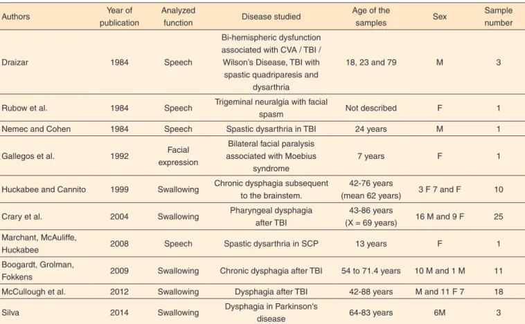
No paper discussed the function of chewing and speech in the treatment of neurological patients (Table 1).
There was a significant difference in the age groups studied among the works, from 7 to 88 years of age. Taking into account
that the process of aging brings physiological changes to the function of swallowing, the elderly population is at risk of dys-phagia(19) as a result. The papers that addressed the swallowing
function focused on age groups that ranged from 42 to 88 years old. Papers that addressed younger age groups referred to the rehabilitation of speech function and facial expression.
The neurological diseases most often addressed in the papers (3 items) were cerebrovascular accident (CVA)(9,15,16,18),
followed by traumatic brain injury (TBI), included in 2 pa-pers(9,11). The other conditions were lesions on the brainstem(14),
Parkinson’s disease(17), trigeminal neuralgia(10), spastic cerebral
palsy (SCP)(13), and bilateral facial paralysis(12). Stroke and TBI
are among the most relevant diseases of the central nervous
Figure 2. Distribution of studies according to their methodology
Table 1. Distribution of papers, according to the authors, year of publication, analyzed function, basic neurological disease, age, gender and sample number of subjects
Authors Year of
publication
Analyzed
function Disease studied
Age of the
samples Sex
Sample number
Draizar 1984 Speech
Bi-hemispheric dysfunction associated with CVA / TBI / Wilson’s Disease, TBI with spastic quadriparesis and
dysarthria
18, 23 and 79 M 3
Rubow et al. 1984 Speech Trigeminal neuralgia with facial
spasm Not described F 1
Nemec and Cohen 1984 Speech Spastic dysarthria in TBI 24 years M 1
Gallegos et al. 1992 Facial
expression
Bilateral facial paralysis associated with Moebius
syndrome
7 years F 1
Huckabee and Cannito 1999 Swallowing Chronic dysphagia subsequent to the brainstem.
42-76 years
(mean 62 years) 3 F 7 and F 10
Crary et al. 2004 Swallowing Pharyngeal dysphagia
after TBI
43-86 years
(X = 69 years) 16 M and 9 F 25
Marchant, McAuliffe,
Huckabee 2008 Speech Spastic dysarthria in SCP 13 years F 1
Boogardt, Grolman,
Fokkens 2009 Swallowing Chronic dysphagia after TBI 54 to 71.4 years 10 M and 1 M 11
McCullough et al. 2012 Swallowing Dysphagia after TBI 42-88 years M and 11 F 7 18
Silva 2014 Swallowing Dysphagia in Parkinson's
disease 64-83 years 6M 3
system that lead to speech and language disorders, and they may cause changes in various stomatognathic system func-tions(20) (Table 1).
With respect to the function of swallowing, most papers stu-died dysphagia in stroke(15,16,18) (60%), followed by Parkinson’s
disease(17) (20%) and brainstem lesion(14) (20%). Therapies were
carried out in around 6 to 20 sessions in total.
DISCUSSION
It is estimated that in patients who experienced a stroke, over 50% have dysphagia, with major complication of aspiration pneumonia. The presence of dysphagia, assessed at the bedside, was associated with increased incidence of lung infection in comparison with patients without dysphagia (33% and 16%, respectively). Dehydration and malnutrition are also common in dysphagia patients, especially those receiving modified thi-ckened liquids or diets(21). In patients with Parkinson’s disease,
the prevalence of oropharyngeal dysphagia occurs between 52% and 82%(22) of cases. Signals of dysphagia in patients
with Parkinson’s disease include increased oral transit time, language festination movement, inadequate control of bolus with premature pharyngeal escape, swallowing in portions, hyolaryngeal complex motion reduction, as well as the base of the tongue, pharynx and epiglottis, and oesophageal dys-motility and reflux. The consequences of dysphagia in these patients may include weight loss, dietary changes and death from aspiration pneumonia(23).
Most papers (80%) used, in speech therapy for swallowing, maneuvers associated with electromyographic biofeedba-ck(14,15,16,18), and only one (20%) used a target goal for functional
training. They showed the patients the normal pattern of swallo-wing and their own pattern, and established a target route for functional training(17). The Mendelsohn maneuver was chosen
by all papers that adopted maneuvers, except for one, which, in addition to Mendelsohn maneuver, used swallowing effort, Masako and Shaker maneuver(14).
The use of a swallowing maneuver, alone, provides the electromyographic recording of their specific physiological effects, thus allowing to examine the use of individual exercises. The Mendelsohn maneuver, used in most studies, was the most appropriate for checking the function of the lifters muscles of the larynx, which can measure the highest peak elevation of larynx during swallowing.
The Mendelsohn maneuver aims to maximize the elevation of the larynx and the opening of the Cervical Esophagus, during swallowing. It consists of voluntarily maintaining, for a few seconds, the elevation of the larynx at its highest point during swallowing. Swallowing with effort, in which the patient should force the ingestion of food, is performed in order to increase the muscle strength of the structures involved, optimizing sending and passing the bolus through the oropharynx. The Masako maneuver is used to increase the movement of the posterior
wall of the pharynx during swallowing. In this maneuver, the patient should undergo swallowing with protruded tongue, caught between the incisors. The Shaker maneuver aims to improve the laryngeal elevation and increase the efficiency of the protective mechanisms of the airways, by working the extrinsic muscles of the larynx. In this maneuver, the patient should be lying without a pillow and lift his head, looking at his feet, without lifting his shoulders(24).
In most studies (80%), the electrodes were placed on submental muscles, the suprahyoideus(14,16,17,18) and in 20% of
the studies, between the hyoid bone and the upper edge of the thyroid cartilage(15).
All studies found increased diet scale level in cases of oro-pharyngeal dysphagia. One study reported that 55% of patients achieved complete oral feeding(15). Another work mentioned
that all patients who underwent complete therapy achieved satisfactory level of oral intake while those who underwent the therapy partially, increased this level by 1 or 2 points on the FOIS scale(18). The management of dysphagia aims to
pro-tect airways and provide safe swallowing, for better quality of life, in addition to maximizing function or compensatory potentials(25) and, hence, patient’s awareness is important to
the completion of the proposed treatment plan.
In one of the studies, researchers conducted two weeks of therapy with biofeedback embedded with conventional thera-py and compared them with weeks without treatment. They were able to show an improvement in the weeks that included treatment and worsening when no biofeedback was applied in cases of dysphagia(16). Thus, treatment continuity is essential
to patient rehabilitation. A longer treatment, with a complete treatment program, can bring greater generalization of the learned pattern.
In the study which compared conventional treatment with conventional therapy associated with electromyographic bio-feedback, greater improvement was noted in patients’ quality of life, in conventional therapy associated with EMG biofee-dback, especially in the long term, thus demonstrating better after-treatment results(17).
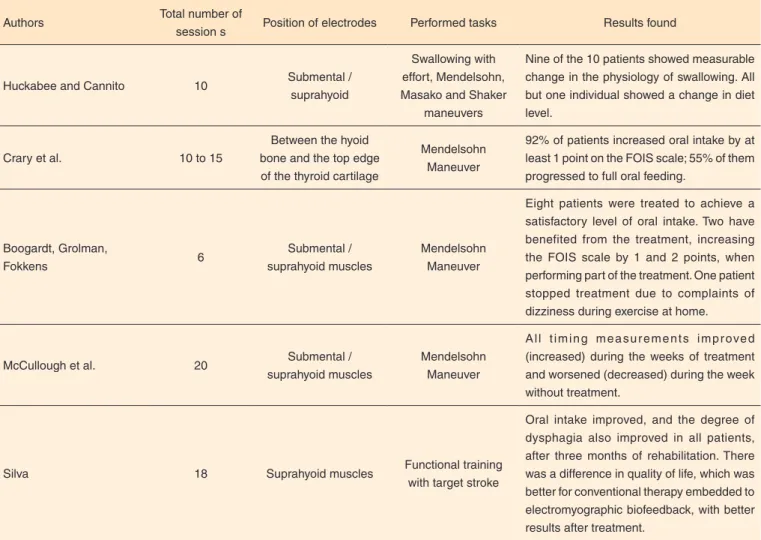
All papers described satisfactory results in the use of electromyographic biofeedback associated with conventional therapy, which suggests that the use of EMG biofeedback method as an adjunct to conventional therapy can facilitate the learning of new neuromuscular patterns for swallowing, and thus provide greater gain for the patient compared with conventional treatment(17) (Table 2).
controlled clinical studies in order to better understand the contribution of this technique(17). In addition to the use of
electromyography, other biofeedback strategies have been described in the literature for swallowing rehabilitation, such as the use of endoscopy. It allows and directs visualization of the oropharyngeal mechanism of swallowing(26), and the use
of equipment, such as the Iowa Oral Performance Instrument (IOPI), can show the amplitude of pressure during tongue resistance exercise(27).
As for studies addressing the function of speech, 40% of them conducted treatment for TBI(9,11), 20% for CVA(9), 20%
for SCP(13) and 20% for trigeminal neuralgia(10).
The most common speech disorder in individuals with neu-rological diseases is dysarthria (60%)(28). Dysarthria is defined
as a neurologic motor impairment of speech, characterized by slowness, weakness, imprecision and / or uncoordinated move-ments of the muscles of speech, which can impair respiration, phonation, resonance, and / or articulation(29). It is one of the
consequences of ECA, when articulation, breathing, voice, rhythm and fluency(30) can be impaired. In TBI, dysarthrias are
present in approximately 45% of cases(31).
In SCP, hyperreflexia and exaggerated increase in muscle
tone occur, with reduction of voluntary movements(13). In
indi-viduals with SCP, pastic dysarthria is characterized by strained vocal quality with possible roughness and reduced pitch va-riation, related to the hypertension of laryngeal musculature(5).
In the publications found, the number of sessions ranged from 10 to 31 in total. In 40% of the studies, the task consisted of reading activities(9,11), 20%, repeating words and syllables,
expanding simple sentences, with increased ability(9). Twenty
percent of the studies used the facial muscle relaxation activity, because of muscular tension caused by spasms, performing relaxation in therapeutic sessions with patients starting in the reclined position, up to the sitting position, ending upright on the chair(10). Twenty percent of studies adopted the production
of isolated speech sounds and then word sounds(13).
The studies used the positioning of the electrodes at various locations. In one study, they were positioned on the suprahyoid muscles(9); in another one, in the frontal region(10); in another,
on the masseter, temporalis and lateral pterygoid(11); and, in the
last study, the electrodes were placed on submental muscles, on suprahyoid muscles and orbicularis(13). The variability of
electrode position was due to different rehabilitation objectives of the studies, as one sought the relaxation of the facial muscles,
Table 2. Description of treatments performed by studies on swallowing, with total number of sessions, placement of electrodes, tasks performed and findings
Authors Total number of
session s Position of electrodes Performed tasks Results found
Huckabee and Cannito 10 Submental /
suprahyoid
Swallowing with effort, Mendelsohn, Masako and Shaker
maneuvers
Nine of the 10 patients showed measurable change in the physiology of swallowing. All but one individual showed a change in diet level.
Crary et al. 10 to 15
Between the hyoid bone and the top edge of the thyroid cartilage
Mendelsohn Maneuver
92% of patients increased oral intake by at least 1 point on the FOIS scale; 55% of them progressed to full oral feeding.
Boogardt, Grolman,
Fokkens 6
Submental / suprahyoid muscles
Mendelsohn Maneuver
Eight patients were treated to achieve a satisfactory level of oral intake. Two have benefited from the treatment, increasing the FOIS scale by 1 and 2 points, when performing part of the treatment. One patient stopped treatment due to complaints of dizziness during exercise at home.
McCullough et al. 20 Submental /
suprahyoid muscles
Mendelsohn Maneuver
A l l t i m i n g m e a s u r e m e n t s i m p r ove d (increased) during the weeks of treatment and worsened (decreased) during the week without treatment.
Silva 18 Suprahyoid muscles Functional training
with target stroke
another, the reduction of spasm and another, the improvement in mandibular elevation, all of them in order to achieve a more intelligible speech.
All studies had results of intelligibility improvement in speech. Two studies described improved tongue mobility and posture and better intraoral pressure(9,11). Other two studies
re-ported that after treatment, the improvements were maintained, getting good generalization of skills learned in the treatment with electromyographic biofeedback(11,13). In one study, the
patient had greater strength in the articulation of words, im-provement in the velopharyngeal function, in intelligibility and awareness of nasality, obtaining a non-nasal voice quality(9).
Another study, in order to reduce muscle spasm to improve speech, obtained the reduction of spasms as a result, and con-sequent improvement in speech intelligibility(10). In another
study, the patient was able to maintain high jaw, adjusting tongue posture in the articulation position(11). The results were
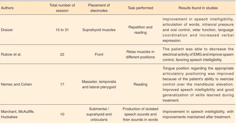
examined through the acoustic analysis of the recording of the patient’s speech, as well as tests and software programs used for quantitative analysis of speech (Table 3).
The analyzed studies show that speech therapy combined with electromyographic biofeedback is effective for neurolo-gical disorders.
In the past years, the treatment provided by speech thera-pies was based on articulatory training of orofacial compo-nents. Nowadays, in addition to articulation training, which is an exercise for accurate production of phonemes, through over-articulation and articulatory compensation, there is the instrumental treatment, which can be accomplished by elec-tromyographic biofeedback; the metronome, which assists
the speed of speech, and devices that provide delayed hearing feedback. These resources have proven to be effective for speech disorders(32).
Electromyographic biofeedback and other biofeedback methods can be effective because they involve constant self--correction. Motor planning and motor control skills are continuously stimulated and beneficial neuronal plasticity is induced. In one study, which used visual biofeedback for indi-viduals with irregularities in the rhythm, the authors found that changes occur in cortical activation when biofeedback is used, highlighting a cortical reorganization induced by this method(33).
There are few studies on the use of electromyographic bio-feedback treatment in neurological patients for improvement of facial expression. In this systematic review of the literature, we found only one paper adopting this type of speech therapy; thus, comparisons cannot be made among studies. The work was performed in a patient with bilateral facial paralysis asso-ciated with Moebius syndrome(12).
Moebius syndrome is a rare, non-progressive disease, cha-racterized by the paralysis of the facial nerve, mostly bilateral, sixth nerve palsy, and it may be associated with injury and other cranial, thoracic abnormalities, and limbs, dental problems, malformation of the tongue and cleft palate(34). The main features are: lack of facial expression; convergent strabismus; inadequate tongue mobility; micrognathia; suction, chewing, swallowing and speech irregularities; clubfoot; syndactyly(35).
There are several types of treatment for facial paraly-sis. Among them, there are treatments with drugs, surgery, botulinum toxin and speech therapy(36,37). Speech therapy is
made through manipulations in the muscles of the face, use
Table 3. Description of treatments performed by speech studies, with total number of sessions, placement of electrodes, task performed and findings
Authors Total number of
session
Placement of
electrodes Task performed Results found in studies
Draizar 15 to 31 Suprahyoid muscles Repetition and
reading
Improvement in speech intelligibility, articulation of words, intraoral pressure and oral control, velar function, language c o o r d i n a t i o n a n d i n c r e a s e d ve r b a l expression.
Rubow et al. 22 Front Relax muscles in
different positions
The patient was able to decrease the electrical activity of EMG and improve spasm control, favoring speech intelligibility.
Nemec and Cohen 17 Masseter, temporalis
and lateral pterygoid Reading
Tongue position regarding the appropriate ar ticulatory positioning was improved because of the patient's ability to exercise control over the mandibular elevation. Improved speech intelligibility and good generalization of skills learned during treatment.
Marchant, McAuliffe,
Huckabee 10
Submental / suprahyoid and
orbicularis
Production of isolated speech sounds and then sounds in words
of physical strength through distal pulse, and stimulation of facial motor zones and points, together with the use of myofunctional exercise(38). One of the treatments for facial
pa-ralysis is the use of EMG biofeedback, referred to as effective for providing the patient with immediate response to muscle activity, promoting facial muscle reeducation, making up for normal activity(36).
Gallegos et al.(12), in a case study, have demonstrated
patient subjective improvement, both with emotional expres-sion, and in their general state of mind. In addition, it was subjectively observed that pronunciation of the phoneme / s / improved, and the patient’s speech had an overall improve-ment (Table 4). The study proved that the treatimprove-ment with electromyographic biofeedback embedded with conventional therapy was effective in a case of facial expression improve-ment in bilateral facial paralysis. Further research is needed to prove this technique in neurological disorders. In addition to the use of electromyographic biofeedback, the use of mirror as a biofeedback tool has also been described in the treatment of facial paralysis(2,38).
In most cases, the results of the selected studies proved sa-tisfactory for speech therapy embedded with electromyographic biofeedback to improve swallowing, speech, and facial expres-sion functions in patients with neurological diseases. However, these works have included a reduced number of individuals. Thus, further studies that include a greater number of subjects are needed to prove such effectiveness.
There were no studies aiming at assessing the use of speech therapy embedded with electromyographic biofeedback in pa-tients with neurological diseases for the phonation and chewing functions. Considering the effectiveness of the results of studies addressing biofeedback electromyographic in swallowing, speech and facial expression functions in individuals with neurological irregularities, further studies should address the other functions of the stomatognathic system, to check for the effectiveness of treatment in these speech disorders.
CONCLUSION
The use of electromyographic biofeedback in orofacial function therapy can produce beneficial effects for the swallo-wing, speech, and facial expression functions of individuals with neurological diseases. In view of the low level of evidence
of the selected studies, further randomized clinical studies with a greater sample size are needed, and they should take into account the specificities of the different types of neurological diseases.
REFERENCES
1. Marchesan IQ, Bianchini EMG. A fonoaudiologia e a cirurgia ortognática. In: Araujo A. Cirurgia ortognática. São Paulo: Santos; 1998. p. 353-62.
2. Dalla Toffola E, Pavese C, Cecini M, Petrucci L, Ricotti S, Bejor M et al. Hypoglossal-facial nerve anastomosis and rehabilitation in patients with complete facial palsy: cohort study of 30 patients followed up for three years. Funct Neurol. 2014;29(3):183-7. 3. Wang J, Fritzsch C, Bernarding J, Holtze S, Brunetti M, Dohle C. A
comparison of neural mechanisms in mirror therapy and movement observation therapy. J Rehabil Med. 2013;45(4):410-34. http:// dx.doi.org/10.2340/16501977-1127
4. Bispo NHM, Whitaker ME, Aferri HC, Neves JDA, Dutka JCR, Pegoraro-Krook MI. Speech therapy for compensatory articulations and velopharyngeal function: a case report. J Appl Oral Sci. 2011;19(6):679-84. http://dx.doi.org/10.1590/S1678-77572011000600023
5. Tomé MC, Oda AL. Intervenção fonoaudiológica nos distúrbios de fala: a origem fonética e a origem neurológica. In: Marchesan IQ, Justino H, Tomé MC. Tratado de especialidades em fonoaudiologia. São Paulo: Guanabara Koogan; 2014.
6. Buzzeti PBMM, Fiorin M, Martinelli NL, Cardoso ACV, Oliveira CMC. Comparação da leitura de escolares com gagueira em duas condições de escuta: habitual e atrasada. Rev CEFAC. 2016;18(1):67-73. http://dx.doi.org/10.1590/1982-0216201618114015
7. Rahal A, Silva MMA, Berrentin-Felix G. Eletromiografia de superfície e biofeedback eletromiográfico. In: Pernambuco LA, Silva HJ, Souza LBR, Magalhães Junior HV, Cavalcanti RVA, organizadores. Atualidades em motricidade orofacial. Rio de Janeiro: Revinter; 2012. p. 49-56.
8. Centre for Evidence-Based Medicine, OCEMB Levels of Evidence. Oxford Centre for evidence-based medicine 2011 levels. Oxford: Centre for Evidence-Based Medicine; 2011 [citado 18 jun 2015]. Disponível em: http://www.cebm.net/index.aspx?o=5653
9. Draizar A. Clinical EMG feedback in motor speech disorders. Arch Phys Med Rehabil. 1984;65(8):481-4.
Table 4. Description of study treatment of facial expression, with total number of sessions, placement of electrodes, tasks performed and findings
Author Total number of
session
Placement of
electrodes Task performed Results found in the study
Gallegos et al. 50 Orbicularis and upper
lip lifter
Performing facial expressions, trying to tighten or relax the
muscles.
10. Rubow RT, Rosenbek JC, Collins MJ, Celesia GG. Reduction of hemifacial spasm and dysarthria following EMG biofeedback. J Speech Hear Disord. 1984;49(1):26-33.
11. Nemec RE, Cohen KC. EMG biofeedback in the modification of hypertonia in spastic dysarthria: case report. Arch Phys Med Rehabil. 1984;65(2):103-4.
12. G a l l eg o s X , M e d i n a R , E s p i n o z a E , B u s t a m a n t e A . Electromyographic feedback in the treatment of bilateral facial paralysis: a case study. J Behav Med. 1992;15(5):533-9. http://dx.doi. org/10.1007/BF00844946
13. Marchant J, McAuliffe MJ, Huckabee M. Treatment of articulatory impairment in a child with spastic dysarthria associated with cerebral palsy. Dev Neurorehabil. 2008;11(1):81-90. http://dx.doi. org/10.1080/17518420701622697
14. Huckabee ML, Cannito MP. Outcomes of swallowing rehabilitation in chronic brainstem dysphagia: a retrospective evaluation. Dysphagia. 1999;14(1):93-109. http://dx.doi.org/10.1007/ pl00009593
15. Crary MA, Carnaby GD, Groher ME, Helseth E. Functional benefits of dysphagia therapy using adjunctive sEMG biofeedback. Dysphagia. 2004;19(3):160-4. http://dx.doi.org/10.1007/s00455-004-0003-8
16. McCullough GH, Kamarunas E, Mann GC, Schmidley JW, Robbins JA, Crary MA. Effects of Mendelsohn Maneuver on measures of swallowing duration post-stroke. Top Stroke Rehabil. 2012;19(3):234-43. http://dx.doi.org/10.1310/tsr1903-234
17. Silva MMA. Biofeedback eletromiográfico como coadjuvante no tratamento das disfagias orofaríngeas em idosos com doença de Parkinson [tese]. Bauru: Faculdade de Odontologia de Bauru, Universidade de São Paulo; 2014.
18. Bogaardt HCA, Grolman W, Fokkens WJ. The use of biofeedback in the treatment of chronic dysphagia in stroke patients. Folia Phoniatr Logop. 2009;61(4):200-5. http://dx.doi.org/10.1159/000227997 19. Wirth R, Dziewas R, Beck AM, Clavé P, Hamdy S, Heppner HJ et
al. Oropharyngeal dysphagia in older persons: from pathophysiology to adequate intervention: a review and summary of an international expert meeting. Clin Interv Aging. 2016;11:189-208. https://dx.doi. org/10.2147/CIA.S97481
20. Talarico TR, Venegas MJ, Ortiz KZ. Perfil populacional de pacientes com distúrbios da comunicação humana decorrentes de lesão cerebral, assistidos em hospital terciário. Rev CEFAC. 2011;13(2):330-9. http://dx.doi.org/10.1590/S1516-18462010005000097
21. González-Fernández M, Ottenstein L, Atanelov L, Christian AB. Dysphagia after stroke: an overview. Curr Phys Med Rehabil Rep. 2013;1(3):187-96. http://dx.doi.org/10.1007/s40141-013-0017-y 22. Rofes L, Arreola V, Almirall J, Cabré M, Campins L, García-Peris
P, Speyer R et al. Diagnosis and management of oropharyngeal dysphagia and its nutritional and respiratory complications in the elderly. Gastroenterol Res Pract. 2011;2011:818979. http://dx.doi. org/10.1155/2011/818979
23. Ciucci MR, Grant LM, Paul Rajamanickam ES, Hilby BL, Blue KV, Jones CA et al. Early Identification and treatment
of communication and swallowing deficits in Parkinson disease. Sem Speech Lang. 2013;34(3):185-202. http://dx.doi. org/10.1055/s-0033-1358367
24. Vose A, Nonnenmacher J, Singer ML, González-Fernández M. Dysphagia management in acute and sub-acute stroke. Curr Phys Med Rehabil Rep. 2014;2(4):197-206. http://dx.doi.org/10.1007/ s40141-014-0061-2
25. Malandraki GA, Rajappa A, Kantarcigil C, Wagner E, Ivey C, Youse K. The intensive dysphagia rehabilitation approach applied to patients with neurogenic dysphagia: a case series design study. Arch Phys Med Rehabil. 2016;97(4):567-74. http://dx.doi.org/10.1016/j. apmr.2015.11.019
26. Thottam PJ, Silva RC, McLevy JD, Simons JP, Mehta DK. Use of fiberoptic endoscopic evaluation of swallowing (FEES) in the management of psychogenic dysphagia in children. Int J Pediatr Otorhinolaryngol. 2015;79(2):108-10. http://dx.doi.org/10.1016/j. ijporl.2014.11.007
27. Steele CM, Bailey GL, Polacco RE, Hori SF, Molfenter SM, Oshalla M, Yeates EM. Outcomes of tongue-pressure strength and accuracy training for dysphagia following acquired brain injury. Int J Speech Lang Pathol. 2013;15(5):492-502. http://dx.doi.org/10.3109/175495 07.2012.752864
28. Jani MP, Gore GB. Occurrence of communication and swallowing problems in neurological disorders: analysis of forty patients. NeuroRehabilitation. 2014;35(4):719-27. http://dx.doi.org/10.3233/ NRE-141165
29. Kwon YG, Do KH, Park SJ, Chang MC, Chun MH. Effect of repetitive transcranial magnetic stimulation on patients with dysarthria after subacute stroke. Ann Rehabil Med. 2015;39(5):793-9. http://dx.doi.org/10.5535/arm.2015.32015;39(5):793-9.5.793
30. López-Hernández MN, Castellanos-Vargas Y, Real-González Y, Armenteros-Herrera N, González-Murgado M, Torriente-Herrera N et al. Intervención neurolingüística en la respiración y la voz en pacientes con lesiones estáticas encefálicas portadores de trastornos disártricos. Rev Ecuat Neurol. 2012;21(1-3):43-8.
31. Bahia MM, Mourão LF, Chun RY. Dysarthria as a predictor of dysphagia following stroke. NeuroRehabilitation. 2016;38(2):155-62. http://dx.doi.org/10.3233/NRE-161305
32. Cho SH, Shin HK, Kwon YH, Lee MY, Lee YH, Lee CH et al. Cortical activation changes induced by visual biofeedback tracking training in chronic stroke patients. NeuroRehabilitation. 2007;22(2):77-84.
33. Angelis ECD, Barros APB. Reabilitação fonoaudiológica das disartrofonias. In: Ortiz KZ. Distúrbios neurológicos adquiridos: fala e deglutição. 2nd ed. São Paulo: Manole; 2010. p. 97-124.
34. Strobel L, Renner G. Quality of life and adjustment in children and adolescents with Moebius syndrome: evidence for specific impairments in social functioning. Res Dev Disabil. 2016;53-54:178-88. http://dx.doi.org/10.1016/j.ridd.2016.02.005
35. Guedes ZC. Möbius syndrome: misoprostol use and speech and language characteristics. Int Arch Otorhinolaryngol. 2014;18(3):239-43. http://dx.doi.org/10.1055/s-0033-1363466
facial periférica [mestrado]. São José dos Campos: Curso de Bioengenharia, Universidade do Vale do Paraíba; 2011.
37. Tessitore A. Paralisia facial: como conduzir e tratar? In: Pernambuco LA, Silva HJ, Souza LBR, Magalhães Jr HV, Cavalcanti RVA. Atualidades em motricidade orofacial. Rio de Janeiro: Revinter; 2012. p. 163-73.


