Arq Neuropsiquiatr 2000;58(1): 174-177
ORBITAL MYOSITIS AND RHEUMATOID ARTHRITIS
CASE REPORT
CLÁUDIA B. PANFILIO*, OTTO HERNÁNDEZ-COSSIO*, OTTO J. HERNÁNDEZ-FUSTES*
ABSTRACT - Orbital myositis implies orbital inflammation confined to one or more of the extraocular muscles. The acute form responds well to high doses of oral corticosteroids tapered gradually, but it may recur or become chronic. We describe a 38 years old female who has been suffering from rheumatoid arthritis for six years. She developed diplopia as a result of a paralysis of the right and left rectus medialis muscle. MRI showed inflammatory process and thickness of the referred muscles. The patient had a total recovery with oral use of 80 mg methylpredinisolone daily. Two months after the first episode she developed a bilateral ophthalmoplegy. The patient improved with oral use of steroids the second time, but a paresis of the left rectus lateralis muscle remained. From the 156 cases we reviewed only three have been related to rheumatic diseases and none has been previously related to rheumatoid arthritis.
KEY WORDS: orbital myositis, ophthalmoplegy, rheumatoid arthritis.
Miosite orbitária e artrite reumatóide: relato de caso
RESUMO - Miosite orbitaria é a inflamação de um ou mais músculos extraoculares. A forma aguda responde ao tratamento com doses altas de esteróides, porém pode-se tornar recorrente ou crônica. Relatamos uma paciente de 38 anos de idade, portadora de artrite reumatoide há 6 anos, que apresentou diplopia e cefaléia de um mês de evolução. A ressonância magnética (RM) de órbitas mostrou processo inflamatório e espessamento dos músculos reto mediais. A paciente apresentou recuperação completa após o uso de 80 mg/dia de metilprednisolona. Dois meses depois, ela apresentou oftalmoplegia bilateral que acometia toda a musculatura ocular extrínseca. Desta vez, a RM foi normal. A paciente melhorou com nova série do esteroide, porém permaneceu com paresia do músculo reto lateral esquerdo como sequela. Revisamos 153 casos de miosite orbitaria publicados; apenas três estavam relacionados com doenças reumatológicas e nenhum deles com artrite reumatóide.
PALAVRAS-CHAVES: miosite orbitária, artrite reumatoide,oftalmoplegia.
Orbital myositis is not a common entity. Nevertheless, it can be caused by several different pathologies. The most common causes of orbital myositis are: endocrine diseases (in particular Grave’s disease)1, orbital tumors, connective tissue disorders, infections2-4, vaccine5, cystic lesions6, rippling muscle disease7 and Brown’s syndrome, which is characterized by paralysis of the superior oblique muscle, always in an unilateral way but very frequently relapsing. In the past almost every inflammatory process of orbit was classified as a pseudotumor but this term has not been more used so frequently8-12. We reviewed 156 cases published in the last 25 years. They were in 36 articles; only three of them appeared in neurologic magazines13-15. Two cases have been related to rheumatic diseases and none to rheumatoid arthritis.
Orbital myositis clinical presentation includes proptosis, conjunctival injection, restriction of eye movements, painful diplopia, periorbital pain, blepharoptosis, orbital hemorrhage, eyelid
*Department of Neurology, Cajuru University Hospital of Pontifical Catholic University of Paraná, Brazil. Aceite: 4-outubro-1999.
175
Arq Neuropsiquiatr 2000;58(1)
echymosis and strabismus12,16,17. The association progressive external ophthalmoplegia and myositis is very rare18. Most of the patients with orbital myositis have a complete recovery when steroids are used15,19-21. Some patients have frequent relapses and some authors have tried therapy with radiation or methotrexate22 without good responses23,24. Computerized tomography scan (CT) shows enlargement of the orbital muscles which strongly suggests the diagnosis25. Before the advent of magnetic resonance imaging (MRI) this syndrome could hardly be separated from Tolosa Hunt syndrome ( an inflammatory process that affects the cavernous sinus, orbital apex, and the superior orbital fissure ) and from other orbit pathologies. Nowadays these differential diagnoses are easily made2,26.
We report on orbital myositis in a patient with rheumatoid arthritis.
CASE
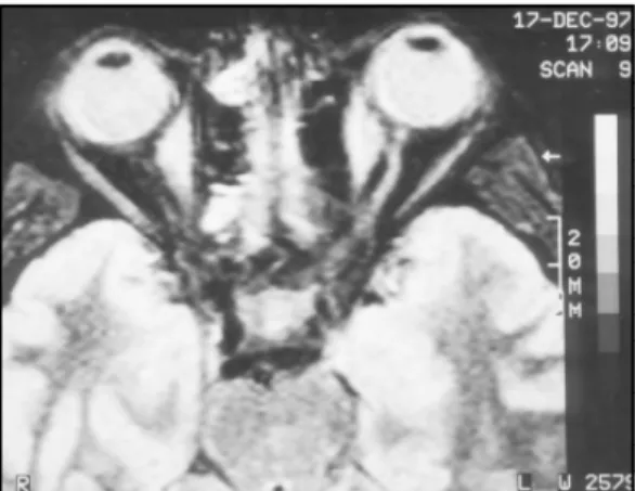
A 38 years old female patient who has had rheumatoid arthritis for the last six years was taking methylprednisolone (10 to 20 mg/day). Her present complaint was headache that started one month ago and worsened day by day, and was accompanied by diplopia. Physical examination showed rheumatoid arthritis hands. Neurological exam showed paralysis of the left rectus medialis muscle and a discrete paresis of the right rectus medialis muscle. MRI showed thickness of the rectus medialis muscle bilaterally (Fig 1). The Cerebrospinal fluid (CSF) was normal. Rheumatoid factor (latex fixation) was positive in the serum. Endocrinological investigation was normal (thyroid function tests). Results were also normal or negative for: serum creatine kinase (CK), aldolase, erythrocyte sedimentation rate, lactic acid dehydrogenase (LDH), serum glutamic oxaloacetic transaminase (SGOT), mucoproteins, serum complement antinuclear antibodies(ANA), C reactive protein, lupus erythematous cells. Diagnosis of orbital myositis was made and methylpredinisolone 80mg dailywas given. Total remission of symptoms occurred.
After approximately two months the patient returned presenting bilateral ophthalmo-plegy that impaired the whole extrinsic mus-culature. MRI, to our surprise, was normal (Fig 2). Electroneuromyography of face and upper members was normal. For the second time we started methylpredinisolone 80 mg daily and the patient improved quite well. However, a paresis of the left rectus medialis muscle remai-ned as a sequel.
DISCUSSION
Orbital myositis was considered a subgroup of the orbital pseudotumor syn-drome in which one or more of the extraocular muscles are primarily infiltra-ted by an inflammatory process. It can be restricted to a single muscle or take the whole orbital musculature causing exoph-thalmus and severe ophthalmoparesys, with uni or bilateral involvement9. Orbital myositis may be acute, subacute or recur-rent. Usually, the acute form has a good response to oral steroids and does not
Fig 1. MRI showing great thickness of the rectus medialis muscle bilaterally (first episode).
176 Arq Neuropsiquiatr 2000;58(1)
leave sequels in spite of its frequent recurrences but, in the same case, if the steroid therapy is discontinued abruptly, myositis can recur17. Most of the cases of orbital myositis do not have their etiology established12,27. Two cases (1.28%) were related to systemic lupus erythematosus28,29, one (0.64%) was related to giant cell myocarditis14, and another one to Crohn disease8 suggesting a possible underlying immunologic mechanism30. Some authors tend to correlate them to previous infectious process and one case have been shown as a paraneoplasic syndrome10. There seem to be a predominance of women against men (2x1) and it is far more frequent in adults9,24.
Rheumatoid arthritis and other connective tissue disorders can cause damage to vasa nervorum and thus produce paralysis of the III, IV, and VI cranial nerves. The frequent vasculopathies that accompany connective tissue disorders can also cause infarcts in the nuclei of the cranial nerves leading to a paralysis of the ocular motility.
Dysthyroid eye disease is a clinical syndrome caused by deposition of mucopolysaccharides and infiltration with chronic inflamatory cells of the orbital tissues, particularly the extraocular muscles. Patients may have clinical or laboratory evidence of thyroid dysfunction, elevated thyroid autoantibodies, or no detectable abnormality outside the orbit.
The primary clinical features are proptosis, lid retraction and lid lag, conjunctival chemosis and episcleral inflammation, and extraocular muscle abnormalities due to restriction of their actions. Resulting symptoms are cosmetic abnormalities, surface irritation which usually responds to artificial tears, and diplopia, which should be treated conservatively (with prisms) in the active stages of the disease and only surgically when the disease has been static for at least 6 months.
Miller-Fisher syndrome can begin by the ophthalmoplegia but ataxia and areflexia are the symptoms that follow. CSF shows increased proteins and normal or slightly elevated cells count. Although we did not collect a second sample of CSF during the second episode, painful ophthalmoplegia was the only symptom our patient had. Tolosa-Hunt syndrome causes painful ophthalmoplegia and involves the III, IV, and VI cranial nerves as well as some trigeminal fibres, and occasionally some periarterial sympathetic fibres and optic nerve. It is known to be caused by periarteritic lesions of the cavernous sinus. Typically it is accompanied by elevation of the erythrocyte sedimentation rate. Myastenic syndrome would seem an improbable diagnosis and electroneu-romyography discarded this possibility.
In our case, in the first episode the, paresis of the ocular convergence had been clearly caused by the thickness of the rectus medialis muscles we found at MRI. However, in the second episode, which was far more severe, MRI was normal.
On the other hand, the narrow relationship between the two episodes strongly suggests that they had been caused by the same pathology. The patient has been followed up for two years and no neoplasia has been detected. In the first case of orbital myositis identified as a paraneoplastic syndrome, neurologic symptoms began more than one year before diagnosis of the lymphoma10. Although the clinical features are frequently suggestive, they are nonspecific and echographic study and/or MRI are required for precise anatomical tissue localization and diagnosis. In some previus articles, ocular muscle biopsy is suggested, but its role is limited to atypical cases or those unresponsive to steroid therapy, particularly to exclude neoplasia9.
We can define our patient after the second episode as having recurrent type orbital myositis. The patient remained with paretic rectus lateralis muscle one year after symptons begun. Like other authors, we believe that gaze restriction is residual.
REFERENCES
177
Arq Neuropsiquiatr 2000;58(1)
2. Casteels I, De Bleecker C, Demaerel P, et al.. Orbital myositis following an upper respiratory tract infection: contribution of high resolution CT and MRI. J Belge Radiol 1991;74:45-47.
3. Cerdá-Nicolás M, Pérez Bacete M, Barberá Alacreu J, Peydró Olaya A. Orbital myositis: an ultrastructural and immunohistochemical study. Arch Neurobiol 1990;53:233-237.
4. Seidenberg KB, Leib ML. Orbital myositis with Lyme disease. Am J Ophthalmol 1990;15:13-16.
5. Thurairajan G, Hope-Ross MW, Situnayake RD, Murray PI. Polyarthropathy, orbital myositis and posterior scleritis: an unusual adverse reaction to influenza vaccine. Br J Rheumatol 1997;36:120-123.
6. Sekhar GC, Lenke BN, Sigh SK. Cystic lesions of the extraocular muscles. Ophthalm Plast Reconstr Surg, 1996;12:199-205. 7. Kormorsky SG, Metha N, Mitsumoto H, Prayson R. Intermitent exotropia associated with rippling muscle disease. J
Neurophthalmol 1995;15:147-151.
8. Durno CA, Ehrlich R, Taylor R, Buncic JR, Hughes P, Griffiths AM. Keeping an eye on Crohn’s disease: orbital myositis as the presenting symptom. Can J Gastroenterol 1997;11:497-500.
9. Hankey GJ, Silbert PL, Edis RH, Nicoll AM. Orbital myositis: a study of six cases. Aust NZ J Med 1987;17:585-591. 10. Harris GJ, Murphy ML, Schmidt EW, Hanson GA, Dotson RM. Orbital myositis as a paraneoplastic syndrome. Arch
Ophthalmol 1994;112:380-386.
11. Orssaud C, Poisson M, Gardeur D. Orbital myositis, recurrence of Whipple’s disease. J Fr Ophtalmol 1992;15:205-208. 12. Slavin ML, Glaser JS. Idiopathic orbital myositis: report of six cases. Arch Ophthalmol 1982;100:1261-1265. 13. Keane JR. Alternating proptosis: a case report of acute orbital myositis defined by the computerized tomographic scan.
Arch Neurol 1977;34:642-643.
14. Klein BR, Hedges TR 3d, Dayal Y, Adelman LS. Orbital myositis and giant cell myocarditis. Neurology 1989;39:988-990. 15. Rohr J, Gauthier G. Acute idiopathic orbital myositis. Rev Neurol (Paris) 1988;144:47-48.
16. Lahoud S, Brownstein S, Barsoum-Homsy M. Inferior oblique myositis presenting as superior oblique muscle palsy. Can J Ophthalmol 1988;23:124-127.
17. Pollard ZF. Acute rectus muscle palsy in children as a result of orbital myositis. J Pediatr 1996;128:230-233.
18. Suoh H, Ibi T, Tashio M, Tanaka F, Mitsuma T, Ohno K. Progressive external ophthalmoplegia and myositis. Intern Med 1993;32:319-322.
19. Dua HS, Smith FW, Singh AK, Forrester JV. Diagnosis of orbital myositis by nuclear magnetic resonance imaging. Br J Ophthalmol 1987;71:54-57.
20. Mannor GE, Rose GE, Moseley IF, Wright JE. Outcome of orbital myositis: clinical features associated with recurrence. Ophthalmology 1997;104:409-414.
21. Mombaerts I, Koornneef L. Current status in the treatment of orbital myositis. Ophthalmology 1997;104:402-408. 22. Shah SS, Lowder CY, Schmitt MA, Wilke WS, Kosmorsky GS, Meisler DM. Low-dose methotrexate therapy for ocular
inflammatory disease. Ophthalmology 1992;99: 1419-1423.
23. Sekhar GC, Mandal AK, Vyas P. Non specific orbital inflammatory diseases. Doc Ophthalmol 1993;84:155-170. 24. Weinstein GS, Dresner SC, Slamovits TL, Kennerdell JS. Acute and subacute orbital myositis. Am J Ophthalmol
1983;96:209-217.
25. Dresner SC, Rothfus WE, Slamovits TL, Kennerdell JS, Curtin HD. Computed tomography of orbital myositis. AJR Am J Roentgenol 1984;143:671-674.
26. Yoritaka A, Kogahara K, Yoshino H, Imai H, Mizuno Y. Clinical and neuroradiological studies on orbital myositis and Tolosa-Hunt syndrome. Rinsho Shinkeigaku 1992;32:593-599.
27. Barnes J. Orbital myositis-a case report. Aust NZ J Ophthalmol 1990;18:251-255.
28. Grimson BS, Simons KB. Orbital inflammation, myositis, and systemic lupus erythematosus. Arch Ophthalmol 1983;101:736-738.
29. Serop S, Vianna RN, Claey M, De Laey JJ. Orbital miositis secondary to systemic lupus erythematous. Acta Ophthalmol (Copenh),1994; 72:520-523.