REVISTA PAULISTA
DE PEDIATRIA
1984-1462/© 2014 Sociedade de Pediatria de São Paulo. Published by Elsevier Editora Ltda. All rights reserved. www.rpped.com.br
DOI of refers to article: http://dx.doi.org/10.1016/j.rpped.2014.05.004
REVIEW ARTICLE
Vitamin D deiciency in pregnancy and its impact
on the fetus, the newborn and in childhood
Marilyn Urrutia-Pereira
a,*, Dirceu Solé
ba Pontiicia Universidade Católica do Rio Grande do Sul (PUC-RS), Porto Alegre, RS, Brazil b Universidade Federal de São Paulo (UNIFESP), São Paulo, SP, Brazil
Received 24 January 2014; accepted 9 May 2014
KEYWORDS
Vitamin D; Pregnancy; Lactation; Fetus; Newborn; Children;
Fetal programming
Abstract
Objective: Vitamin D deiciency (VDD) in pregnant women and their children is an im -portant health problem with severe consequences for the health of both. Thus, the ob-jectives of this review were to reassess the magnitude and consequences of VDD during pregnancy, lactation and infancy, associated risk factors, prevention methods, and to explore epigenetic mechanisms in early fetal life capable of explaining many of the
non-skeletal beneits of vitamin D (ViD).
Data source: Original and review articles, and consensus documents with elevated level of evidence for VDD-related clinical decisions on the health of pregnant women and their
children, as well as articles on the inluence of ViD on epigenetic mechanisms of fetal
programming of chronic diseases in adulthood were selected among articles published on PubMed over the last 20 years, using the search term VitD status, in combination with
Pregnancy, Offspring health, Child outcomes, and Programming.
Data synthesis: The following items were analyzed: ViD physiology and metabolism, risk factors for VDD and implications in pregnancy, lactation and infancy, concentration
cu-toff to deine VDD, the variability of methods for VDD detection, recommendations on
ViD replacement in pregnant women, the newborn and the child, and the epigenetic
inluence of ViD.
Conclusions: VDD is a common condition among high-risk pregnant women and their
chil-dren. The routine monitoring of serum 25(OH)D3 levels in antenatal period is mandatory.
Early preventive measures should be taken at the slightest suspicion of VDD in pregnant women, to reduce morbidity during pregnancy and lactation, as well as its subsequent impact on the fetus, the newborn and the child.
© 2014 Sociedade de Pediatria de São Paulo. Published by Elsevier Editora Ltda. All rights reserved.
*Corresponding author.
PALAVRAS-CHAVE
Vitamina D; Gestação; Lactação; Feto;
Recém-nascido; Criança;
Programação fetal
Deiciência de vitamina D na gravidez e o seu impacto sobre o feto, o recém-nascido e na infância
Resumo
Objetivo: Deiciência de vitamina D (DVD) nas gestantes e seus ilhos é problema de saúde, com consequências graves à saúde de ambos. Assim, esta revisão visou reavaliar a magnitude e as consequências da DVD na gestação, lactação e infância, fatores de risco
associados, métodos de prevenção, além de explorar os mecanismos epigenéticos na vida
fetal capazes de explicar benefícios não-esqueléticos da vitamina D (ViD).
Fonte de dados: Selecionaram-se artigos originais, de revisão e consensos com nível
ele-vado de evidência para decisões clínicas relacionadas à DVD na saúde das gestantes e seus ilhos e artigos sobre sua ação sobre os mecanismos epigenéticos da programaçāo
fetal de doenças crônicas na vida adulta, publicados no PubMed nos últimos 20 anos, empregando-se VitD status, e em combinaçāo com Pregnancy, Offspring health, Child outcomes e Programming.
Síntese dos dados: Abordou-se isiologia, metabolismo, fatores de risco para a DVD e implicações na gravidez, lactação e infância, concentração de corte para deinir DVD, variabilidade de métodos na sua detecção, recomendações sobre a reposição de ViD nas
gestantes, no recém-nascido e na criança, bem como sobre ter as inluências epigenéti
-cas da ViD.
Conclusões: DVD é frequente entre gestantes de alto risco e seus ilhos. Monitorar
rotineiramente os níveis séricos de 25(OH)D3 no período antenatal é imperativo. Medidas preventivas precoces devem ser instituídas à menor suspeita de DVD na gestante, para
reduzir morbidades durante a gestação e a lactação, bem como seu posterior impacto
sobre o feto, o recém-nascido e na infância.
© 2014 Sociedade de Pediatria de São Paulo. Publicado por Elsevier Editora Ltda. Todos os direitos reservados.
Introduction
Vitamin D deficiency (VDD) is identified as a public health
problem in many countries, and pregnant women have been identified as a high-risk group, among whom the prev-alence of VDD ranges between 20 and 40%.1
While it is acknowledged that vitamin D (ViD) supple -mentation is effective in preventing the VDD, many chil-dren are born with this deficiency, raising questions as to how and why VDD affects the pregnancy, the fetus and the newborn’s health.2
The increase in the number of studies on this subject
shows conflicting results on the association between 25(OH)
D levels in pregnancy and adverse effects on maternal and
fetal health, both skeletal and non-skeletal (autoimmune
diseases, cardiovascular diseases, diabetes and certain types
of cancer through “fetal imprinting”).3 Thus, it is advisable to review VDD in mothers and their children so that strate-gies can be implemented to prevent VDD in pregnancy and lactation, in order to prevent its impact on the fetus, the newborn and in childhood, aiming at a possible reduction in the future development of chronic diseases in adulthood.
Method
PubMed database was used for the selection of the arti-cles used in this review, and the evaluated search period comprised the last 20 years. The following search terms
were used: VitD status alone and in combination with the words: Pregnancy, Offspring health, Child outcomes, Programming. Among the identified studies, case reports and intervention studies without randomization were excluded. Original articles, review articles and consensus-es with high level of evidence for clinical decisions relat-ed to VDD regarding the health of pregnant women and their children were selected. Moreover, we selected arti-cles that evaluated the influence of ViD in the epigenetic mechanisms of fetal programming of chronic diseases in adulthood, focusing on the latest works. The most rele-vant articles according to the objectives of this review were therefore chosen.
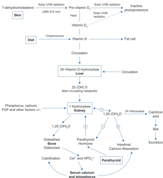
Physiology and vitamin D metabolism
There are two sources of ViD for humans. An exogenous one is provided by the diet in the form of vitamins D2 and D3. In the endogenous production, cholecalciferol
(D3), the main source of ViD, is synthesized in the skin by the action of ultraviolet B (UVB) radiation through the
photolysis of 7-dehydrocholesterol and transformed into
vitamin D3. Sufficient exposure to sunlight or UVB radia
-tion is up to 18IU/cm2 in 3 hours. This process takes place in two phases: the first one occurs in the deep layers of the dermis and consists in the photo conversion of 7-dehydrocholesterol into pre-vitamin D or
In the second phase, there is a chemical isomerization depending on body temperature, and pre-vitamin D slowly and progressively turns into vitamin D3, which has high
affinity for the ViD carrier protein (DBP), and the pre-vita -min D, with lower binding affinity, remains in the skin.4
Upon reaching the skin capillary network, ViD is transport -ed to the liver and binds with DBP, where it starts its met-abolic transformation.4
The two types of ViD undergo complex processing to be metabolically active.5 Initially, the pre-hormone is hydrox-ylated in the liver at the carbon 25 position through the action of vitamin D-25-hydroxylase 1a (1-OHase), which
constitutes an enzyme system dependent on cytochrome
P-450 (CYP27B) present in liver microsomes and mitochon
-dria, and originates 25-hydroxyvitamin D (25(OH)D), the
most abundant circulating form of ViD.4 Its mean blood
con-centration is 20-50ng/mL (50-125nmol/L) and it has an
average life of approximately 3-4 weeks.4 It is estimated that its circulating pool is in dynamic equilibrium with
reserves of 25(OH)D (muscle and adipose tissue), which
makes blood levels a reliable indicator of the state of the ViD reserves in the body.4 Under normal circumstances, the
percentage of conversion into 25(OH)D is low, with a distri -bution of almost 50% in the fat and muscle compartments. When there is excess intake of ViD, most of it is stored in the fatty deposits.4
As 25(OH)D has low biological activity, it is transported
to the kidney where it undergoes the second hydroxyl-ation, and then the active forms are obtained: calcitriol
(1a
-
dihydroxyvitamin D) (1.25(OH)2D) and 24.25-dihy
-droxyvitamin D (24.25(OH)2D), through the respective
action of enzymes 1
-
OHase and vitamin D-
24-hydroxylaseSkin
Diet
Parathyroid
Serum calcium and phosphorus
Vitamin D3
Vitamin D
25 (OH) D Main circulating metabolite
Circulation 7-dehydrocholesterol
Phosphorus, calcium, FGF and other factors +/–
Calcitroic acid
Bile
Intestinal Calcium Absorption
Excretion
Pre-vitamin D3 Inactive
photoprotectors
Fat cell
Circulation Solar UVB radiation
(290-315 nm)
Chylomicrons
1,25 (OH)2D
1,25 (OH)2D
24 hidroxylase –
+
Heat Solar UVBradiation Solar UVB radiation
25-Vitamin D-hydroxylase
Liver
Osteoblast
Bone
Osteoclast
Calcification Ca2+ and HPO 4
2–
Parathyroid Hormone 1 Hydroxylase
Kidney
– +
Figure 1 Synthesis and metabolism of vitamin D as well as its action on the regulation of levels of calcium, phosphorus and bone
metabolism (Adapted from Holick MF.8).
(24
-
OHase) present in mitochondria of cells of the proxi -mal convoluted tubule.5DBP and 25(OH)D are filtered by the glomerulus and
absorbed in the proximal tubule by low-density lipoprotein
receptors, which regulate the uptake of the 25(OH)D-DBP
complex within the tubule cells and the subsequent
hydrox-ylation to 1.25(OH)2D.4
1-OHase is also found in other tissues that express ViD
receptors, such as the placenta, colon, activated
mononu-clear cells and osteoblasts, which could produce 1.25(OH)2D with local autocrine or paracrine function.6
Several factors regulate the levels of 1.25(OH)2D:
1-OHase, whose hydroxylation is activated by the parathor
-mone (PTH), and calcitonin, which is inhibited by serum levels of calcium, phosphorus and 1.25(OH)2D itself, and whose average life is 15 days.6
Blood levels of phosphorus have a direct action, without
the intervention of PTH, and hypophosphatemia increases the production of 1.25(OH)2D.
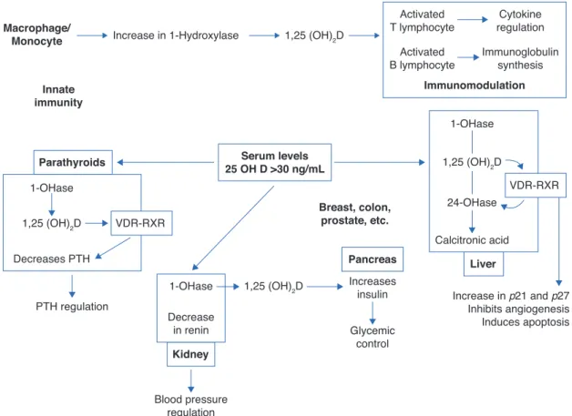
Thus, in addition to the main action of ViD in maintaining physiological levels of calcium and phosphorus capable of allowing metabolism, neuromuscular transmission and bone mineralization, the presence of ViD receptors in bone, bone marrow, cartilage, hair follicle, adipose tissue, adrenal gland, brain, stomach, small intestine, distal kidney tubule,
colon, pancreas (B cells), liver, lung, muscle, activated B
and T lymphocytes, heart cells, vascular smooth muscle cells, gonads, prostate, retina, thymus and thyroid glands has been described, which reinforce such diverse and
important ViD functions (Fig. 2).5 Figure 3 summarizes the mechanisms involved in the control of serum calcium and phosphorus levels.7
Risk factors for ViD deiciency
The main source of ViD for children and adults is exposure to sunlight, so the main cause of VDD is the decrease of its endogenous production. Any factor that affects the
trans-mission of UVB radiation or interferes with its skin penetra
-tion will determine the reduc-tion of 25(OH)D.4 Among these risk factors are:
• Use of sunscreen with a protection factor of 30 reduces
the synthesis of ViD in the skin, above 95%
• Individuals with darker skin have natural sun protection,
as melanin absorbs UVB radiation, and thus they need
3-5 times longer sun exposure to synthesize the same amount of ViD than individuals with light skin
• Skin aging as well as age decrease the capacity of the skin to produce ViD due to lower availability of 7-dehy-drocholesterol
• Skin damage such as burns decrease ViD production
Parathyroids
Pancreas Serum levels
25 OH D >30 ng/mL
Kidney
Liver Macrophage/
Monocyte
Breast, colon, prostate, etc. Innate
immunity
Increase in 1-Hydroxylase
Immunomodulation
Activated T lymphocyte
Cytokine regulation
Activated B lymphocyte
Immunoglobulin synthesis 1,25 (OH)2D
1-OHase
1-OHase
Calcitronic acid
Increase in p21 and p27 Inhibits angiogenesis Induces apoptosis Increases
insulin
Glycemic control
24-OHase 1,25 (OH)2D
1,25 (OH)2D
1-OHase
Decrease in renin 1,25 (OH)2D
Decreases PTH
PTH regulation
Blood pressure regulation VDR-RXR
VDR-RXR
Figure 2 Non-skeletal functions of 1.25-dihydroxyvitamin D (Adapted from Holick MF.8).
VDR, vitamin D receptor; RXR, target region; 1-OHase, 25-hydroxyvitamin D-1a-hydroxylase; 25 (OH) D: 25-hydroxyvitamin D; 1,25
(OH) 2D, 1,25-dihydroxyvitamin D; 24-OHase, 25-hydroxyvitamin D-24-hydroxylase; p21 and p27, genes involved in the control of
• Atmospheric contamination and overcast may act as sun-screen
• The season of the year and the time of the day influence dramatically on the skin production of ViD
The second cause is the reduced intake of ViD, as few
foods contain high quantities of it (blue fish, egg yolks).
The intake of the vitamin can be increased with fortified products such as dairy products, although the amount of ViD they provide may be insufficient for an adequate state of ViD.8
Obesity can also be associated to VDD, because being a fat-soluble vitamin, ViD is sequestered by body fat. Another factor is the malabsorption of fats, as it occurs
with the use of bile acid chelating agents (cholestyr
-amine), in cystic fibrosis, celiac disease and Crohn’s dis -ease, among others.8 Also, anticonvulsants,
glucocorti-coids and drugs used in HIV treatment can lead to VDD by
increasing the hepatic expression of cytochrome P-450
and the catabolism of 25(OH)D. In severe liver failure,
chronic granulomatous disease, certain lymphomas and primary hypoparathyroidism, patients have increased
metabolism of 25(OH)D into 1.25 (OH)2D, and thus a high
risk of VDD.8
Vitamin D deiciency in pregnancy and fetal
programming
During fetal life, the body tissues and organs go through critical development periods that coincide with periods of rapid cell division.9 Fetal programming is a process through
which a stimulus or insult, during a certain development period, would have effects throughout life.10 This term is used to describe the mechanisms that determine fetal adaptation to changes that accompany the gene-environ-ment interaction during specific periods of fetal develop-ment.9
It has been demonstrated that nutritional and environ-mental exposures during these sensitive periods of life may influence fetal growth and the development of physiologi-cal functions of organs and systems. Permanent changes in many physiological processes of this programming can mod-ify the expression patterns of genes, with consequent
influ-ence on phenotypes and functions (epigenetic mecha
-nisms).11
Thus, the closer to fertilization these changes take place, the greater the potential for epigenetic changes and their correspondence in newborns to occur in response to
environmental changes. These changes in placenta/ embryo/fetus provide a plausible explanation for the con -cept of fetal origin of adult diseases.12
It is currently recognized that nutrition in early life and other environmental factors play a key role in the patho-genesis and predisposition to diseases, which seem to prop-agate to subsequent generations. Epigenetic modifications establish a link with the nutritional status during critical periods of development and cause changes in gene expres-sion that can lead to the development of disease pheno-types.13
Recent evidence indicates that nutrients can modify the immune and metabolic programming during sensitive peri-ods of fetal and postnatal development. Thus, modern diet patterns could increase the risk of immune and met-abolic dysregulation associated with the increase of a wide range of noncommunicable diseases.11 Among these nutrients, ViD is emphasized, and its effects on fetal pro-gramming and gene regulation might explain why it has been associated with many health benefits throughout life.8,14,15
There seems to be a window of early development in life that can shape the nature of the immune response in adult-hood, and thus early life factors that predispose individuals to chronic lung disease would not be limited to the post-na-tal period, as evidence indicates that there are intrauter-ine effects such as maternal smoking, diet and ViD that influence the development of the lung and the subsequent development of asthma and chronic obstructive pulmonary disease.16,17
As much of the reprogramming that occurs during child-hood may go unnoticed until adultchild-hood, the better under-standing of the interaction between genetics and epi-genetics in critical time windows of development would improve our capacity to determine individual susceptibil-ity to a wide range of diseases.13 Although these epigene-tic changes appear to be potentially reversible, little is known about the rate and extent of improvements in response to positive environmental changes, including nutrition, and to what extent they depend on the duration of exposure to a deficient maternal environment also remains unknown.18
Thus, it can be observed that, in spite of all this new range of information, maternal nutrition has received lit-Figure 3 Regulatory mechanisms of serum levels of calcium
and phosphorus. Adapted from Ross AC et al.7
PTH, parathyroid hormone; Ca2+, calcium ion; PO 4
3–, phosphate ions; F, increase; f, decrease.
Effects on metabolism
Vitamin D
F 1-a-hydroxylase
activity
F calcitriol
Intestinal reabsorption F of Ca2+ and PO
4 3–
F of Ca2+
F PO43–
f renal PO43–
reabsorption F renal Ca2+
reabsorption Effects on renal function F PTH secretion
F of Ca2+
Normal PO43–
F of Ca2+
F PO43–
F bone turnover
Effects on bone
tle attention in the context of implementation of
effec-tive prevention goals (MDG, Millennium Development Goals). This could be attributed to the lack of a solid and
strong foundation to justify the enormous effort required to improve the nutritional status of all women of repro-ductive age.19 To elucidate the true role of nutritional epi-genetics13,14 in fetal programming of pregnant women, especially those with VDD, would allow the use of effec-tive prevention measures to improve maternal and fetal health and prevent the development of future chronic dis-eases.
Vitamin D and calcium metabolism
in pregnancy
During pregnancy and lactation, significant changes in cal-cium and ViD metabolism occur to provide for the needs required for fetal bone mineralization. In the first
trimes-ter, the fetus accumulates 2-3mg/day of calcium in the
skeleton, which doubles in the last trimester.1
The pregnant woman’s body adapts to the fetal needs and increases calcium absorption in early pregnancy, reach-ing a peak in the last trimester.1 The transfer is counterbal-anced by increased intestinal absorption and decreased urinary excretion of calcium.
Plasma levels of 1.25(OH)2D increase in early pregnancy, reaching a peak in the third trimester and returning to nor-mal during lactation. The stimulus for increased synthesis
of 1.25(OH)2D is unclear, considering that PTH levels do not
change during pregnancy.1
A potent stimulus to placental transfer of calcium and
placental synthesis of ViD is the PTH-related peptide (PTHrP), produced in the fetal parathyroid and placental
tissues, which increases the synthesis of ViD.1 The PTHrP can reach the maternal circulation and it acts through the
PTH/PTHrP receptor in the kidney and bones, being a medi
-ator in the increase of 1.25(OH)2D and helping in the
regu-lation of calcium and PTH levels in pregnancy.1
Other signals involved in the regulation process include prolactin and the placental lactogen hormone, which increase intestinal calcium absorption, reduce urinary
calcium excretion and stimulate the production of PTHrP
and 1.25(OH)2D. Moreover, the increase in the maternal
blood levels of calcitonin and osteoprotegerin protects the mother’s skeleton from excessive calcium resorp-tion.1
Additionally, during lactation, there is a relative estro-gen deficiency, caused by elevated levels of prolactin,
which determines bone resorption and suppression of PTH levels. PTHrP levels are elevated and act as a substitute for PTH, while maintaining urinary calcium absorption and
bone resorption.1
Implications of vitamin D deiciency
in pregnancy
Recent studies emphasize the importance of non-classical roles of ViD during pregnancy and in the placenta and cor-relate VDD in pregnancy with preeclampsia, insulin
resis-tance, gestational diabetes, bacterial vaginosis and increased frequency of cesarean delivery.20
ViD supplementation reduces the risk of preeclampsia. Studies in women with preeclampsia have shown low uri-nary excretion of calcium, low ionized calcium levels, high
levels of PTH and low levels of 1.25(OH)2D.21 An association
between maternal VDD (<50nmol/L) and increased risk of gestational diabetes (OR:2.66, 95% CI: 1.01 to 7.02),22 as well as the fact that VDD is an independent risk factor for bacterial vaginosis in pregnancy23 have also been docu-mented. A recent randomized and controlled study showed
that supplementation with 4,000IU/d during pregnancy was
associated with reduced risk of combined morbidities, such as maternal infections, cesarean section and preterm deliv-ery.21-24
A prospective study showed that cesarean delivery is four
times more common in women with VDD (<37.5nmol/L) when compared to women with normal levels of ViD (OR: 3.84, 95% CI: 1.71 to 8.62).25
Implications of vitamin D deiciency
in lactation and childhood
Adequate levels of ViD are also important for the health of the fetus and the newborn, and poor skeletal mineraliza-tion in utero due to VDD can be manifested in the newborn as congenital rickets, osteopenia or craniotabes.1
Maternal VDD is one of the main risk factors for VDD in childhood, as in the first 6-8 weeks of life newborns depend on the ViD transferred across the placenta while in the womb. This association is linear,26 and the 25(OH)D levels of the newborn correspond to 60-89% of maternal values .2
These levels decrease on the 8th week and, therefore, exclusively breastfed infants have an increased risk of VDD,
as human milk has a low concentration of ViD (approxi
-mately 20-60IU/L; 1.5-3% of the maternal level). This con -centration is not sufficient to maintain optimal levels of ViD, especially when exposure to sunlight is limited,27 and may induce seizures caused by hypocalcemia and dilated cardiomyopathy.1
Observational studies have shown that low levels of ViD during pregnancy and VDD in childhood are related to the increase in other non-skeletal manifestations,2 such as a higher incidence of acute lower respiratory tract infections and recurrent wheezing in the first five years of life.28
Contradictory results are observed in relation to the increased risk of allergic diseases such as asthma, eczema and rhinitis in the presence of VDD.29 However, a cohort study showed increased asthma and eczema among
chil-dren whose mothers had high serum levels of 25(OH)D
during pregnancy.30
Japanese schoolchildren that received ViD
supplementa-tion (1,200IU/d) had a 42% reducsupplementa-tion in the incidence of
type A Influenza.1 A cohort study showed that
supplementa-tion with 2,000IU/d of ViD during the first year of life was
associated with a reduction in the incidence of type I dia-betes during a 30-year follow up.31
Vitamin D deiciency, insuficiency
and suficiency
The cutoff point to define the ViD status based on the
val-ues of 25(OH)D is debatable. There are currently two crite -ria:
• The Committee of the Institute of Medicine (IOM, USA)32
considers values lower than 20ng/mL (50nmol/L) as indi
-cators of VDD, with 10ng/mL (25nmol/L) being consid
-ered severe VDD, and 10-19ng/mL (25-49nmol/L) being considered ViD insufficiency. ViD levels <10ng/mL are
associated with rickets and osteomalacia in adults and
children. Between 10-19ng/mL, there is increased rate
of bone resorption and increased risk of secondary hypo-parathyroidism. Thus, the IOM recommends a threshold
level of 20ng/mL as adequate to maintain bone health at
all ages
• The Endocrine Society (USA) proposes VDD in the pres
-ence of ViD levels inferior to 20ng/mL and ViD insuffi
-ciency between 20-30ng/mL (50-75nmol/L).33 In clini
-cal practice, a patient would have sufficient levels
when the concentration of 25(OH)D were greater than 30ng/mL. Several authors support this concentration
cutoff for musculoskeletal health and mineral
metabo-lism (prevention of rickets and osteomalacia, elevated PTH levels, osteoporotic fractures and falls among the elderly)34
The main differences between the IOM32 and the Endocrine Society33 are the overall health endpoints. The IOM makes recommendations to ensure skeletal health and suggests there is lack of evidence to support recommenda-tions of potential non-skeletal benefits of ViD, considering
that individuals with levels inferior to 20ng/mL are not
deficient, as 97% of individuals with these levels have ade-quate bone health.32
The Endocrine Society33 considers that serum levels
superior to 30ng/mL bring greater benefits to health in general, when compared to a level of 20ng/mL, and that
skeletal health is not guaranteed with levels inferior to
30ng/mL; these data are supported by three supposed
observations:
• The increase in PTH reaches a plateau when serum 25(OH)D is ≥30ng/mL;
• There is a decrease in the risk of fractures in individuals
with levels ≥30ng/ml;
• Calcium absorption is maximal for serum levels of 30ng/
mL.
Detection method
Serum levels of 25(OH)D are the best indicators of ViD
status; however, methodological issues limit comparisons between studies, as well as the adoption of cutoffs to define hypovitaminosis D.4
Considering the method employed, it is important to ask:
1) Does the method quantify the actual level of ViD? and 2) Are these results reproducible and comparable between laboratories?4
Liquid Chromatography Coupled with Mass Spectrometry
(LC-MS/MS) was recommended as the preferred method by
the National Diet and Nutrition Survey.34 In general, all available methods are valid to detect severe VDD. As for moderate deficiency, there is a risk of error, which can be reduced by considering the reference values of each labo-ratory; however, for research studies, the methods employed should be standardized.6
Recommendations
If the main source of ViD comes from sunlight exposure, it is difficult to establish generalized requirements for the intake, especially due to the many variables associated with its deficiency.6
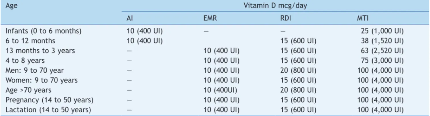
Table 1 shows the different recommended daily doses of
ViD. Although the IOM recommends a daily intake of 200IU
of ViD, this was insufficient to keep concentrations of
25(OH)D above 50nmol/L.35 On the other hand, while
rec-ognizing that the exclusion of habitual sunlight exposure is a risk for VDD, it is unknown what level of exposure is safe and sufficient to maintain adequate levels of ViD.35 Table 2 shows the different contents of ViD-fortified and non-ViD-fortified foods available for consumption in the
United States. In Brazil, such data are scarce and most
often do not reflect the content of all available processed foods.
In most countries, the monitoring of serum levels of
25(OH)D during pregnancy is not performed; however, it is
recommended that women with one or more risk factors for VDD be monitored in early and mid-pregnancy.36 Consequently, the risk of VDD during pregnancy would be reduced, as well as the negative effects on the mother and the fetus; however, the appropriate dose of ViD supplemen-tation for pregnant women to prevent VDD remains unknown.
Few studies have evaluated ViD supplementation in preg-nancy, as well as the optimal levels to be offered. Several factors hinder the observation of an adequate
dose-re-sponse between low 25(OH)D levels and clinical outcomes: lack of data with extreme serum 25(OH)D levels and the wide variety of studied subjects (diversity of location, lat -itude, season, ethnicity, body mass index, type of diet, lifestyle, skin pigmentation, family history of metabolic complications in pregnancy, physical activity and method
used to quantify 25(OH)D).1
A meta-analysis of studies carried out in adults on ViD
supplementation (2,000IU/d) and bone health showed that for each 1IU of vitamin D3 ingested, there is a corre
-sponding increase of 0.016nmol/L in serum levels of 25(OH)D.37 Despite the limited evidence on the effects of ViD supplementation in pregnancy and the outcomes in the mother’s health and perinatal and early childhood
effects, ViD supplementation (800-1,000IU/d) was accom
-panied by a protective effect in newborns with low birth weight.9,38
The Canadian Academy of Pediatrics (CAP)37recommends
supplementation with 2.000IU/d during pregnancy and lac
-tation.38 According to the American College of Obstetricians and Gynecologists,38 in the presence of VDD diagnosed during pregnancy, there should be supplementation with
Studies have shown that maternal exposure during
preg-nancy to serum levels of 25(OH)D superior to 75nmol/L had
no effect on the intelligence and psychological health of the children or on their cardiovascular system, but it could increase the risk of atopic diseases.30
In summary, ViD serum levels in pregnancy are a major concern, and the prevention of VDD in pregnant women and their newborns is vital and urgent.
Vitamin D recommendations for the newborn
and children
The Canadian Academy of Pediatrics defines ViD needs during
the first year of life as 200IU/d for preterm newborns and 400IU/d for other children. However, would the weight gain
observed in the first year of life be accompanied by increased
needs of ViD in a weight-dependent mode?38 Moreover, the CAP also recommends that infants and children be exposed to sunlight for short periods – probably less than 15 minutes.38
The American Academy of Pediatrics recommends that children who are exclusively breastfed should receive
sup-plementation with 400IU/day of ViD soon after birth and
continue to receive during their development up to adoles-cence.5 Concerned about the bone health of premature infants, they recommend biochemical monitoring of their
25(OH)D levels during hospitalization, and recommend 200-400IU/d of ViD, both during hospitalization and after dis -charge.39,40 Recently, the IOM recommended 400IU/d for
children younger than one year and 600IU/d for children
aged between 1-8 years.32
Conclusion
VDD in pregnant women and their children is a major health problem, with potential adverse consequences for overall health. Prevention strategies should ensure the ViD suffi-ciency in women during pregnancy and lactation. Evidence-based interventions to improve maternal and fetal nutri-tion, such as for ViD, are accompanied by a decrease of the impact on the health of their children.41
The ambiguities between the definitions of ViD status, combined with a lack of consistency in recommendations
related to incorporation of routine testing of 25(OH)D lev -els in the prenatal period, especially in women with risk factors for VDD, dose and gestational age for the start of ViD supplementation, universal cutoffs for normal ViD val-ues, lack of education about the benefits of ViD and the need for adequate sunlight exposure represent important barriers to the advance of the implementation of ViD sup-plemental guides, in order to improve this important health problem in pregnant women and their children in the short term. Large-scale studies in different geographical loca-tions are necessary to identify the true role of ViD on the health of pregnant women and the “fetal imprinting” of their children.
Table 1 Dietary reference intakes and maximum tolerable intakes of vitamin D in different stages of life - IOM, 2010.32
Age Vitamin D mcg/day
AI EMR RDI MTI
Infants (0 to 6 months) 10 (400 UI) — — 25 (1,000 UI)
6 to 12 months 10 (400 UI) 15 (600 UI) 38 (1,520 UI)
13 months to 3 years — 10 (400 UI) 15 (600 UI) 63 (2,520 UI)
4 to 8 years — 10 (400 UI) 15 (600 UI) 75 (3,000 UI)
Men: 9 to 70 year — 10 (400 UI) 20 (800 UI) 100 (4,000 UI)
Women: 9 to 70 years — 10 (400 UI) 15 (600 UI) 100 (4,000 UI)
Age >70 years — 10 (400UI) 20 (800 UI) 100 (4,000 UI)
Pregnancy (14 to 50 years) — 10 (400 UI) 15 (600 UI) 100 (4,000 UI) Lactation (14 to 50 years) — 10 (400 UI) 15 (600 UI) 100 (4,000 UI)
AI, intake adequate; EMR, estimated mean requirement; RDI, Recommended daily intake; MIT, maximum tolerable intake level;
between brackets, the corresponding value in international units (IU).
Table 2 Dietary sources of vitamins D2 and D3. 7
Source Vitamin D content
Salmon
Wild – 100g 600-1000UI de Vit D3
Bred in captivity – 100g 100-250UI Vit D3 ou D2
Canned – 100g 300-600UI Vit D3
Canned sardines – 100g ~300 UI Vit D3
Canned Horsetail – 100g ~250 UI Vit D3 Canned tuna – 100g ~230 UI Vit D3
Cod-liver oil (1 tbsp) ~400-1000 UI Vit D3 Fresh Shiitake mushroom – 100g ~100 UI Vit D3 Dried Shiitake mushroom – 100g ~1600 UI Vit D3
Egg yolk ~20 UI Vit D3 ou D2
Fortiied foods
Fortiied milk – 240mL ~100 UI Vit D3
Orange juice – 240mL ~100 UI Vit D3
Conlicts of interest
The authors declare no conflicts of interest.
References
1. Mulligan ML, Felton SK, Riek AE, Bernal-Mizrachi C. Implications
of vitamin D deiciency in pregnancy and lactation. Am J Obstet
Gynecol. 2010;202:429.e1-9.
2. Dawodu A, Wagner CL. Prevention of vitamin D deiciency in
mothers and infants worldwide - a paradigm shift. Paediatr Int
Child Health. 2012;32:3-13.
3. Souberbielle JC, Body JJ, Lappe JM, Plebani M, Shoenfeld Y, Wang
TJ, et al. Vitamin D and musculoskeletal health, cardiovascular disease, autoimmunity and cancer: recomendations for clinical practice. Autoimmun Rev. 2010;9:709-15.
4. Chicote CC, Lorencio FG; Comité de Comunicación de la Sociedad Española de Bioquímica Clínica y Patología Molecular. Vitamina D: una perspectiva actual. Barcelona: Comité de Comunicación de la Sociedad Española de Bioquímica Clínica y Patología Molecular; 2013.
5. Wagner CL, Greer FR; American Academy of Pediatrics Section on Breastfeeding; American Academy of Pediatrics Committee
on Nutrition. Prevention of rickets and vitamin D deiciency in
infants, children, and adolescentes. Pediatrics. 2008;122:1142-52.
6. Masvidal Aliberch RM, Ortigosa Gómez S, Baraza Mendoza MC, Garcia-Algar O. Vitamin D: pathophysiology and clinical
applicability in paediatrcs. An Pediatr (Barc).
2012;77:279.e1-279.e10.
7. Institute of Medicine (US) Committee to Review Dietary
Reference Intakes for Vitamin D and Calcium; Ross AC, Taylor
CL, Yaktine AL, Del Valle HB. Overview of vitamin D. Washington:
National Academies Press; 2011.
8. Holick MF. Vitamin D deiciency. N Engl J Med. 2007;357:266-81.
9. Cunninghan S, Cameron IT. Consequences of fetal growth restriction during childhood and adult life. Curr Obstet Gynecol. 2003;13:212-7.
10. Kim YJ. In utero programming of chronic disease. J Womens
Med. 2009;2:48-53.
11. Amarasekera M, Prescott SL, Palmer SL. Nutrition in early life, imune-programming and allergies: the role of epigenetics. Asian Pac J Allergy Immunol. 2013;31:175-82.
12. McMillen IC, MacLaughlin SM, Muhlhausler BS, Gentili S, Dufield
JL, Morrison JL. Developmental origins of adults health and disease: the role of periconceptional and fetal nutrition. Basic Clin Pharmacol Toxicol. 2008;102:82-9.
13. Jang H, Serra C. Nutrition, Epigenetics, and Diseases. Clin Nutr
Res. 2014;3:1-8.
14. Hossein–Nezhad A, Holick MF. Optimize dietary intake of
Vitamin D: an epigenetic perspective. Curr Opin Clin Nutr Metab Care. 2012;15: 567-79.
15. Hossein–Nezhad A, Holick MF. Vitamin D for health: a global
perspective. Mayo Clin Proc. 2013;88:720-55
16. Hykema MN, Blacuire MJ. Intrauterine effects of maternal
smoking on sensitization asthma and chronic obstructive pulmonary disease. Proc Am Thorac Soc. 2009;6:660-2.
17. Sharma S, Chhabra D, Kho AT, Hayden LP, Tantisira KG, Weiss ST.
The genomic origins of asthma. Thorax. 2014;69:481-4. 18. Hambidge KM, Krebs NF, Westcott JE, Garces A, Goudar SS,
Kodkany BS, et al. Preconception maternal nutrition: a multi-site randomized controlled trial. BMC Pregnancy Childbirth. 2014;14:111.
19. Shrimpton R: Global policy and programme guidance on maternal nutrition: what exists, the mechanisms for providing
it, and how to improve them? Paediatr Perinat Epidemiol. 2012;26 (S1):315-25.
20. Kaushal M, Magon. Vitamin D in pregnancy: a metabolic outlook. Indian J Endocrinol Metab. 2013;17:76-82.
21. Tauield PA, Ales KL, Resnick LM, Druzin ML, Gerther JM, Laragh JH. Hypocalciuria in preeclampsia. N Engl J Med. 1987: 316:
715-8.
22. Zhang C, Qiu C, Hu FB, David RM, van Dam RM, Bralley A, et al.
Maternal plasma 25-hydroxyvitamn D concentration and the risk for gestacional diabetes mellitus. PLoS ONE. 2008;3:e3753. 23. Bodnar LM, Krohn MA, Simhan HN. Maternal vitamin D deiciency
is associated with bacterial vaginose in the irst trimester of
pregnancy. J Nutr. 2009;139:1157-61.
24. Wagner CL, McNeil R, Hamilton SA, Winkler J, Rodriguez Cook
C, Warner G, et al. A randomized trial of vitamin D supplementation in 2 community health center networks in South Carolina. Am J Obstet Gynecol. 2013;208:137e1-13. 25. Merewood A, Mehta SD, Chen TC, Bauchner H, Holick MF.
Association between vitamin D deiciency and primary cesarean
section. J Clin Endocrinol Metab. 2009;94:940-5.
26. Hillman LS, Haddad JG. Human perinatal vitamin D metabolism I: 25- Hydroxyvitamin D in maternal and cord blood. J Pediatr.
1974;84:742-9.
27. American Academy of Pediatrics. Policy statement - ultraviolet radiation: a hazard to children and adolescents. Pediatrics [serial on the Internet]. 2011;104:328 [accessed 29 February
2011]. Available from: http://pediatrics.aappublications.org/ content/early/2011/02/28/peds.2010-3501.abstract.
28. Camargo CA, Ingham T, Wickens K, Thadhani R, Silvers KM, Epton MJ, et al. Cord-blood 25-hydroxyvitamin D levels and risk of respiratory infection, wheezing, and asthma. Pediatrics. 2011;127:180-7.
29. Devereux G, Litonjua AA, Turner SW, Craig LC, McNeill G, Martindale S, et al. Maternal vitamin D intake during pregnancy and early childhood wheezing. Am J Clin Nutr. 2007;85:853-9. 30. Gale CR, Robinson SM, Harvey NC, Javaid MK, Jiang B, Martyn
CN, et al. Maternal vitamin D status during pregnancy and child outcomes. Eur J Clin Nutr. 2008;62:68-77.
31. Hyppönen E, Läärä E, Reunanen A, Järvelin MR, Virtanen SM.
Intake of vitamin D and risk of type 1 diabetes: a birth-cohort study. Lancet. 2001;358:1500-3.
32. Ross AC, Manson JE, Abrams SA, Aloia JF, Brannon PM, Clinton SK, et al. The 2011 report on dietary reference intakes for calcium and vitamin D from the institute of medicine: what clinicians need to know. J Clin Endocrinol Metab. 2011;96:53-8. 33. Holick MF, Binkley NC, Bischoff-Ferrari HA, Gordon CM, Hanley
DA, Heaney RP, et al. Evaluation, treatment, and prevention of vitamin D deiciency: an Endocrine Society clinical practice
guideline. J Clin Endocrinol Metab. 2011;96:1911-30.
34. De la Hunty A, Wallace AM, Gibson S, Viljakainen H, Lamberg-Allardt C, Ashwell M. UK Food Standards Agency
Workshop Consensus Report: the choice of method for measuring 25-hydroxyvitamin D to estimate vitamin D
status for the UK national diet and nutrition survey. Br J
Nutr. 2010;104:612-9.
35. Institute of Medicine (US) Committee to Review Dietary
Reference Intakes for Vitamin D and Calcium; Ross AC, Taylor
CL, Yaktine AL, Del Valle HB. Dietary reference intakes for
calcium and vitamin D. Washington: National Academies Press; 2011.
36. Ponsonby AL, Lucas RM, Lewis RM, Halliday J. Vitamin D status
during pregnancy and aspects of offspring. Nutrients. 2010; 2:389-407.
37. Thorne-Lyman A, Fawzi WW. Vitamin D during pregnancy and maternal, neonatal and infant health outcomes: a systematic review and meta-analysis. Paediatr Perinat Epidemiol. 2012;26
38. Canadian Paediatric Society. Vitamin D supplementation: recommendations for Canadian mothers and infants. Paediatr
Child Health. 2007;12;583-9.
39. ACOG Committee on Obstetric Practice. ACOG committee opinion No. 495: Vitamin D: screening and supplementation during pregnancy. Obstet Gynecol. 2011;118:197-8.
40. Abrams SA; Committee on Nutrition. Calcium and vitamin D requirements of enterally fed preterm infants. Pediatrics. 2013;131:e1676-83.
41. Bhutta ZA, Das JK, Rizvi A, Gaffey MF, Walker N, Horton S, et al.
Evidence-based interventions for improvement of maternal and
child nutrition: what can be done and at what cost? Lancet.