VIEIRA RAC ETAL.
518 REV ASSOC MED BRAS 2014; 60(6):518-519
IMAGE IN MEDICINE
Giant sclerosing papilloma mimicking locally advanced breast
carcinoma
P
APILOMA ESCLEROSANTEGIGANTEMIMETIZANDO CARCINOMA DEMAMA LOCALMENTE AVANÇADORENÉ ALOISIODA COSTA VIEIRA1*, SILVIA MARIA PRIOLIDE SOUZA SABINO2, GUSTAVO ZUCCA MATTHES1,
ANAPAULA HIDEMI UEMA WATANABE2, LUCAS FARIA ABRAHAO-MACHADO3
1Department of Mastology and Reconstructive Surgery, Barretos Cancer Hospital – Pio XII Foundation, Barretos, SP. 2Department of Radiology Breast Image Division, Barretos Cancer Hospital – Pio XII Foundation, Barretos, SP. 3Department of Pathology, Barretos Cancer Hospital – Pio XII Foundation, Barretos, SP.
Study conducted at Barretos Cancer Hospital -- Pio XII Foundation, Barretos, SP
*Correspondence
Address: Rua Antenor Duarte Villela, 1331 Bairro Dr Paulo Prata Postal Code: 14784 – 400 Barretos – SP [email protected] http://dx.doi.org/10.1590/1806-9282.60.06.007
Conflict of interest: none
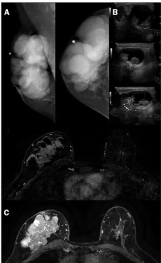
The patient was a 54-year-old Brazilian woman presen-ting a progressive mass in the right breast. The clinical exam showed a 9 x 8 cm tumor and a hardened axillary lymph node. It was clinically considered a T3N1M0 breast tumor (Figure 1A). Mammography showed multiple oval formations, occupying the whole breast (Figure 2A). Ul-trasonography showed the presence of multiple cysts, many of which containing vegetating lesions with inten-se vascular flow (Figure 2B) and abinten-sence of axillary lesion. Magnetic resonance imaging showed multiple oval cysts associated with vegetative lesions, a 4.7cm infiltrative area near the pectoral muscle (Figure 2C), and normal enlar-ged lymph node. As findings highly suspicious of malig-nancy were noted, radiological staging was performed. Abdominal ultrasound, bones scan and thoracic
radio-graphy showed absence of metastatic disease.
FIGURE 1 Clinical exam. (A) lump in right breast; (B) marked area showing clinical localization; (C) open biopsy.
FIGURE 2 Radiologic findings. (A) Mammography: multiple round image in the whole right breast; (B) breast ultrasound: cystic mass with intense vascular flow; (C) MRI findings: infiltrative solid mass with intense early enhancement and washout kinetic curve associated with multiple cysts occupying the right breast. A
A
B
B
C
GIANTSCLEROSINGPAPILLOMAMIMICKINGLOCALLYADVANCEDBREASTCARCINOMA
REV ASSOC MED BRAS 2014; 60(6):518-519 519
The core biopsy showed a benign complex papillary le-sion. Since the radiologic and pathologic divergence did not allow a definitive diagnosis of malignancy, an ultrasound-gui-ded stereotactic needle biopsy was scheduled. The open biopsy was performed in a vegetative intracystic lesion (Figures 1B and 1C) and pathologic findings showed a papillary neopla-sia with atypical cells. Due to atypical findings and the neces-sity to evaluate the whole lesion,1-4 the patient underwent a
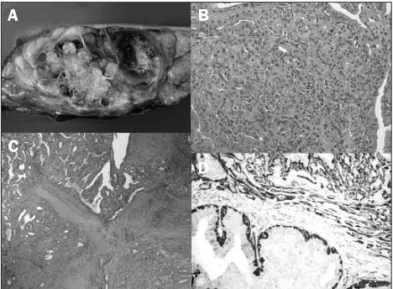
simple mastectomy with sentinel lymph node dissection. No reconstructive surgery was considered because of the lesion size and tumor characteristics. The macroscopic assessment showed a 7.5 x 6.0 cm solid-cyst lesion, with a 3.8 cm solid component (Figure 3A). The microscopy revealed a sclerosing papilloma harboring ductal carcinoma in situ in about 30% of the lesion (Figures 3B and 3C), with free margins and absen-ce of lymph node metastasis. Immunohistochemistry for myoepithelial cells was performed in order to exclude foci of invasion in the periphery of the lesion (Figure 3D).
FIGURE 3 Pathologic findings. Macroscopic finding: (A) Gross examination showed a large solid-cystic tumor. Microscopic findings: (B) papillary neoplasia with sclerotic stroma (HE, 40x); (C) areas containing carcinoma in situ (HE, 200x); (D) Immunohistochemistry positive for myoepithelial cells (Calponin, 200x).
Mammary extensive papillomatous lesions represent a clinical challenge, especially when observing a highly sus-picious malignant tumor based on clinical and radiologi-cal findings.5 As core biopsy showed a benign lesion, an
open biopsy in the vegetative intracystic lesion was perfor-med to improve material sampling. So, when a definitive diagnosis of malignancy cannot be done because of discor-dant findings, sampling limitations of a core biopsy5 or
open biopsy, or limited sensibility of breast images,5,6
re-section of the entire lesion is mandatory1-4,6 due to high
as-sociation with malignancy.1,2,4 The open biopsy was an
at-tempt to improve the pathological results that were hindered by limitation of diagnostic procedures and dis-cordant findings. Also, the indication for diagnostic mas-tectomy,7 as seen in this case, is a fact that must be
tho-roughly discussed with the patient.
R
EFERENCES1. Sydor MK, Wilson JD, Hijaz TA, Massey HD, Paredes ESS. Underestimation of the presence of breast carcinoma in papillary lesions initially diagnosed at core-needle biopsy. Radiology 242(1):58-62.
2. Liberman L, Tornos C, Huzjan R, Bartella L, Morris EA, Dershaw DD. AJRIs surgical excision warranted after benign, concordant diagnosis of papilloma at percutaneous breast biopsy? ARJ. 2006; 186(5):1328-1334.
3. Ueng SH, Mezzeti T, Tavassoli FA. Papillary neoplasms of the breast. Arch Pathol Lab Med. 2009; 133(6):893-907.
4. Youk JH, Kin EK, Kwak JY, Son EJ. Atypical papilloma diagnosed by sonographically guided 14-gauge core needle biopsy of breast mass. AJR. 2010;194(5):1397-492.
5. Lam WWM, Chu MCW, Tang APY, Tse G, Ma TKF. Role of radiologic features in the management of papillary lesions of the breast. AJR 2006;186(5):1322-1327.
6. Eliada R, Chong H, Lylmarni S, Goldberg F,Muradai S. Papilllary lesions of the breast: MRI, ultrasound, and mammographic appearances. AMJ J Roentgenol.2012; 183(2): 264-71.
7. Fenoglio C, Raffaele L. Sclerosing papillary proliferations in the female breast. A benign lesion often mistaken for carcinoma. Cancer 1974; 33(3): 691-700.
A
C
B