188
C
ASER
EPORT1Ph.D., Professor at the Federal University of Juiz de Fora (UFJF), Juiz de Fora/MG, Brazil; Visiting Professor at University La Sapienza, Rome, Italy. 2Specialist title from University La Sapienza, Rome, Italy.
3Professor at University La Sapienza, Rome, Italy.
4Undergraduate student at University La Sapienza, Rome, Italy.
5Ph.D., Professor at the Federal University of Juiz de Fora (UFJF), Juiz de Fora/MG, Brazil
Work conducted at University La Sapienza, Rome, Italy, and the Federal University of Juiz de Fora (UFJF), Juiz de Fora/MG, Brazil.
Electrophysiological findings in Oguchi disease
Alterações eletrofisiológicas na doença de Oguchi
Regina Halfeld Furtado de Mendonça
1, Stefania Abbruzzese
2, Rocco Plateroti
3, Pasquale Plateroti
4, Eliana Lucia
Ferreira
5Os autores declaram não haver conflitos de interesse
Recebido para publicação em: 7/8/2011 - Aceito para publicação em: 24/10/2012
A
BSTRACTTo describe the electrophysiological alterations in a very rare case of Oguchi's disease. A 17-year-old italian girl complaining of night blindness underwent complete ophthalmological exams, including electrophysiological tests. Rod responses were nondetectable in full-field electroretinogram (ERG). The photopic ERG funtions, including the 30 Hz flicker ERG response was normal, while the scotopic b-wave was diminished in amplitude. The electrooculography (EOG) ratios within the normal range were 208% in the right eye and 222% in the left eye. The Mizuo-Nakamura phenomenon was present. The electrophysiological tests are important tools in Oguchi's disease diagnosis. In the present case, it's clear the non correspondance between EOG and ERG. Considering the rod function, the normal EOG ratio contrast with non-detectable rod ERG responses. More studies are necessary to understand the compless electrofuntional mecanism of the disease helping to understand the origin of the light-sensitive component of the EOG.
Keywords: Night blindness; Oguchi's disease; Electroretinography; Electrooculography; Mizuo-Nakamura phenomenon; Case reports
R
ESUMODescrever as alterações eletrofuncionais em um caso raríssimo da Doença de Oguchi. Paciente do sexo feminino, italiana de 17 anos de idade se queixava de cegueira noturna. A resposta escotópica de bastonetes, do ERG era não registrável. A resposta escotópica ao estímulo branco forte demonstrava uma diminuição de amplitude da onda B. As respostas ao flicker de 30Hz e ao EOG eram dentro dos limites da normalidade. Era presente o fenômeno de Mizuo-Nakamura. Os exames eletrofuncionais são muito importan-tes no diagnóstico de certeza da doença de Oguchi. É nítida, no presente caso, a discordância entre EOG e ERG. Considerando a função dos bastonetes, as respostas normais do EOG contrastam com a ausência de respostas dos bastonetes em condições escotópicas no ERG. Mais estudos são necessários para entender o complexo mecanismo eletrofuncional dessa doença e melhor definir a origem dos componentes sensíveis à luz do EOG.
Descritores: Cegueira noturna; Doença de Oguchi; Eletrorretinografia; Eletro-oculografia; Fenômeno de Mizuo-Nakamura; Relatos de casos
189
I
NTRODUCTIONT
here are three forms of stationary night blindness (SNB): congenital stationary night blindness (CSNB), fundus albipunctatus and Oguchi disease.Oguchi disease is a rare congenital, autosomal recessive form of SNB characterised by a peculiar greyish or yellow-greenish discoloration of the fundus1, which reverts to normal
after prolonged dark adaptation (Mizuo-Nakamura phenomenon)2.
Patients with Oguchi disease can be classified into two types depending on the shape of the adaptation curve1. In type I, rod
adaptation is markedly slow. Function is fully recovered after hours of dark adaptation and the curve is normal or slightly altered. In type II there is no rod adaptation, retinal changes are less clear and the Mizuo phenomenon may be absent.
Electrofunctional changes are very important in the definitive diagnosis of Oguchi disease. Electroretinography and electrooculography are useful in understanding the complex mechanism of the disease.
The aim of this paper is to describe the electrofunctional changes in a very rare case of Oguchi disease.
Case report
A white, female, 17-year-old Italian patient presented with complaints of night blindness. Her medical history showed no systemic diseases or previous health conditions. The patient underwent a complete ophthalmic examination including electrofunctional tests at University La Sapienza, Rome, in the year 2007. A full-field electroretinogram was performed using Metrovision equipment (MONPAK3 Moniteur Ophtalmologique - Electrophysiologie visuelle). Standard ISCEV stimuli were used: rod response (dim white light under scotopic conditions), maximum response (strong white light under scotopic conditions), oscillatory potential, cone response (strong white light under photopic conditions), and 30 Hz flicker. A slow-oscillation electrooculogram was then performed using the Biomedica Mangoni BM6000-MAXI device.The patient was pre-adapted for a period of 15 minutes.
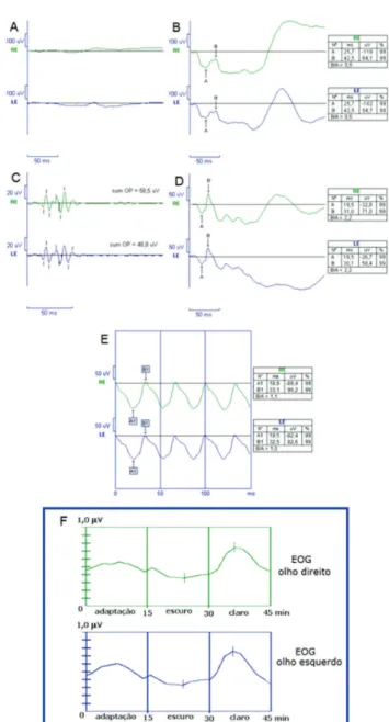
Visual acuity was 20/20 in both eyes. Biomicroscopy and tonometry were normal. On fundus examination, the metallic yellow appearance was noted, with vessels of intense colour (Fi-gure 1). On ERG, rod response (dim white light under scotopic conditions) was unrecordable (Figure 2-A). The maximum response (strong white light under scotopic conditions) showed a decrease in b-wave amplitude (Figure 2-B). The oscillatory potential was altered (Figure C). Photopic response (Figure 2-D) and the response to the 30 Hz flicker (Figure 2-E) were within normal limits. Electrooculography (EOG) response was 208% in the right eye and 222% in the left eye (Figure 2-F). The Mizuo-Nakamura phenomenon was observed.
Figure 1. Retinography showing the metallic yellow appearance with vessels of intense colour, typical of Oguchi disease
Figure 2. A) Rod response (dim white light under scotopic conditions) was unrecordable. B) The maximum response (strong white light under scotopic conditions) showed a decrease in b-wave amplitude (Figure 2-B). C) Oscillatory potential response. D) and E) The photopic response and the response to the 30 Hz flicker were within normal limits. F) EOG within normal limits in both eyes
Rev Bras Oftalmol. 2013; 72 (3): 188-90 Alterações eletrofisiológicas na doença de Oguchi
D
ISCUSSIONOguchi disease is a very rare form of SNB. Few cases have been described in Brazil3. Perhaps the most important feature of
the electrical response in Oguchi disease is the lack of correspondence between visual sensitivity and b-wave amplitu-de; even though the scotopic curve tends to normalise after 4 hours of dark adaptation, b-wave amplitude remains very low 4.
On fundus examination, the typical metallic yellow appearance of Oguchi disease was noted.5,6 The Mizuo-Nakamura
phenomenon was present2,5,6 and rod adaptation was markedly slow,
190
examination may be unrelated to the dark adaptation curve4.
On ERG, rod response (dim white light under scotopic conditions) was unrecordable and the maximum response (strong white light under scotopic conditions) showed a decrease in b-wave amplitude. This is consistent with the literature, which reports that under scotopic conditions, a-wave amplitude is normal and b-wave amplitude is markedly decreased or absent7. Responses
within normal limits to the 30 Hz flicker confirm that the cones are fully functional, which is common in Oguchi disease4.
The EOG within normal limits is in line with some previously-reported cases4, but not with others8. Histological
examination shows that the rods are present and that there is secondary adaptation, indicating that, under certain circumstances, these rods can be functional. It is also believed that the rods may have difficulty converting light energy into nerve impulses due to the lower concentration or absence of photosensitive pigments9. In any case, the normal EOG response
confirms the hypothesis that the light-sensitive phase of the EOG may not depend only on rod function.
Electrofunctional tests are very important in the definitive diagnosis of Oguchi disease. In this case, the mismatch between EOG and ERG was clear. With regard to rod function, normal EOG responses contrast with the absence of rod response in the ERG under scotopic conditions. Further studies are needed to understand the complex electrofunctional mechanism of the disease and to better determine the origin of the light-sensitive components of the EOG.
R
EFERÊNCIAS1. Krill AE. Congenital stationary night-blindness. In: Krill AE. Krill's hereditary retinal and choroidal disease. Maryland, USA: Harper & Row; 1977. v. 2. p. 391-420.
2. Mizuo G. On new discovery in dark adaptation in Oguchi's disease. Acta Soc Ophthalmol Jpn. 1913;17:1148-50.
3. Goulart DG, Myai C, Atique D, Takahashi WY, Aihara T. Doença de Oguchi: relato de caso e revisão bibliográfica. Arq Bras Oftalmol. 2002;65(6):669-73
4. Carr RE, Ripps H. Rhodopsin kinetics and rod adaptation in Oguchi's disease. Invest Ophthalmol Vis Sci. 1967;6(4):426-36.
5. Yamamoto S, Hayashi M, Takeuchi S, Shirao Y, Kita K, Kawasaki K. Normal S cone electroretinogram b-wave in Oguchi's disease. Br J Ophthalmol. 1997;81(12):1043-5. Comment in Br J Ophthalmol. 1997;81(12):1027.
6. Yoshii M, Murakami A, Akeo K, Nakamura A, Shimoyama M, Ikeda Y, et al. Visual function and gene analysis in a family with Oguchi's disease. Ophthalmic Res. 1998;30(6):394-401.
7. Carr RE, Gouras P. Oguchi's disease. Arch Ophthalmol. 1965;73:646-56. 8. Miyake Y, Horiguchi M, Suzuki S, Kondo M, Tanikawa A. Electro-physiological findings in patients with Oguchi's disease. Jpn J Ophthalmol. 1996;40(4):511-9.
9. François J, Verriest G, De Rouck A. La maladie d'Oguchi. Ophthalmologica. 1956;131(1):1-40.
Corresponding author:
Regina Halfeld Furtado de Mendonça Universidade Federal de Juiz de Fora Faculdade de Educação Física e Desportos Grupo de Pesquisa em Inclusão,
Movimento e Ensino à Distância Campus Universitário S/N Bairro: Martelos
CEP: 36036-900 - Juiz de Fora (MG), Brazil. Tel: 55(32)2102-3283
E-mail: [email protected]
Rev Bras Oftalmol. 2013; 72 (3): 188-90