Although most postoperative complications are related to the graft, the native lung can also be a source of complications, such as infec-tious processes and pneumothorax.(3,4) A unique complication, which occurs particularly in COPD patients and can cause graft dysfunction, is native lung hyperinflation.(5,6)
Native lung hyperinflation is a common radiological finding in the immediate postopera-tive period, usually resolves within a few months and is not associated with worse disease progres-sion.(7) However, progressive hyperinflation can cause mediastinal shift with compression, restric-tion and disorders of ventilarestric-tion of the graft.
Introduction
Lung transplantation is a well-established treatment for patients with advanced COPD. Currently, COPD is the main indication for lung transplantation, accounting for 36% of all indi-cations in the literature.(1) Worldwide, most of the procedures performed consist of double lung transplantation. However, single lung transplan-tation still accounts for 32%. In this category, COPD is responsible for 50% of indications. In Brazil, there are no centralized data on indica-tions or types of procedures performed, although, nationwide, 50% of the procedures consist of double lung transplantation, and COPD also constitutes the leading cause of transplantation, corresponding to 28.2%.(2)
Lung hyperinflation after single lung
transplantation to treat emphysema*
Hiperinsuflação pulmonar após transplante unilateral por enfisema
Marcos Naoyuki Samano, Jader Joel Machado Junqueira, Ricardo Henrique de Oliveira Braga Teixeira, Marlova Luzzi Caramori,
Paulo Manuel Pêgo-Fernandes, Fabio Biscegli Jatene
Abstract
Despite preventive measures, lung hyperinflation is a relatively common complication following single lung transplantation to treat pulmonary emphysema. The progressive compression of the graft can cause mediastinal shift and respiratory failure. In addition to therapeutic strategies such as independent ventilation, the treatment consists of the reduction of native lung volume by means of lobectomy or lung volume reduction surgery. We report two cases of native lung hyperinflation after single lung transplantation. Both cases were treated by means of lobectomy or lung volume reduction surgery.
Keywords: Lung transplantation; Postoperative complications; Pulmonary emphysema; Pneumonectomy.
Resumo
Apesar das medidas de prevenção, a hiperinsuflação pulmonar é uma complicação relativamente comum após transplantes unilaterais por enfisema. Quando progressiva, pode comprimir o pulmão transplantado, gerando desvio mediastinal e insuficiência respiratória. Além de estratégias terapêuticas como a ventilação independente, o tratamento consiste na redução volumétrica do pulmão nativo, seja por meio de cirurgia redutora, seja por lobec-tomia. São relatados dois casos de hiperinsuflação do pulmão nativo após transplante pulmonar, tratados com redução volumétrica do pulmão por meio de lobectomia ou cirurgia redutora.
Descritores: Transplante de pulmão; Complicações pós-operatórias; Enfisema pulmonar; Pneumonectomia.
* Study carried out at the Instituto do Coração, Hospital das Clínicas da Faculdade de Medicina da Universidade de São Paulo – InCor/ HC-FMUSP, Heart Institute/University of São Paulo School of Medicine Hospital das Clínicas – São Paulo, Brazil.
Correspondence to: Marcos Naoyuki Samano. Avenida Dr. Enéas Carvalho Aguiar, 44, 2º andar, bloco 2, sala 9, Cerqueira César, CEP 05403-000, São Paulo, SP, Brasil.
Tel 55 11 3069-5248. Fax: 55 11 3069-5351. E-mail: marcos.samano@incor.usp.br Financial support: None.
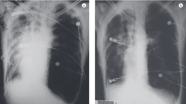
After 7 months on a waiting list, the patient underwent single right lung transplantation, in which the duration of ischemia was 210 min. Immunosuppression was performed with azathio-prine, cyclosporine and methylprednisolone. The patient was referred to the ICU and remained intubated for 5 days. However, he developed respiratory failure and required reintubation. There was progressive native lung hyperinfla-tion, with mediastinal shift and worsening of the respiratory pattern. Independent ventilation was employed but did not result in radiological or clinical improvement (Figure 1).
The patient underwent left lung volume reduction surgery to prevent compression of the graft. A linear cutting stapler was used, and multiple wedge resections were performed. Approximately 20% of the parenchyma in the lung apex was removed. The mechanical suture line was reinforced by interposing bovine pericardial tissue, as previously described by Cooper.(8)
There was radiological improvement, with expansion of the graft and normalization of the diaphragmatic contour, as well as improvement in pulmonary function (Figure 1). However, the patient developed dehiscence of the anastomosis of the anterior wall of the right main bronchus, together with bronchopleural fistula. The conse-Here, we report two cases of native lung
hype-rinflation after single lung transplantation. Both cases were associated with progressive pulmo-nary dysfunction and were treated by means of procedures to reduce native lung volume.
Case report
Case 1
A 41-year-old male smoker (20 pack-years) presented with a six-year history of progressive dyspnea, a diagnosis of centrilobular emphy-sema and a history of right bullectomy. The emphysema worsened, and the patient needed home oxygen therapy. In addition, the patient experienced dyspnea on minimal exertion (func-tional class IV, in accordance with the New York Heart Association criteria).
Physical examination revealed satisfactory general health, weight loss (BMI, 13.4 kg/m2), gastrostomy (for the purpose of feeding), dyspnea and diffuse wheezing upon pulmonary auscul-tation. Spirometry showed an FEV1 of 15% and an FVC of 42%. Quantitative perfusion scinti-graphy presented values of 33% and 67% in the left and right lungs, respectively. Radiographic and tomographic findings were consistent with severe bilateral emphysema.
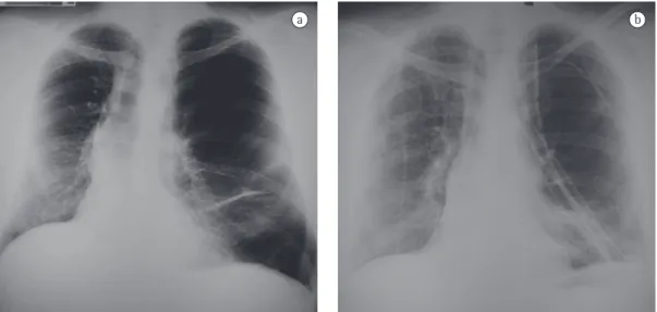
the breathing pattern. Postoperative pulmonary function testing showed an FEV1 of 81%, FVC of 76%, TLC of 99% and RV of 150%. Four years after surgery, the patient was in good health and was fully able to perform his habitual activities (Figure 2).
Discussion
Single lung transplantation is a well-esta-blished therapeutic option for the treatment of respiratory failure resulting from emphysema.(9) This procedure makes it possible to maximize the use of organs (a single donor for two recipients), as well as allowing relatively minor surgical trauma and satisfactory postoperative pulmo-nary function. However, maintaining the native lung can cause hyperinflation, with consequent mediastinal shift and compression of the graft.
One group of authors conducted a retros-pective analysis of 27 patients undergoing single lung transplantation for emphysema and defined two risk factors for the occurrence of native lung hyperinflation. Hyperinflated patients with severe COPD (FEV1 < 15% and RV > 200% of predicted) are candidates for hyperinflation. The second factor is associated with acute graft dysfunction, there being a need for prolonged mechanical ventilation and high positive end-expiratory pressure. Patients with pulmonary hypertension are also at increased risk for developing primary graft dysfunction, quent empyema and sepsis led to his death on
postoperative day 16.
Case 2
A 46-year-old male nonsmoker presented with a nine-year history of progressive dyspnea and a diagnosis of talcosis and pulmonary emphy-sema. The patient was being treated with home oxygen therapy, with maximal oxygen uptake of 19 mL/kg per min. Pulmonary function testing showed an FEV1 of 13%, FVC of 25%, TLC of 110% and RV of 298%. After 2 months on a waiting list, the patient underwent single right lung transplantation, in which the duration of ischemia was 240 min. Immunosuppression was performed with cyclosporine, azathioprine and prednisone. The patient remained intubated for 28 h and stayed in the hospital for 17 days.
Three years after transplantation, there was progressive deterioration of pulmonary function, with clear signs of lung hyperinflation (FEV1, 69%; FVC, 71%; TLC, 136%; and RV, 278%). At the time, the patient experienced dyspnea on minimal exertion.
Reduction of native lung volume by means of left lower lobectomy was chosen because scinti-graphy revealed lower perfusion in the left lung base (22%) than in the right lung base (78%). In addition, we considered the lower likelihood of prolonged air leak related to lobectomy. The patient underwent lobectomy. There was a favo-rable response and immediate improvement in
a b
Despite the adoption of preventive measures in single lung transplantation for COPD, such as preferential right lung transplantation and early extubation, lung hyperinflation is a rela-tively common and unpredictable complication. Although there is a tendency toward double transplantation, even in COPD patients, single transplantation remains the treatment of choice for COPD worldwide, and transplantation teams should be on the alert for this type of problem. In our experience, the result of the reduction of native lung volume by means of lung volume reduction surgery or lobectomy was satisfactory in both cases. The first patient had complications, but those were unrelated to the procedure.
Other methods still in the experimental phase have been considered for patients with diffuse pulmonary emphysema and for native lungs of patients undergoing lung transplantation. Chief among those methods is the creation of spira-cles, by installing an intrapulmonary tube that exits the lung through a small opening in the chest wall, allowing direct air exchange between the pulmonary parenchyma and the environ-ment.(13)
Although there is a tendency toward double lung transplantation, COPD continues to be a major indication for single lung transplanta-tion. Therefore, the incidence of native lung hyperinflation remains a morbidity factor in this patient population. Despite preventive mecha-nisms and ventilation strategies, hyperinflation can develop, and the reduction of native lung volume by means of lobectomy or lung volume reduction surgery can be effective in the treat-ment of this type of complication.
References
1. Christie JD, Edwards LB, Aurora P, Dobbels F, Kirk R, Rahmel AO, et al. Registry of the International Society for Heart and Lung Transplantation: twenty-fifth official adult lung and heart/lung transplantation report--2008. J Heart Lung Transplant. 2008;27(9):957-69.
2. Samano MN, Minamoto H, Junqueira JJ, Yamaçake KG, Gomes HA, Mariani AW, et al. Bronchial complications following lung transplantation. Transplant Proc. 2009;41(3):921-6.
3. Murray JG, McAdams HP, Erasmus JJ, Patz EF Jr, Tapson V. Complications of lung transplantation: radiologic findings. AJR Am J Roentgenol. 1996;166(6):1405-11. 4. Venuta F, Boehler A, Rendina EA, De Giacomo T, Speich
R, Schmid R, et al. Complications in the native lung after single lung transplantation. Eur J Cardiothorac Surg. 1999;16(1):54-8.
requiring mechanical ventilation with positive end-expiratory pressure, which can lead to acute native lung hyperinflation.
The diagnosis of acute lung hyperinfla-tion is based on a group of radiological signs (mediastinal shift and flattening of the ipsila-teral diaphragm), as well as on clinical signs of hemodynamic or respiratory instability, there being need for different ventilation strate-gies (use of nitric oxide or use of independent lung ventilation). Since there is a difference in compliance between the native lung and the graft, hyperinflation results in severe ventilation/ perfusion mismatch, with preferential venti-lation of the native lung and perfusion of the graft, causing respiratory failure and hemody-namic instability in the postoperative period. To prevent this from happening, early extubation is recommended whenever possible. One group of authors(10) recommended certain measures to prevent native lung hyperinflation: giant bullae should be resected; the VC should be greater in the donor lung than in the recipient lung; in cases of homogenous emphysema with symme-tric distribution, right transplantation should be preferred, allowing expansion of the native lung toward the diaphragm rather than toward the mediastinum; early extubation should be attempted and positive end-expiratory pressure in the native lung should be avoided; and, after transplantation, the patient should be placed in the lateral decubitus position on the native lung side. Once hyperinflation has become establi-shed, independent ventilation with the use of a double-lumen orotracheal tube is one alterna-tive. Minimal ventilation (5 mL/kg, 2-6 breaths/ min) should be maintained in the native lung. However, the need for frequent bronchoscopies to maintain the orotracheal tube in an adequate position complicates this ventilation strategy.
emphysema: predictors for native lung hyperinflation. J Heart Lung Transplant. 1998;17(2):192-201.
10. Le Pimpec-Barthes F, Debrosse D, Cuenod CA, Gandjbakhch I, Riquet M. Late contralateral lobectomy after single-lung transplantation for emphysema. Ann Thorac Surg. 1996;61(1):231-4.
11. Kapelanski DP, Anderson MB, Kriett JM, Colt HG, Smith CM, Mateos M, et al. Volume reduction of the native lung after single-lung transplantation for emphysema. J Thorac Cardiovasc Surg. 1996;111(4):898-9.
12. Fitton TP, Bethea BT, Borja MC, Yuh DD, Yang SC, Orens JB, et al. Pulmonary resection following lung transplantation. Ann Thorac Surg. 2003;76(5):1680-6. 13. Saad Junior R, Dorgan Neto V, Botter M, Stirbulov R,
Rivaben JH, Gonçalves R. Therapeutic application of collateral ventilation with pulmonary drainage in the treatment of diffuse emphysema: report of the first three cases. J Bras Pneumol. 2009;35(1):14-9. 5. Malchow SC, McAdams HP, Palmer SM, Tapson
VF, Putman CE. Does hyperexpansion of the native lung adversely affect outcome after single lung transplantation for emphysema? Preliminary findings. Acad Radiol. 1998;5(10):668-93.
6. Mal H, Brugière O, Sleiman C, Rullon I, Jebrak G, Groussard O, et al. Morbidity and mortality related to the native lung in single lung transplantation for emphysema. J Heart Lung Transplant. 2000;19(2):220-3.
7. Weill D, Torres F, Hodges TN, Olmos JJ, Zamora MR. Acute native lung hyperinflation is not associated with poor outcomes after single lung transplant for emphysema. J Heart Lung Transplant. 1999;18(11):1080-7.
8. Cooper JD. Technique to reduce air leaks after resection of emphysematous lung. Ann Thorac Surg. 1994;57(4):1038-9.
9. Yonan NA, el-Gamel A, Egan J, Kakadellis J, Rahman A, Deiraniya AK. Single lung transplantation for
About the authors
Marcos Naoyuki Samano
Attending Physician. Department of Thoracic Surgery, Instituto do Coração, Hospital das Clínicas da Faculdade de Medicina da Universidade de São Paulo – InCor/HC-FMUSP, Heart Institute/University of São Paulo School of Medicine Hospital das Clínicas – São Paulo, Brazil.
Jader Joel Machado Junqueira
Resident in General Surgery. Hospital das Clínicas da Faculdade de Medicina da Universidade de São Paulo – HC-FMUSP, University of São Paulo School of Medicine Hospital das Clínicas – São Paulo, Brazil.
Ricardo Henrique de Oliveira Braga Teixeira
Attending Physician. Department of Pulmonology, Instituto do Coração, Hospital das Clínicas da Faculdade de Medicina da Universidade de São Paulo – InCor/HC-FMUSP, Heart Institute/University of São Paulo School of Medicine Hospital das Clínicas – São Paulo, Brazil.
Marlova Luzzi Caramori
Attending Physician. Department of Pulmonology, Instituto do Coração, Hospital das Clínicas da Faculdade de Medicina da Universidade de São Paulo – InCor/HC-FMUSP, Heart Institute/University of São Paulo School of Medicine Hospital das Clínicas – São Paulo, Brazil.
Paulo Manuel Pêgo-Fernandes
Associate Professor. Department of Cardiorespiratory Diseases, Faculdade de Medicina da Universidade de São Paulo – FMUSP, University of São Paulo School of Medicine – São Paulo, Brazil.
Fabio Biscegli Jatene