* Study carried out at the Pavilhão Pereira Filho of the Irmandade Santa Casa de Misericórdia de Porto Alegre – ISCMPA, Santa Casa Hospital at Porto Alegre – Porto Alegre, Brazil.
1. Thoracic Surgeon on the Lung Transplant Team. Irmandade Santa Casa de Misericórdia de Porto Alegre – ISCMPA, Santa Casa Hospital at Porto Alegre – Porto Alegre, Brazil.
2. Adjunct Professor of Thoracic Surgery. Fundação Faculdade Federal de Ciências Médicas de Porto Alegre – FFFCMPA, Federal Foundation School of Medical Sciences of Porto Alegre – Porto Alegre, Brazil.
3. Clinician on the Lung Transplant Team. Irmandade Santa Casa de Misericórdia de Porto Alegre – ISCMPA, Santa Casa Hospital at Porto Alegre – Porto Alegre, Brazil. 4. Pulmonologist on the Lung Transplant Team. Irmandade Santa Casa de Misericórdia de Porto Alegre – ISCMPA, Santa Casa Hospital at Porto Alegre – Porto Alegre, Brazil.
5. Assistant Professor of Thoracic Surgery. Fundação Faculdade Federal de Ciências Médicas de Porto Alegre – FFFCMPA, Federal Foundation School of Medical Sciences of Porto Alegre – Porto Alegre, Brazil.
6. Adjunct Professor of Pulmonology. Universidade Federal do Rio Grande do Sul – UFRGS, Federal University of Rio Grande do Sul – Porto Alegre, Brazil. 7. Thoracic Surgeon. Santo Antônio Children’s Hospital, Porto Alegre, Brazil.
Correspondence to: Cristiano Feijó Andrade. Santa Casa de Porto Alegre, Hospital da Criança Santo Antônio, Avenida Independência, 155, 3º andar, Centro, CEP 90020-090, Porto Alegre, RS, Brasil
Tel 55 51 32148674. E-mail: [email protected] Submitted: 13 May 2007. Accepted, after review: 6 August 2007.
Spencer Marcantônio Camargo1, José de Jesus Peixoto Camargo2,
Sadi Marcelo Schio3, Leticia Beatriz Sánchez4, José Carlos Felicetti5,
José da Silva Moreira6, Cristiano Feijó Andrade7
Abstract
Objective: To evaluate post-operative complications in living lobar lung transplant donors. Methods: Between September of 1999 and
May of 2005, lobectomies were performed in 32 healthy lung transplant donors for 16 recipients. The medical charts of these donors were retrospectively analyzed in order to determine the incidence of post-operative complications and alterations in pulmonary function after lobectomy. Results: Twenty-two donors (68.75%) presented no complications. Among the 10 donors presenting complications, the most
frequently observed complication was pleural effusion, which occurred in 5 donors (15.6% of the sample). Red blood cell transfusion was necessary in 3 donors (9.3%), and 2 donors underwent a second surgical procedure due to hemothorax. One donor presented pneumothorax after chest tube removal, and one developed respiratory infection. There were two intra-operative complications (6.25%): one donor required bronchoplasty of the middle lobe; and another required lingular resection. No intra-operative mortality was observed. Post-operative pulmonary function tests demonstrated an average reduction of 20% in forced expiratory volume in one second (p < 000.1) compared to pre-operative values. Conclusions: Lobectomy in living lung transplant donors presents high risk of post-operative complications and
irreversible impairment of pulmonary function. Careful pre-operative evaluation is necessary in order to reduce the incidence of complications in living lobar lung transplant donors.
Keywords: Lung transplantation; Living donors; Pneumonectomy; Postoperative complications.
Resumo
Objetivo: Avaliar as complicações pós-operatórias imediatas de doadores vivos de lobos pulmonares para transplante. Métodos: Entre setembro de 1999 e maio de 2005 foram realizadas lobectomias em 32 doadores saudáveis para transplante pulmonar em 16 receptores. Os prontuários médicos destes doadores foram analisados retrospectivamente para verificar a incidência de complicações pós-operatórias e as alterações da função pulmonar após a lobectomia. Resultados: Vinte e dois doadores (68,75%) não apresentaram complicações. Entre os 10 casos que apresentaram alguma complicação o derrame pleural foi a mais freqüente, ocorrendo em 5 deles (15,6% da amostra). Três doadores (9,3%) necessitaram de transfusão de hemácias e, em 2 casos, foi necessária nova intervenção cirúrgica devido a hemotórax. Um doador apresentou pneumotórax após a retirada do dreno de tórax e houve um caso de infecção respiratória. Ocorreram duas intercor-rências intra-operatórias (6,25%): em um doador foi realizada broncoplastia do lobo médio; em outro, foi necessária a ressecção da língula. Não houve mortalidade cirúrgica nesta série. As provas de função pulmonar do pós-operatório demonstraram uma redução média de 20% no volume expiratório forçado no primeiro segundo (p < 000,1), em comparação com os valores verificados antes da cirurgia. Conclusões: A lobectomia em doadores pulmonares vivos para transplante apresenta elevado risco de complicações pós-operatórias e resulta em perda definitiva da função pulmonar Uma cuidadosa avaliação pré-operatória faz-se necessária para reduzir a incidência de complicações nos doadores vivos de lobos pulmonares para transplante.
has been described, albeit without mortality.(6,7) The
present study is the first in the Brazilian literature aimed at demonstrating the results and difficul-ties encountered by a lung transplantation team in managing the treatment of living lobar lung trans-plant donors.
Methods
This study was approved by the Ethics in Research Committee of the Santa Casa Hospital at Porto Alegre (Protocol no. 1.114/05), located in Porto Alegre, Brazil.
Between September of 1999 and May of 2005, 32 lobectomies were performed for lung trans-plant into 16 recipients. Surgical procedures were performed in the Pereira Filho Ward of the Santa Casa Hospital at Porto Alegre. Medical charts, including surgical and anesthetic history, were analyzed for all 32 donors. Of the 32 donors, 28 (87.5%) were monitored after hospital discharge for a period of 10 to 78 months (mean period of 36 months). Four of the donors (12.5%) were lost to follow-up due to a change of address and could not be monitored. Even in cases of failure involving the death of the recipient, follow-up evaluations of donors were made through periodic visits or telephone contact. At approximately 60 days after the surgery, the donors were re-evaluated through additional chest X-rays. The 28 donors were submitted to postop-erative spirometry testing within 3 to 68 months after the procedure (mean, 27 months; median, 24 months).
Sixteen patients between 6 and 29 years of age (mean, 12.4 years) underwent bilateral lobar lung transplantation. Recipients were evaluated by a multidisciplinary team composed of the psycholo-gists, nutritionists, physiotherapists, social workers, pulmonologists and intensivists on the trans-plantation team. Transtrans-plantation was indicated for patients in an advanced stage of disease and with little chance of surviving until a compatible deceased donor could be located. Transplantation was indicated in cases of cystic fibrosis (8 cases) and bronchiolitis obliterans (8 cases). All recipients were in use of supplemental oxygen when transplanta-tion was recommended, and there were no cases of second transplantation in this group.
The potential donors were first evaluated as to blood type and lung size in relation to the
respec-Introduction
Due to the high mortality of patients on lung transplant waiting lists and the lack of deceased donors, the use of living lung transplant donors has come to be viewed as a realistic option.
Since the time of the first successful isolated lung transplantation,(1) the number of conditions
for which lung transplant is recommended and the number of patients on waiting lists have increased progressively. However, the number of deceased donors has not increased in the same proportion. This difference between the number of donors and the number of recipients gives a patient on a lung transplant waiting list a better chance of dying than of receiving a viable lung.(2) As an alternative
to obtaining viable deceased donors, using organs from living donors has become a possibility.(3) With
this new source of organs, an increase in kidney, liver and lung transplantations has been observed. In the case of kidney transplantation, it corresponds to more than 50% of the procedures performed every year.(4,5) In particular, the use of living donors
for different types of transplantation creates an unique scenario in medicine, in which the treatment of a patient with an advanced disease also exposes a healthy individual (the donor) to a considerable risk of morbidity and mortality.(6,7) Recently, this scenario
has been widely discussed in literature, principally after the news of deaths among living donors of kidneys and livers for transplantation.(8)
The use of living donor lung lobes for a trans-plantation in two children with terminal lung disease was first described in 1992.(3) Subsequently,
the use of living donors in lung transplantation continued, stimulated principally by the fact that, due to a lack of deceased donors, mortality among patients on waiting lists was high.(9) This modality
of treatment has recently been used in various facil-ities as an efficacious alternative for patients with progressive disease, who might die while waiting for a compatible deceased donor.(10-15) As a
peculi-arity of this technique, 2 healthy donors are needed, preferably relatives compatible with the recipient in terms of blood type and organ size.(9) Each of
being implanted, another team initiated the lobec-tomy on the second donor.
The surgical technique used in the lobectomy of the donors was first described by Starnes et al.(9)
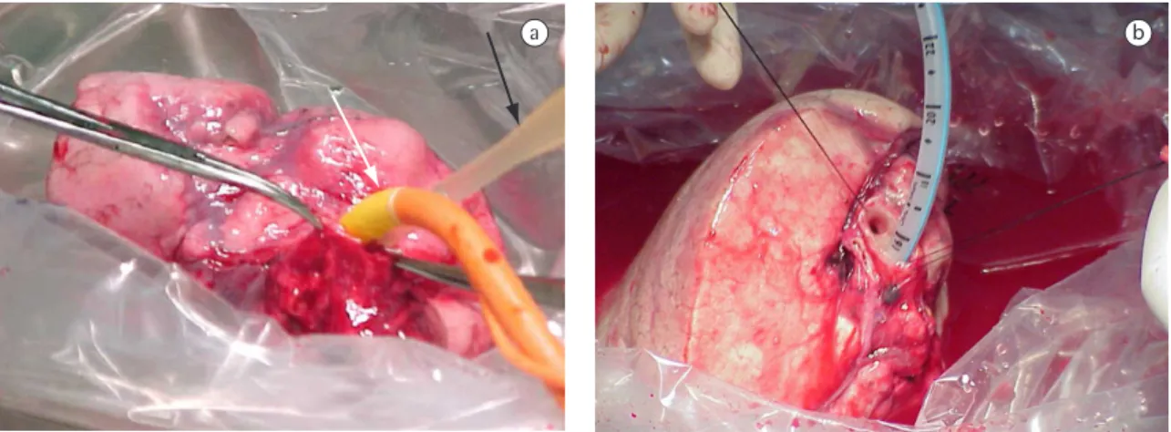
The donors were anesthetized and intubated with double-lumen tubes for single-lung ventilation. In each donor, an epidural catheter was inserted for post-operative analgesia, after which a poste-rolateral thoracotomy was performed. After the cavity had been inspected and the pulmonary liga-ment freed, the pericardium was opened in order to expose the lower pulmonary vein, together with a margin of left atrial muscle. The fissures, when incomplete, were freed using mechanical staplers, allowing access to the pulmonary artery and dissec-tion of the lower lobe artery, with special attendissec-tion to the branches of the upper segment and left middle lobe, as well as to the arterial branches of the left lingula (Figure 1). After vessel and bronchus dissection, 5000 IU of intravenous heparin were given. After 3 minutes of pulmonary ventilation, vascular clamps were placed on the lower pulmo-nary artery and left atrium of the recipient. After tive recipients. All potential donors evaluated were
relatives of the recipients. The compatible donors were submitted to evaluation by the multidis-ciplinary teams and were informed regarding the planned procedures, as well as regarding the morbidity and mortality risks involved. In the case of the 3 underage donors (9.3%), authorization was obtained from the juvenile court.
The selected donors were then clinically evalu-ated and submitted to simple X-ray and computed tomography scan of the chest with volume measurement of lower lobes, together with elec-trocardiogram, Doppler echocardiogram, renal function tests, hepatic function tests and serology, as well as evaluation of pulmonary function using spirometry.(16-18) In order to analyze the
bron-chial tree, all potential donors were submitted to fiberoptic bronchoscopy, which, in one case, deter-mined the choice of the side of the lobectomy. Also, for the 10 first transplantations, pulmonary arte-riograms were performed in the donors on the side chosen for lobectomy, in order to identify altera-tions in the origin of the segmental arteries that might cause difficulties during surgery. However, in unfavorable situations, the identification of the arteries was not always possible. In view of this, and since performing an arteriogram increases morbidity, the use of this test was subsequently abandoned.
When no contra-indication to donation was found, the choice of donors was finalized.
When the anatomy of both lower lobar bronchi was favorable, the choice of the side for lobectomy was made based on the lung size of each donor in relation to that of the intended recipient, using, to that end, the lobe lung volumes of the donors in comparison with those of the recipients. A history of pulmonary disease or chest trauma on one of the sides was also a decisive factor in choosing the side for lobectomy.(16,17) Table 1 summarizes the data
related to the 32 donors.
Lobectomies were performed in the same surgical ward in which the recipient was hospital-ized. After scintigraphic measurements had been taken (to determine the degree of perfusion), we removed the donor lobe that was compatible in size with the recipient lobe presenting the lowest perfusion. In most cases, this procedure permitted the first lobe to be implanted without the use of extracorporeal circulation. While the first lobe was
Table 1 - Characteristics of the 32 donors.
Characteristics n (%) Gender
female 18 (56.2) male 14 (43.7) Race
Caucasian 30 (93.7)
Black 2 (6.2)
Age bracket (years)
10-20 6 (18.7) 21-30 5 (15.6) 31-40 9 (28.1) 41-50 8 (25.0) 51-60 4 (12.5) Smoking status
nonsmoker 26 (81.2) smoker 6 (18.7) Relationship
whereas that of the left atrium was made using 5-0 prolene sutures. In both cases, continuous suture was employed. The bronchi were closed with sutures 4-0, with interrupted suture. The thoracoto-mies were then closed. Two chest tubes were left in place. The donors were extubated and sent to the intensive care unit for recovery.
All data were compiled in a spreadsheet (Microsoft Excel®). Student’s t-test was used to
compare the groups in which pre-operative and post-operative spirometry tests were performed Data are represented as mean ± standard deviation from the mean. Statistical significance was set at p < 0.05.
Results
Sixteen patients were submitted to bilateral lobar transplantation. Of those, 10 recovered well and were discharged in good condition. There were 6 deaths in the immediate post-operative period: 4 cases due to acute failure of the organ and 2 due to sepsis. One patient died 15 months after trans-plantation, due to generalized infection, after having presented acute intolerance to immunosuppressive agents. The one-year survival rate was 62.5%, and the three-year survival rate was 56%.
Of the 32 lobar lung donors, 22 (68.7%) presented no complications. The mean length of the hospital stay was 6.7 days, and the mean duration of chest tube use was 3.25 days. Of the 10 donors (31.3%) who presented complications, 5 were right lower lobe donors, and 5 were left lower lobe donors. There was no significant bleeding during the surgeries (mean, 127 mL), nor there was any need for intra-operative transfusion. Nor was there any intra-operative mortality, although there were two intra-operative complications. In one case, the artery of the lingula was connected to allow a suffi-cient margin for the anastomosis of the lower lobar artery of the recipient. In the other case, the margin was not sufficient to allow the closure of the lower lobar bronchus stump without impinging upon the middle lobar bronchus. The latter was sectioned and re-inserted into the intermediate bronchus. Three donors (9.37%) required blood transfusion in the intensive care unit due to bleeding through the chest tube. Of those, two underwent a second surgical procedure on post-operative day 5 due to a retained pleural clot. Video-assisted thoracoscopies the division of the atrium and the lower pulmonary
vein, the lobar bronchus was divided. The sectioning of the bronchus was performed without clamping, which required care so that no impairment of the middle lobar bronchus would occur. At the same time, it allowed a minimal distal margin, therefore avoiding the separation of the bronchi of the upper segment and basal pyramid, in order not to impair the anastomosis in the bronchus of the recipient. An oblique bronchotomy typically allows an adequate margin for closing the proximal bronchial stump without infringing upon the distal margin required to perform the anastomosis in the recipient.
After lobectomy, the lobe was placed in a basin immersed in cold saline solution and ventilated with 100% oxygen through an infant tracheal tube, receiving, at the same time, an infusion of 2 L of pulmonary preservation (Perfadex®; Vitrolife,
Kungsbacka, Sweden), administered initially antero-gradely (pulmonary artery) and, later, retroantero-gradely (pulmonary veins), using a small-bore cannula. Preservation was considered ideal when the drained solution was clear and the parenchyma had a homogeneous whitish color (Figures 2a and 2b). The closure of the pulmonary arteries of the donors was made using 6-0 prolene sutures (Prolene®;
Ethicon, Johnson & Johnson, Cincinnati, OH, USA),
Figure 1 - Separation of left lung lobes using a mechanical
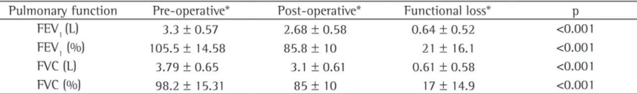
values obtained, there were significant decreases in mean forced expiratory volume in one second (20%; range 3-40%) and forced vital capacity (17%). Ninety days after lobectomy, all donors reported having returned to the activities they had engaged in before surgery (Table 2) In the chest X-ray results, there were no other alterations other than those expected for the corresponding pulmo-nary resections.
Discussion
In recent years, the use of living donors in transplantations has steadily increased, principally for kidney and liver transplants. The main critique of this method of treatment is that it exposes a healthy individual, the donor, to a risk of morbidity and mortality, a risk that cannot be ignored. A review of the records of 16,395 kidney and hepatic donors compiled by the United Network of Organ Sharing showed that there were two deaths among the kidney donors and one death among the liver donors.(8) In addition, 2 liver donors had to be put
on liver transplant waiting lists.
Living donor lung transplantation is still in the incipient phase, principally if compared to that of living donor kidney transplantation, which now were performed, with clot evacuation and lavage
of the cavity. Another donor presented a sudden episode of dyspnea on post-operative day 8, and a pneumothorax was diagnosed on the side on which the lobectomy had been performed. A chest tube was inserted, and there was complete re-expansion of the lung. The chest tube was left in place for three days. Prior to the removal of the chest tube, the bronchus was re-evaluated through fiberoptic bron-choscopy, and there was no evidence of fistula.
The other complications which occurred in this group were 5 pleural effusions (15.6%). Three cases were managed conservatively; in other 2 cases initial thoracentesis was necessary due to dyspnea complaint. One donor presented fever, and X-ray images showed pulmonary consolidation during hospitalization. That donor was treated with intra-venous antibiotic therapy (ampicillin and sulbactam). It was necessary to leave the chest tube in place for more than seven days in only one case, due to persistent air leakage. In that case, the chest tube was removed on post-operative day 9. There were no cases of bronchial aspiration or empyema in the immediate or late post-operative period.
As can be seen in Table 2, the post-operative pulmonary function tests (performed in 28 of the donors) showed that, in relation to the pre-operative
a b
Figure 2 - a) Initiation of the perfusion with saline solution in the lobar artery (black arrow) and ventilation of the left
When compared to conventional lobectomy for the treatment of lung neoplasms, lobectomy in a lobar donor has certain features that can increase the risk of complications.(6) In lobectomies for the
use of the lung lobe to transplantation, the vascular structures and the bronchus must be resected with a distal margin sufficient to allow implant in the recipient without impairing the bronchus and the vascularization of the lung of the donor.(6) That
implies proximal dissection of the structures, allowing a smaller margin for the closure of the artery and of the bronchus, favoring small alveolar lesions at the fissures, which can often be the cause of the prolonged air leakage. Taking into account the possibility of the middle lobar bronchus origi-nating in opposition to the superior bronchial segment, the distal bronchial margin, in some cases, can only be maintained through amputation of the middle lobar bronchus. When possible, it is re-inserted in the intermediate bronchus. If that is not possible, the middle lobar bronchus must be removed.(6,7) In the present study, there was only
one case in which bronchoplasty of the middle lobe was necessary. Bilobectomy, which can occur in up to 1.5% of the cases,(6,7) was not necessary in any of
the donors. Other complications described, such as bronchial fistula, empyema and severe arrhythmia, were not observed in this study.(6) These
complica-tions might have been avoided by taking care not to advance the vascular clamp too far into the left atrium when sectioning the pulmonary vein and by closing the bronchial stump without excessive devascularization.
The occurrence of minor or major complica-tions in 10 donors (31.25% of the sample) was higher than found by Bowdish et al. (19.8%),(7)
lower than described by Bataffarano et al. (61.3%),(6) comparable to the indices reported in the
literature(20,21) and comparable to that observed at
accounts for half of the procedures performed.(4)
Living donor lung transplantation presents a viable option for recipients with diseases in rapid evolu-tion and with no hope of receiving an organ from a deceased donor in a timely fashion.
As previously stated, for lung transplantation, 2 donors are needed for each recipient, which makes this type of transplantation unique in terms of organ transplantation. In the literature, the complications in lobar lung transplant donors have been associ-ated with morbidity rates of 19.8 to 61.3%.(6,7,15,19)
Such morbidity includes minor complications, such as pleural effusions and atelectasis, and more severe complications, such as bronchial fistulas and atrial fibrillation, in which implantation of a pacemaker is necessary. To date, there have been no reports of deaths in living lung transplant donors.
It is important that the relatives selected as donors be carefully evaluated in an interview with doctors and psychologists, in order to make them aware of the risks involved, and principally in order to iden-tify any possible pressure from other members of the family, creating a situation in which the dona-tion is not absolutely voluntary and altruist. In such cases, the use of this donor must be ruled out for a reason that is exclusively medical, thus fostering family harmony.
Lobectomy, as it is traditionally performed for the treatment of neoplastic and inflamma-tory diseases, is not a procedure free of morbidity and mortality.(20-22) Although complications
related to bronchial fistulas are more often seen in pneumonectomies, they can also occur in lobectomies.(22-24) Persistence of air leakage for more
than seven days occurs in approximately 15% of pulmonary resections,(22,25) Mild pleural effusion at
the base of the cavity, which frequently occurs after lobectomy, can usually be managed in a conservative fashion.(26)
Table 2 - Pre-operative and post-operative pulmonary function of 28 lung donors.
Pulmonary function Pre-operative* Post-operative* Functional loss* p FEV1 (L) 3.3 ± 0.57 2.68 ± 0.58 0.64 ± 0.52 <0.001 FEV1 (%) 105.5 ± 14.58 85.8 ± 10 21 ± 16.1 <0.001 FVC (L) 3.79 ± 0.65 3.1 ± 0.61 0.61 ± 0.58 <0.001 FVC (%) 98.2 ± 15.31 85 ± 10 17 ± 14.9 <0.001
7. Bowdish ME, Barr ML, Schenkel FA, Woo MS, Bremner RM, Horn MV, et al. A decade of living lobar lung transplantation: perioperative complications after 253 donor lobectomies. Am J Transplant. 2004;4(8):1283-8.
8. Ellison MD, Mc Bride MA, Edwards LB, Taranto SE, Barr ML, Trotter JF, et al. Living organ donation: mortality and early complications among 16,395 living donors in the U.S [abstract]. Am J Transplant. 2003;3(Suppl 5):283.
9. Starnes VA, Barr ML, Cohen RG. Lobar transplantation. Indications, technique, and outcome. J Thorac Cardiovasc Surg. 1994;108(3):403-10; discussion 410-1.
10. Starnes VA, Barr ML, Cohen RG, Hagen JA, Wells WJ, Horn MV, et al. Living-donor lobar lung transplantation experience: intermediate results. J Thorac Cardiovasc Surg. 1996;112(5):1284-90; discussion 1290-1.
11. Starnes VA, Bowdish ME, Woo MS, Barbers RG, Schenkel FA, Horn MV, et al. A decade of living lobar lung transplantation: recipient outcomes. J Thorac Cardiovasc Surg. 2004;127(1):114-22.
12. Bowdish ME, Pessotto R, Barbers RG, Schenkel FA, Starnes VA, Barr ML. Long-term pulmonary function after living-donor lobar lung transplantation in adults. Ann Thorac Surg. 2005;79(2):418-25.
13. Mendeloff EN, Huddleston CB, Mallory GB, Trulock EP, Cohen AH, Sweet SC, et al. Pediatric and adult lung transplantation for cystic fibrosis. J Thorac Cardiovasc Surg. 1998;115(2):404-13; discussion 413-4.
14. Camargo JJ; Grupo de Transplante Pulmonar da Santa Casa de Porto Alegre, RS, Brasil. [Lung transplant in children] [Article in Portuguese]. J Pediatr (Rio J). 2002;78(Suppl 2):S113-22. 15. Date H, Aoe M, Nagahiro I, Sano Y, Andou A, Matsubara H, et
al. Living-donor lobar lung transplantation for various lung diseases. J Thorac Cardiovasc Surg. 2003;126(2):476-81. 16. Bowdish ME, Barr ML. Living lobar lung transplantation.
Respir Care Clin N Am. 2004;10(4):563-79.
17. Watson TJ, Starnes VA. Pediatric lobar lung transplantation. Semin Thorac Cardiovasc Surg. 1996;8(3):313-25. 18. Schenkel FA, Horn MV, Woo MS, Barr ML, Starnes VA. Screening
potential donors for living lobar lung transplantation. J Heart Lung Transplant. 2003;22(suppl. 1):S86-S87. 19. Prager LM, Wain JC, Roberts DH, Ginns LC. Medical and
psychologic outcome of living lobar lung transplant donors. J Heart Lung Transplant. 2006;25(10):1206-12.
20. Stéphan F, Boucheseiche S, Hollande J, Flahault A, Cheffi A, Bazelly B, et al. Pulmonary complications following lung resection: a comprehensive analysis of incidence and possible risk factors. Chest. 2000;118(5):1263-70.
21. Deslauriers J, Ginsberg RJ, Piantadosi S, Fournier B. Prospective assessment of 30-day operative morbidity for surgical resections in lung cancer. Chest. 1994;106(6 Suppl): 329S-330S.
22. Sanchez PG, Vendrame GS, Madke GR, Pilla ES, Camargo JJ, Andrade CF, et al Lobectomy for treating bronchial carcinoma: analysis of comorbidities and their impact on postoperative morbidity and mortality. J Bras Pneumol. 2006;32(6):495-504.
23. Vester SR, Faber LP, Kittle CF, Warren WH, Jensik RJ. Bronchopleural fistula after stapled closure of bronchus. Ann Thorac Surg. 1991;52(6):1253-7; discussion 1257-8. 24. Asamura H, Naruke T, Tsuchiya R, Goya T, Kondo H,
Suemasu K. Bronchopleural fistulas associated with lung cancer operations. Univariate and multivariate analysis of
our facilities in routine lung resections performed for other ends.(22)
No differences were detected in number or severity of the complications in relation to the side on which the lobectomy was performed, whereas Bowdish et al. found a statistically significant difference for this variable.(7) This discrepancy can be attributed
to anatomical differences principally related to the position of the middle lobar bronchus. In the series studied, 5 complications occurred in donors of right lobes and 5 in donors of left lobes, all of whom presented similar types of complication.
Two of the most renowned living donor lung transplantation teams have reported one-year survival rates of 63.7 and 70%, respectively, and a three-year survival rates of 54%.(6,11) These rates are
similar to those found in the present study.
The present study is the first to describe this type of transplantation performed in Brazil. The number of cases is still small, and a more consistent analysis of the results can only be made through long-term follow-up evaluation of larger patient samples. We found that lobectomy in living lung transplant donors presents a high risk of post-oper-ative complications, and that it results in permanent loss of lung function in donors. Whether that implies future complications, only the monitoring of these individuals for a longer period will be able to clarify.
References
1. Unilateral lung transplantation for pulmonary fibrosis. Toronto Lung Transplant Group. N Engl J Med. 1986;314(18):1140-5.
2. Langone AJ, Helderman JH. Disparity between solid-organ supply and demand. N Engl J Med. 2003;349(7):704-6. 3. Starnes VA, Lewiston NJ, Luikart H, Theodore J, Stinson EB,
Shumway NE. Current trends in lung transplantation. Lobar transplantation and expanded use of single lungs. J Thorac Cardiovasc Surg. 1992;104(4):1060-5; discussion 1065-6. 4. Nathan HM, Conrad SL, Held PJ, McCullough KP, Pietroski
RE, Siminoff LA, et al. Organ donation in the United States. Am J Transplant. 2003;3 Suppl 4:29-40.
5. ABTO - Associação Brasileira de Transplante de Órgãos [homepage on the Internet]. São Paulo: Associação Brasileira de Transplante de Órgãos. [cited 2007 May 13] Registro Brasileiro de Transplantes - Ano XI nº2. Available from: http://abto.org.br/profissionais/rbt/anoXI_n2/RBT_ANO_XI_ N2_final.pdf
26. Ponn RB. Complications of Pulmonary Resection. In: Shields TW, LoCicero J, Ponn RB, Rusch VW, editors. General Thoracic Surgery. 6th ed. Philadelphia: Lippincott Williams
& Wilkins; 2004. p. 554-86. risk factors, management, and outcome. J Thorac Cardiovasc
Surg. 1992;104(5):1456-64.