Cellular composition of induced sputum in healthy adults*
Composição celular do escarro induzido em adultos saudáveisTiago Neves Veras, Emilio Pizzichini, Leila John Marques Steidle, Cristiane Cinara Rocha, Pablo Moritz, Márcia Margarete Menezes Pizzichini
Abstract
Objective: To establish reference values for cellularity in induced sputum samples collected from healthy adults.
Methods: Induced sputum samples were obtained from 88 healthy adult never-smokers (39 males). The mean age was 36 years (range, 18-68 years). The participants had been residing in the city of Florianópolis, Brazil (a medium-sized non-industrial city) for at least two years. After the samples had been processed, we obtained total and differential cell counts. Results: The mean total cell count was 4.8 ± 4.2 × 10⁶ cells/g. There was a predominance of macrophages (mean, 77.5 ± 14.7%) and neutrophils (mean, 23.4 ± 14.3%). Eosinophils were virtually absent (mean, 0.1 ± 0.3%). Lymphocytes and bronchial epithelial cells were scarce. Neither age nor atopy had any effect on the total or differential cell counts. Conclusions: In the induced sputum of this healthy adult population, macrophages and neutrophils predominated. However, the proportion of neutrophils was lower than that reported in previous studies, which suggests that reference values might vary depending on geographic location.
Keywords: Sputum; Reference values; Brazil.
Resumo
Objetivo: Estabelecer valores de referência para a celularidade de amostras de escarro induzido coletadas de indivíduos adultos saudáveis. Métodos: O escarro induzido foi obtido de 88 adultos saudáveis que nunca fumaram (39 homens) com média de idade de 36 anos (variação: 18-68 anos) residentes há pelo menos dois anos em Florianópolis, uma cidade brasileira não industrial e de tamanho médio. As amostras foram processadas, e foi realizada a contagem total e diferencial das células. Resultados: A média da contagem celular total foi de 4,8 ± 4,2 × 10⁶ células/g. Houve predomínio de macrófagos (média de 77,5 ± 14,7%) e de neutrófilos (média de 23,4 ± 14,3%). Os eosinófilos estiveram virtualmente ausentes (média de 0,1 ± 0,3%). A proporção de linfócitos e de células broncoepiteliais foi pequena. Não houve efeito da idade ou de atopia sobre a contagem celular total ou diferencial. Conclusões: Nesta população de indivíduos saudáveis, macrófagos e neutrófilos foram as células predominantes no escarro induzido. Contudo, a proporção de neutrófilos foi inferior à previamente relatada, sugerindo que os valores de normalidade podem variar de acordo com o local onde ele é amostrado.
Descritores: Escarro; Valores de referência; Brasil.
* Study carried out at the Federal University of Santa Catarina, Florianópolis, Brazil.
Correspondence to: Márcia M. M. Pizzichini. Departamento de Clínica Médica, Universidade Federal de Santa Catarina, NUPAIVA, Hospital Universitário, Campus Universitário, Trindade, CEP 88040-970, Florianópolis, SC, Brasil.
Tel. 55 47 3234-7711. E-mail: [email protected] Financial support: None.
Methods
A total of 99 healthy adult never-smokers who had been residing in Florianópolis, a medium-sized non-industrial city located in southern Brazil, for at least two years were recruited through advertisements in the local media and posters displayed at the local university. We applied the following exclusion criteria: presenting with respiratory symptoms (nasal or pulmonary); having a history of respiratory diseases, including asthma and allergic rhinitis; having a history of occupational exposure to dust, chemical materials, or smoke; and having experience symptoms of respiratory infection in the last four weeks. All subjects had normal spirometry results (FEV1 > 80% of predicted, FEV1/FVC ratio ≥ 80%, and normal methacholine responsiveness—the provocative concentration of methacholine causing a 20% fall in FEV1 [PC20] being > 8.0 mg/mL). Of the 99 subjects evaluated, 27 were atopic, as indicated by the presence of positive skin test reactivity to one or more common allergens. The study was approved by the Human Research Ethics Committee of the Universidade Federal de Santa Catarina (UFSC, Federal University of Santa Catarina; Protocol no. 084/84). All volunteers gave written informed consent.
This was a single-visit, cross-sectional study. All subjects were evaluated in the research laboratory of the UFSC Center for Airway Inflammation Research. After having been screened for the inclusion and exclusion criteria, the subjects with normal spirometry results and normal methacholine responsiveness underwent sputum induction and skin testing for the investigation of atopy.
The characteristics of the volunteers were documented with a structured questionnaire. Spirometry was performed in accordance with the American Thoracic Society guidelines.(17) The
predicted reference values were those established by Crapo et al.(18) Methacholine challenge was
performed in accordance with the method described by Juniper et al.,(19) and the results
are expressed as PC20 in cumulative units. Skin testing was performed by a modified puncture technique(20) with the use of 14 extracts of
common inhalant allergens, 1 negative control (glycerol), and 1 positive control (histamine). Test results were considered positive when the papule size was 3 mm greater than was that of
Introduction
Nearly two decades ago,(1,2) the introduction
of induced sputum to assess airway inflammation provided a breakthrough in the understanding of the pathophysiology of airway diseases. Currently, there is sufficient evidence that induced sputum is the most comprehensive method for noninvasive examination of airway inflammation because of its properties (reliability, reproducibility, and responsiveness).
(3-5) In addition, the results of recent studies
have consistently shown that induced sputum is an important tool for the determination of the phenotype of asthma,(6-8) COPD,(7) and other
airway diseases, as well as for the study of the effect of treatment of these diseases.(9-11) The
method has been important for the study of the pathogenesis of obstructive(12) and infectious(13)
respiratory diseases. In contrast, few studies have examined the cellular composition of induced sputum in healthy subjects.
Knowledge of normal values for induced sputum measurements, especially as regards the proportion of eosinophils and neutrophils (the two cell types most commonly used for characterizing the inflammatory response of the airways), is essential to understand the result of this test. The use of induced sputum, together with pulmonary function tests, makes it possible to monitor the pattern of inflammation more appropriately in specific respiratory conditions.
(14,15) However, total and differential cell counts
in induced sputum samples should be delimited by normal reference values.
To date, only three studies,(14-16) all of which
involved large samples of healthy subjects, have provided reference values for total and differential cell counts in induced sputum. The only difference among the results of those studies was in the proportion of neutrophils.
and post-albuterol FEV1) are expressed as means and standard deviations or ranges (minimum and maximum). Because of the non-Gaussian distribution of the total cell counts and of some cellular components (eosinophils and lymphocytes), sputum cytology results are expressed as means and standard deviations, as medians and interquartile ranges, and as the 10th and 90th percentiles. Differences among groups were analyzed by ANOVA or unpaired t-test. Values of p < 0.05 were considered statistically significant. All tests were two-tailed. Data were analyzed with the Statistical Package for the Social Sciences, version 18.0 for Windows (SPSS Inc., Chicago, IL, USA).
Results
Sputum induction produced an adequate sample in 88 participants (39 males). The mean age was 36 years (range, 18-68 years). The success rate of sputum induction was 88.9%. We excluded 11 sputum samples because of insufficient sputum volume for evaluation or lack of cell viability. In the remaining samples, the quality of sputum obtained was satisfactory, as indicated by a mean viability of 77.5 ± 11.1%, with a median (90th percentile) of 79.0% (94.0%). Tables 1 and 2 show the total and differential cell counts, the distribution of eosinophils and neutrophils, expressed as absolute and percentage values, as well as the the negative control. Atopy was defined as the
presence of one or more positive test results. Sputum was induced and processed by the method described by Pizzichini et al.(3)
In brief, the procedure was initiated 15 min after the administration of 200 µg of inhaled albuterol, through the inhalation of increasing concentrations of saline (3%, 4%, and 5%), each inhaled for 7 min consecutively or until there was a decrease in FEV1≥ 20% in relation to baseline values. Saline nebulization was performed with a Fisoneb ultrasonic nebulizer (Fisons, Pickering, Ontario, Canada), with an output rate of 0.87 mL/min and particles presenting a median aerodynamic mass diameter of 5.58 µm. After each inhalation period, FEV1 was measured to ensure the safety of the test. If there was a decrease in FEV1≥ 10% in relation to baseline values, the saline concentration was not increased. Samples were considered appropriate if total and differential cell counts could be obtained from material with cell viability of at least 50% and contamination by oropharyngeal squamous cells lower than 20%.
The sample size was estimated on the basis of previous calculations.(14-16) Categorical variables
(gender, race, atopy, and induction success) are expressed as absolute frequencies and percentages. Continuous variables were initially evaluated in terms of the criteria of normality. Variables with a continuous distribution (age, weight, height, baseline FEV1, FEV1/FVC ratio,
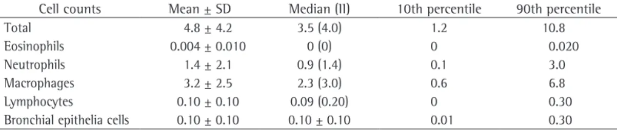
Table 1 - Total and differential cell counts (× 106 cells/g) in induced sputum samples collected from healthy adult subjects.
Cell counts Mean ± SD Median (II) 10th percentile 90th percentile
Total 4.8 ± 4.2 3.5 (4.0) 1.2 10.8
Eosinophils 0.004 ± 0.010 0 (0) 0 0.020
Neutrophils 1.4 ± 2.1 0.9 (1.4) 0.1 3.0
Macrophages 3.2 ± 2.5 2.3 (3.0) 0.6 6.8
Lymphocytes 0.10 ± 0.10 0.09 (0.20) 0 0.30
Bronchial epithelia cells 0.10 ± 0.10 0.10 ± 0.10 0.01 0.30
II: interquartile range.
Table 2 - Differential non-squamous cell counts in induced sputum samples from healthy subjects in the study. Differential cell counts Mean ± SD Median (II) 10th percentile 90th percentile
Eosinophils 0.1 ± 0.3 0 (0) 0 0.5
Neutrophils 23.4 ± 14.3 21.7 (21.0) 6.9 41.3
Macrophages 68.4 ± 14.7 71.0 (17.9) 47.9 86.0
Lymphocytes 3.1 ± 2.4 3.0 (3.0) 0 6.5
Bronchial epithelial cells 5.2 ± 7.9 2.8 (4.9) 0.5 13.3
eosinophils and lymphocytes, are similar to those in the literature.(14,15) The percentage of healthy
adults excluded from the sample evaluated is also similar to that reported in previous studies.
(14,15) However, the proportion and the absolute
number of neutrophils found in our study were lower than those previously reported. Knowledge of reference values for sputum cellularity, especially as regards neutrophils and eosinophils, is important for understanding and interpreting the meaning of inflammatory subtypes—neutrophilic, eosinophilic, mixed granulocytic, and paucigranulocytic—which have often been used for determining the phenotype of asthma.(21)
The lower proportion of neutrophils found in the present study cannot be attributed to technical aspects, because the sputum induction and processing methods employed are similar to those used in previous studies.(14,15,22) Nor does
it seem to be due to bias in the selection of participants, because we found that neither age nor atopy had any effect on the total or differential cell counts. We speculate that this lower proportion of neutrophils was caused by local variations, such as environmental pollution.
The present study was not designed to investigate the effect of environmental pollution on airway secretions. Instead, it was designed to establish normal values for induced sputum cellularity in healthy adults not exposed to environmental pollution, because all participants had been residing in a non-industrial city for at least two years. We cannot rule out previous household or environmental exposure with certainty. However, none of the study subjects upper limits for each cell type. Macrophages
and neutrophils predominated. Eosinophils were absent from the sputum of most subjects (85%). There were few lymphocytes and bronchial epithelial cells. We also examined cellularity by age bracket and by the presence or absence of atopy (Tables 3 and 4, respectively). Although eosinophil counts were higher in atopic subjects, the difference was not clinically relevant or statistically significant. Age had no effect on the distribution of total or differential cell counts, although macrophages predominated in the younger age group (18-29 years).
Discussion
The objective of the present study was to establish reference values for induced sputum cellularity in a sample of healthy Brazilian never-smokers residing in a non-industrial city. Our results, showing that sputum from healthy subjects is characterized by a predominance of macrophages and neutrophils and by a lack of
Table 3 - Total and differential cell counts in induced sputum samples from healthy subjects in the study by age bracket.a
Cell counts Age bracket, years
18-29 30-39 40-49 ≥50
(n = 39) (n = 12) (n = 13) (n = 24)
Total 4.1 ± 3.2 5.3 ± 4.3 5.2 ± 3.7 5.8 ± 5.6
Eosinophils 0.10 ± 0.30 0.08 ± 0.30 0.20 ± 0.40 0.08 ± 0.30
Neutrophils 20.2 ± 14.5 29.0 ± 14.8 28.1 ± 11.1 23.1 ± 14.8
Macrophages 73.0 ± 14.8 62.3 ± 11.2 64.5 ± 10.6 66.0 ± 16.5
Lymphocytes 3.1 ± 2.5 3.3 ± 2.5 3.0 ± 2.1 3.0 ± 2.6
Bronchial epithelial cells 4.3 ± 5.2 5.3 ± 4.4 4.2 ± 4.6 5.0 ± 6.0 aValues expressed as mean ± SD.
Table 4 - Total and differential cell counts in induced sputum samples from healthy subjects in the study by presence or absence of atopy.a
Cell counts Atopy
Present Absent (n = 27) (n = 61)
Total 3.5 ± 3.0 5.4 ± 4.6
cell counts to investigate airway inflammation in asthma. Thorax. 1992;47(1):25-9.
2. Vianna EO. Induced sputum cell counts in medical practice. J Bras Pneumol. 2008;34(11):889-90.
3. Pizzichini E, Pizzichini MM, Efthimiadis A, Evans S, Morris MM, Squillace D, et al. Indices of airway inflammation in induced sputum: reproducibility and validity of cell and fluid-phase measurements. Am J Respir Crit Care Med. 1996;154(2 Pt 1):308-17. 4. Efthimiadis A, Pizzichini MM, Pizzichini E, Dolovich
J, Hargreave FE. Induced sputum cell and fluid-phase indices of inflammation: comparison of treatment with dithiothreitol vs phosphate-buffered saline. Eur Respir J. 1997;10(6):1336-40.
5. Brightling CE, Monteiro W, Ward R, Parker D, Morgan MD, Wardlaw AJ, et al. Sputum eosinophilia and short-term response to prednisolone in chronic obstructive pulmonary disease: a randomised controlled trial. Lancet. 2000;356(9240):1480-5.
6. Simpson JL, McElduff P, Gibson PG. Assessment and reproducibility of non-eosinophilic asthma using induced sputum. Respiration. 2010;79(2):147-51. 7. Boorsma M, Lutter R, van de Pol MA, Out TA, Jansen
HM, Jonkers RE. Repeatability of inflammatory parameters in induced sputum of COPD patients. COPD. 2007;4(4):321-9.
8. Moritz P, Steidle LJ, Felisbino MB, Kleveston T, Pizzichini MM, Pizzichini E. Determination of the inflammatory component of airway diseases by induced sputum cell counts: use in clinical practice. J Bras Pneumol. 2008;34(11):913-21.
9. Green RH, Brightling CE, Woltmann G, Parker D, Wardlaw AJ, Pavord ID. Analysis of induced sputum in adults with asthma: identification of subgroup with isolated sputum neutrophilia and poor response to inhaled corticosteroids. Thorax. 2002;57(10):875-9. 10. Jayaram L, Pizzichini MM, Cook RJ, Boulet LP, Lemière
C, Pizzichini E, et al. Determining asthma treatment by monitoring sputum cell counts: effect on exacerbations. Eur Respir J. 2006;27(3):483-94.
11. Leigh R, Pizzichini MM, Morris MM, Maltais F, Hargreave FE, Pizzichini E. Stable COPD: predicting benefit from high-dose inhaled corticosteroid treatment. Eur Respir J. 2006;27(5):964-71.
12. Costa C, Rufino R, Traves SL, Lapa E Silva JR, Barnes PJ, Donnelly LE. CXCR3 and CCR5 chemokines in induced sputum from patients with COPD. Chest. 2008;133(1):26-33.
13. Almeida AS, Lago PM, Boechat N, Huard RC, Lazzarini LC, Santos AR, et al. Tuberculosis is associated with a down-modulatory lung immune response that impairs Th1-type immunity. J Immunol. 2009;183(1):718-31. 14. Belda J, Leigh R, Parameswaran K, O’Byrne PM, Sears
MR, Hargreave FE. Induced sputum cell counts in healthy adults. Am J Respir Crit Care Med. 2000;161(2 Pt 1):475-8.
15. Spanevello A, Confalonieri M, Sulotto F, Romano F, Balzano G, Migliori GB, et al. Induced sputum cellularity. Reference values and distribution in normal volunteers. Am J Respir Crit Care Med. 2000;162(3 Pt 1):1172-4. 16. Thomas RA, Green RH, Brightling CE, Birring SS, Parker
D, Wardlaw AJ, et al. The influence of age on induced sputum differential cell counts in normal subjects. Chest. 2004;126(6):1811-4.
had been exposed to environmental pollution in the last two years.
Environmental exposure results from contact with a variety of gaseous and particulate pollutants, among which the most harmful to human health seems to be particulate matter. Repeated inhalation of particulate pollutants can result in a moderate level of oxidative stress and a moderate degree of inflammatory response in the airways and lungs.(23) In recent years, various
studies have investigated the effect of acute and chronic exposure of the lungs to environmental pollution. Studies using bronchoalveolar lavage(24) or induced sputum have shown that
healthy subjects exposed to ozone(25-27) or diesel
exhaust(27) have an inflammatory response
characterized by an increase in neutrophils and IL-6. Changes in induced sputum can be observed as early as six hours after exposure.
(24) In addition, it has been shown that healthy
workers who are exposed to traffic pollution on a daily basis have sputum neutrophilia.(28)
Occupational exposure should also be considered a major modulator of the cellular composition of induced sputum.(29) Although it is reasonable to
assume that the low proportion of neutrophils in our sample is associated with lower exposure to environmental pollution, this hypothesis needs to be confirmed or refuted in studies specifically designed to investigate this aspect.
Finally, it is important to mention that, in 85% of the study subjects, there were no eosinophils in the induced sputum samples. This result confirms previous reports(14,15) and
supports the concept that the presence of eosinophils in sputum is a robust and reliable marker of eosinophilic inflammation. Our results also suggest that the current cut-off value of 3.0% for eosinophilic inflammation can be lowered.
In summary, the results of the present study show that, in this population of healthy subjects, there was a predominance of macrophages and neutrophils in induced sputum samples. However, the proportion of neutrophils was lower than that reported in previous studies, which suggests that reference values might vary by geographic location.
References
24. Stenfors N, Nordenhäll C, Salvi SS, Mudway I, Söderberg M, Blomberg A, et al. Different airway inflammatory responses in asthmatic and healthy humans exposed to diesel. Eur Respir J. 2004;23(1):82-6.
25. Nightingale JA, Rogers DF, Hart LA, Kharitonov SA, Chung KF, Barnes PJ. Effect of inhaled endotoxin on induced sputum in normal, atopic, and atopic asthmatic subjects. Thorax. 1998;53(7):563-71.
26. Fahy JV, Liu J, Wong H, Boushey HA. Cellular and biochemical analysis of induced sputum from asthmatic and from healthy subjects. Am Rev Respir Dis. 1993;147(5):1126-31.
27. Nordenhäll C, Pourazar J, Blomberg A, Levin JO, Sandström T, Adelroth E. Airway inflammation following exposure to diesel exhaust: a study of time kinetics using induced sputum. Eur Respir J. 2000;15(6):1046-51. 28. Burgess JL, Fleming JE, Mulenga EM, Josyula A, Hysong
TA, Joggerst PJ, et al. Acute changes in sputum IL-10 following underground exposure to diesel exhaust. Clin Toxicol (Phila). 2007;45(3):255-60.
29. Fireman E. Induced sputum and occupational diseases other than asthma. Curr Opin Allergy Clin Immunol. 2009;9(2):93-6.
17. Standardization of Spirometry, 1994 Update. American Thoracic Society. Am J Respir Crit Care Med. 1995;152(3):1107-36.
18. Crapo, RO, Morris AH, Gardner RM. Reference spirometric values using techniques and equipment that meet ATS recommendations. Am Rev Respir Dis. 1981;123(6):659-64.
19. Juniper EF, Cockcroft DW Hargreave FE. Histamine and Methacholine Inhalation Test: A Laboratory Tidal Breathing Protocol. Lund: Astra Draco AB; 1994. 20. Pepys J, Coombs RR, Lachmann PJ. Skin test in diagnosis.
In: Gell PG, Coombs RR, Lachmann PJ, editors. Clinical Aspects of Immunology. Oxford: Blackwell; 1975. p. 55-80.
21. Simpson JL, Scott R, Boyle MJ, Gibson PG. Inflammatory subtypes in asthma: assessment and identification using induced sputum. Respirology. 2006;11(1):54-61. 22. Belda J, Giner J, Casan P, Sanchis J. Induced sputum
in asthma: study of validity and repeatability [Article in Spanish]. Arch Bronconeumol. 1997;33(7):325-30. 23. Nobutomo K. Air pollution and cytological changes in
sputum. Lancet. 1978;1(8063):523-6.
About the authors
Tiago Neves Veras
Pediatric Pulmonologist. Hospital Infantil Jeser Amarante Faria, Joinville, Brazil.
Emilio Pizzichini
Professor of Pulmonology. Federal University of Santa Catarina, Florianópolis, Brazil.
Leila John Marques Steidle
Pulmonologist. Federal University of Santa Catarina University Hospital, Florianópolis, Brazil.
Cristiane Cinara Rocha
Research Nurse. Núcleo de Pesquisa em Asma e Inflamação das Vias Aéreas – NUPAIVA, Center for Research on Asthma and Airway Inflammation – Federal University of Santa Catarina, Florianópolis, Brazil.
Pablo Moritz
Pulmonologist. Federal University of Santa Catarina University Hospital, Florianópolis, Brazil.
Márcia Margarete Menezes Pizzichini