TSC disorder is associated with mutations in the TSC1 or TSC2 gene.(1,2)
Although there is still no definitive treatment for LAM, new perspectives, with different targets, have been studied. Rapamycin is one of the examples, because of its ability to inhibit mammalian target of rapamycin, a protein that promotes cell proliferation. A recent randomized trial of patients with moderate to severe impairment revealed that FEV1 stabilized over a one-year period of rapamycin use.(3) Tetracycline and its analogues, such as doxycycline, inhibit matrix metalloproteinases (MMPs) and have therefore been tested as treatments for LAM, because MMPs, especially MMP-2, are increased in patients with LAM, promoting extracellular Pulmonary lymphangioleiomyomatosis
(LAM) is a rare disease that affects women of reproductive age and is characterized by proliferation of atypical smooth muscle cells (LAM cells, which test positive for alpha-actin antigen and for monoclonal antibody HMB-45) around small airways, blood vessels, and lymphatic vessels, leading to bronchial and vascular obstruction, together with cyst formation. The disease can occur in isolation or in association with tuberous sclerosis complex (TSC), which is a sporadic or autosomal dominant neurocutaneous disorder characterized by hamartomas of the skin, eyes, kidneys, heart, and central nervous system, as well as by convulsive seizures and mental retardation. The
Evolution of pulmonary function after treatment with
goserelin in patients with lymphangioleiomyomatosis*
Evolução da função pulmonar após tratamento com goserelina em pacientes com linfangioleiomiomatose
Bruno Guedes Baldi, Pedro Medeiros Junior, Suzana Pinheiro Pimenta, Roberto Iglesias Lopes, Ronaldo Adib Kairalla, Carlos Roberto Ribeiro Carvalho
Abstract
In the atypical smooth muscle cells that are characteristic of lymphangioleiomyomatosis (LAM), there are estrogen and progesterone receptors. Therefore, anti-hormonal therapy, despite having produced controversial results, can be considered a treatment option. The objective of this retrospective study was to evaluate hormonal and spirometric data for nine women with LAM after one year of treatment with goserelin. The mean increase in FEV1 and FVC was 80 mL and 130 mL, respectively. There was effective blockage of the hormonal axis. It is still not possible to exclude a potential beneficial effect of the use of gonadotropin-releasing hormone analogues in LAM patients, which underscores the need for randomized trials.
Keywords: Lymphangioleiomyomatosis; Spirometry; Goserelin.
Resumo
Nas células musculares lisas atípicas características da linfangioleiomiomatose (LAM) encontram-se receptores de estrogênio e progesterona, de modo que o tratamento anti-hormonal pode ser considerado uma opção, mas ainda com resultados controversos. O objetivo deste trabalho foi avaliar retrospectivamente parâmetros hormonais e espirométricos em nove mulheres com LAM após o tratamento com goserelina por um ano. Houve um aumento médio de 80 mL e 130 mL, respectivamente, em VEF1 e CVF, assim como bloqueio hormonal efetivo. Ainda não se
pode excluir um potencial efeito favorável da utilização de análogos de hormônio liberador de gonadotrofina em pacientes com LAM, reforçando a necessidade de ensaios randomizados.
Descritores: Linfangioleiomiomatose; Espirometria; Gosserrelina.
* Study carried out in the Department of Pulmonology, Instituto do Coração – InCor, Heart Institute – and in the Department of Urology, University of São Paulo School of Medicine Hospital das Clínicas, São Paulo, Brazil.
Correspondence to: Bruno Guedes Baldi. Endereço: Avenida Dr. Enéas de Carvalho Aguiar, 44, 5º andar, CEP 05403-900, São Paulo, SP, Brasil.
Tel. 55 11 3069-5695. Fax: 55 11 3069-5695. E-mail: [email protected] Financial support: None.
specializing in the evaluation of lung diseases. The study was approved by the local research ethics committee, and all of the patients gave written informed consent.
For each patient, we obtained data regarding the following: age at diagnosis; presence of dyspnea; history of pneumothorax, chylothorax, and renal angiomyolipoma; and smoking history. In order to assess goserelin-induced hormone suppression, we determined the levels of progesterone, FSH, and estradiol before treatment initiation and at the end of the treatment period.
Spirometry was performed in accordance with the criteria established by the Brazilian Thoracic Association, and the equations devised by Pereira et al. for the Brazilian population were used for calculating predicted values. (16,17) None of the patients had chylothorax or pneumothorax at the time of spirometry.
Post-bronchodilator FEV1, FVC, and FEV1/ FVC ratio were measured before treatment initiation and at the end of the treatment period. The results are expressed as means and standard deviations. In addition, variations in FEV1 and FVC were calculated to investigate the role of treatment in the deterioration of lung function over time. The statistical analysis was performed with the Statistical Package for the Social Sciences, version 15.0 for Windows (SPSS Inc., Chicago, IL, USA). Student’s t-test was used for comparing pre- and post-treatment data. Medians and interquartile ranges were calculated for the levels of estradiol, progesterone, and FSH. The Wilcoxon test was used for comparing pre- and post-treatment data. Values of p < 0.05 were considered statistically significant.
The mean patient age at diagnosis was 37.9 years (range, 19-47 years), and four patients (44.4%) were former smokers. Of the nine patients studied, six (66.7%) had a history of pneumothorax, two (22.2%) had a history of chylothorax, eight (88.9%) complained of dyspnea, and five (55.6%) had renal angiomyolipoma.
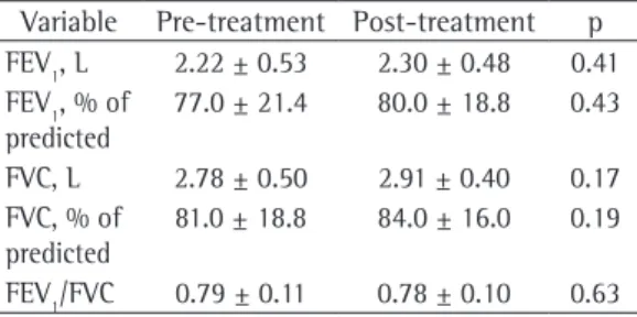
Table 1 shows the spirometric values, which are expressed as means and standard deviations. None of the patients had a positive bronchodilator response in the spirometric test performed before antihormone therapy. After one year of treatment with goserelin, the mean increase in FEV1 was 80 mL and the mean matrix degradation.(4) Lung transplantation and
combined heart-lung transplantation remain as options for advanced cases.(5)
More than 25 years ago, it was demonstrated that hormones are involved in the pathogenesis of LAM. Brentani et al. reported the presence of estrogen, progesterone and glucocorticoids receptors in the lung tissue of patients with LAM.(6) Another study confirmed estrogen and progesterone receptor positivity in angiomyolipoma.(7) Yu et al. confirmed that estradiol and tamoxifen stimulate LAM-associated angiomyolipoma cell growth in culture.(8) On the basis of this association between hormones and LAM, antihormone therapy can be considered a treatment option. Oophorectomy, as well as the administration of tamoxifen, progesterone, gonadotropin-releasing hormone (GnRH) agonists, and GnRH analogues, has been tested, producing controversial results, because there have been no randomized clinical trials of these treatment modalities.(9-14) In a retrospective study of 35 LAM patients receiving various hormone therapy regimens, 10-year survival was 90%.(11) However, other studies have shown a decrease in FEV1 even after hormonal manipulation.(12,14) In one retrospective study, treatment with goserelin, compared with the use of progesterone, showed a trend toward slowing the decline in FEV1 over time.(15) To date, because of the rarity of the disease and the lack of standard treatment protocols, there have been no studies comparing treatment options in an appropriate manner.
The potential beneficial effect of GnRH analogues in patients with LAM is based on their ability to reduce luteinizing hormone levels and follicle-stimulating hormone (FSH) levels, suppressing ovarian function. The primary objective of the present study was to evaluate the effect that treatment with goserelin (a GnRH analogue) has on FEV1 in patients with LAM. A secondary objective was to determine whether there is effective blockade of the hypothalamic-pituitary axis.
dependent on the route of administration, the absorption of the drug, the interval between doses, and the effect of the drug on the hypothalamic-pituitary axis. Disease progression has been observed in all LAM patients treated with buserelin, which is typically administered via nasal spray. Nasal administration can be characterized by inadequate drug absorption and failure to inhibit GnRH pulse secretion, resulting in ineffective blockade of the hypothalamic-pituitary axis.(19,20) In the study conducted by Harari et al., triptorelin was administered i.m., in a single dose once every three months, and, although it promoted suppression of ovarian function, it did not prevent the decline in lung function.(14) In contrast, there was no fall in FEV
1 in our study, which might be explained by the fact that our patients had higher baseline FEV1 than did those in the Harari et al. study (2.22 vs. 1.9 L), and that we initiated the treatment at an earlier stage, when disease progression can still be prevented.
The annual decline in FEV1 is an important parameter analyzed in the follow-up of patients with LAM. In a case series of 32 patients, conducted in the United Kingdom, the mean annual decline in FEV1 was found to be 118 mL, regardless of menopausal status or hormone therapy.(9) In another study, in which 275 patients treated with progesterone were evaluated, the mean annual decline in FEV1 was 75 mL (1.7% of predicted), whereas, in a nonrandomized prospective study of 10 patients treated with triptorelin, there was a mean annual decline of 156 mL (4.9% of predicted).(12,14) In the present study, there was a mean annual increase in FEV1 of 80 mL (2.5% of predicted) in LAM patients treated with goserelin. However, this difference was not significant, probably because of the small size of the patient sample, which is a well-recognized limitation of studies evaluating rare diseases.
In conclusion, although the use of GnRH analogues might have a beneficial effect in patients with LAM, their use remains increase in FVC was 130 mL. However, the
difference was not statistically significant. The mean FEV1/FVC ratio did not vary. Of the nine patients evaluated, five showed increased FEV1, three showed no variation in FEV1, and only one showed reduced FEV1.
Table 2 shows the hormone levels, which are expressed as medians and interquartile ranges. After one year of treatment with goserelin, there was effective hormonal blockade, with a significant reduction in the median levels of FSH (to values below the lower limit of normality for adult women), estradiol, and progesterone (to values below the lower limit of normality for adult women and within the range of reference values for postmenopausal women). The medication was well tolerated by all patients, and there were no significant adverse effects.
The GnRH agonists goserelin, buserelin, and triptorelin have previously been evaluated as treatments for LAM.(14,18-20) Rossi et al. described the case of a patient in whom treatment with goserelin yielded favorable results in many parameters.(18) Conversely, in studies conducted by Radermecker et al. and by Harari et al., the use of buserelin and triptorelin, respectively, did not prevent the decline in lung function.(14,19)
Our hypothesis is that the functional response to treatment with GnRH analogues is dependent on the specific agent used and on whether the agent is administered at an earlier or at a more advanced stage of LAM. That response is also Table 1 - Spirometric parameters before and after treatment with goserelin in the nine patients evaluated.a
Variable Pre-treatment Post-treatment p FEV1, L 2.22 ± 0.53 2.30 ± 0.48 0.41 FEV1, % of
predicted
77.0 ± 21.4 80.0 ± 18.8 0.43
FVC, L 2.78 ± 0.50 2.91 ± 0.40 0.17 FVC, % of
predicted
81.0 ± 18.8 84.0 ± 16.0 0.19
FEV1/FVC 0.79 ± 0.11 0.78 ± 0.10 0.63
aValues expressed as mean ± SD.
Table 2 - Hormone levels before and after treatment with goserelin in the nine patients evaluated.a
Hormone Pre-treatment Post-treatment p FSH, IU/L 4.7 (1.7-8.2) 1.7 (0.6-5.8) 0.034 Progesterone, ng/mL 1.3 (0.5-4.3) 0.3 (0.3-0.4) 0.018 Estradiol, pg/mL 64.0 (48.4-96.0) 15.0 (11.0-26.1) 0.008
in lymphangioleiomyomatosis and benign metastasizing leiomyoma. N Engl J Med. 1981;305(4):204-9.
11. Chu SC, Horiba K, Usuki J, Avila NA, Chen CC, Travis WD, et al. Comprehensive evaluation of 35 patients with lymphangioleiomyomatosis. Chest. 1999;115(4):1041-52.
12. Taveira-DaSilva AM, Stylianou MP, Hedin CJ, Hathaway O, Moss J. Decline in lung function in patients with lymphangioleiomyomatosis treated with or without progesterone. Chest. 2004;126(6):1867-74.
13. Schiavina M, Contini P, Fabiani A, Cinelli F, Di Scioscio V, Zompatori M, et al. Efficacy of hormonal manipulation in lymphangioleiomyomatosis. A 20-year-experience in 36 patients. Sarcoidosis Vasc Diffuse Lung Dis. 2007;24(1):39-50.
14. Harari S, Cassandro R, Chiodini I, Taveira-DaSilva AM, Moss J. Effect of a gonadotrophin-releasing hormone analogue on lung function in lymphangioleiomyomatosis. Chest. 2008;133(2):448-54.
15. Medeiros Jr P, Kairalla RA, Pereira CA, Lopes RI, Chaves CN, Araujo FL, et al. GnRH analogs X progesterone-lung function evolution in two treatment cohort groups in lymphangioleiomyomatosis (LAM). Am J Respir Crit Care Med 2003;167(7):A953.
16. Sociedade Brasileira de Pneumologia e Tisiologia. Diretrizes para testes de função pulmonar. J Pneumol. 2002;28(Suppl 3):S1-S82.
17. Pereira CA, Sato T, Rodrigues SC. New reference values for forced spirometry in white adults in Brazil. J Bras Pneumol. 2007;33(4):397-406.
18. Rossi GA, Balbi B, Oddera S, Lantero S, Ravazzoni C. Response to treatment with an analog of the luteinizing-hormone-releasing hormone in a patient with pulmonary lymphangioleiomyomatosis. Am Rev Respir Dis. 1991;143(1):174-6.
19. Radermecker M, Broux R, Corhay JL, Limet R, Radermecker M. Failure of buserelin-induced medical castration to control pulmonary lymphangiomyomatosis in two patients. Chest. 1992;101(6):1724-6.
20. de la Fuente J, Páramo C, Román F, Pérez R, Masa C, de Letona JM. Lymphangioleiomyomatosis: unsuccessful treatment with luteinizing-hormone-releasing hormone analogues. Eur J Med. 1993;2(6):377-8.
controversial. Our findings underscore the need for large, multicenter, randomized trials of these agents in order to determine their true impact on the progression of the disease.
References
1. Johnson SR, Cordier JF, Lazor R, Cottin V, Costabel U, Harari S, et al. European Respiratory Society guidelines for the diagnosis and management of lymphangioleiomyomatosis. Eur Respir J. 2010;35(1):14-26.
2. Medeiros Jr P, Carvalho CR. Linfangioleiomiomatose pulmonar. J Bras Pneumol. 2004;30(1):66-77.
3. McCormack FX, Inoue Y, Moss J, Singer LG, Strange C, Nakata K, et al. Efficacy and Safety of Sirolimus in Lymphangioleiomyomatosis. N Engl J Med. 2011[Epub ahead of print].
4. Moses MA, Harper J, Folkman J. Doxycycline treatment for lymphangioleiomyomatosis with urinary monitoring for MMPs. N Engl J Med. 2006;354(24):2621-2. 5. Kpodonu J, Massad MG, Chaer RA, Caines A, Evans A, Snow
NJ, et al. The US experience with lung transplantation for pulmonary lymphangioleiomyomatosis. J Heart Lung Transplant. 2005;24(9):1247-53.
6. Brentani MM, Carvalho CR, Saldiva PH, Pacheco MM, Oshima CT. Steroid receptors in pulmonary lymphangiomyomatosis. Chest. 1984;85(1):96-9. 7. Logginidou H, Ao X, Russo I, Henske EP. Frequent
estrogen and progesterone receptor immunoreactivity in renal angiomyolipomas from women with pulmonary lymphangioleiomyomatosis. Chest. 2000;117(1):25-30. 8. Yu J, Astrinidis A, Howard S, Henske EP. Estradiol and
tamoxifen stimulate LAM-associated angiomyolipoma cell growth and activate both genomic and nongenomic signaling pathways. Am J Physiol Lung Cell Mol Physiol. 2004;286(4):L694-700.
9. Johnson SR, Tattersfield AE. Decline in lung function in lymphangioleiomyomatosis: relation to menopause and progesterone treatment. Am J Respir Crit Care Med. 1999;160(2):628-33.
About the authors
Bruno Guedes Baldi
Attending Physician. Department of Pulmonology, Instituto do Coração – InCor, Heart Institute – University of São Paulo School of Medicine Hospital das Clínicas, São Paulo, Brazil.
Pedro Medeiros Junior
Collaborating Physician. Department of Pulmonology, Instituto do Coração – InCor, Heart Institute – University of São Paulo School of Medicine Hospital das Clínicas, São Paulo, Brazil.
Suzana Pinheiro Pimenta
Collaborating Physician. Department of Pulmonology, Instituto do Coração – InCor, Heart Institute – University of São Paulo School of Medicine Hospital das Clínicas, São Paulo, Brazil.
Roberto Iglesias Lopes
Graduate Student. Department of Urology, University of São Paulo School of Medicine Hospital das Clínicas, São Paulo, Brazil.
Ronaldo Adib Kairalla
Assistant Professor. Department of Pulmonology, Instituto do Coração – InCor, Heart Institute – University of São Paulo School of Medicine Hospital das Clínicas, São Paulo, Brazil.
Carlos Roberto Ribeiro Carvalho