Association between paracoccidioidomycosis and tuberculosis:
reality and misdiagnosis*
Reynaldo Quagliato Júnior1, Tiago de Araújo Guerra Grangeia2,
Reinaldo Alexandre de Carvalho Massucio2, Eduardo Mello De Capitani1,
Sílvio Moraes de Rezende1, Alípio Barbosa Balthazar3
Abstract
Objective: To evaluate the frequency of the real association between paracoccidioidomycosis (PCM) and tuberculosis (TB) as well as the rate of previous TB misdiagnosis in individuals with PCM among the patients treated in the Pulmonology Division of the State University of Campinas Hospital das Clínicas, Campinas, Brazil. Methods: A retrospective study of 227 adult patients with PCM (chronic form) treated between 1980 and 2005. Results: Of the 227 patients studied, 36 (15.8%) had been previously treated for TB. However, only 18 (7.9%) presented positive sputum smear microscopy results. The remaining 18 (7.9%) neither presented positive sputum smear microscopy nor showed improvement after receiving specific anti-TB treatment. Conclusion: Although the existence of an association between PCM and TB has been documented in the literature, misdiagnosis is common due to the superimposition of and the similarity between their clinical and radiographic presentations, thereby warranting the need for bacteriological diagnosis before initiating specific treatment.
Keywords: Paracoccidioidomycosis; Tuberculosis, pulmonary; Diagnosis, differential.
* Study carried out in the Department of Pulmonology of the Universidade Estadual de Campinas – UNICAMP, State University at Campinas – School of Medical Sciences, Campinas (SP) Brazil.
1. PhD and Assistant Professor in the Department of Pulmonology of the Universidade Estadual de Campinas – UNICAMP, State University at Campinas – School of Medical Sciences, Campinas (SP) Brazil.
2. Resident in Pulmonology at the Universidade Estadual de Campinas - UNICAMP, State University at Campinas – School of Medical Sciences, Campinas (SP) Brazil.
3. Attending physician in the Department of Pulmonology of the Universidade Estadual de Campinas – UNICAMP, State University at Campinas – School of Medical Sciences, Campinas (SP) Brazil.
Introduction
The combination of paracoccidioidomycosis (PCM) and tuberculosis (TB) has long been recognized by clinicians. The diseases can occur simultaneously or sequentially. Data in the literature indicate that the frequency of this combination ranges from 5.5
to 19%.(1,2) A drop in cellular immunity seems to be
the principal trigger for both diseases.(3,4) Deficient
production of certain cytokines, such as interleukin
(IL)-12, IL-23, and interferon gamma (IFN-γ), as
well as of their receptors, predisposes patients to TB
and to PCM.(5)
Since the clinical presentation of these diseases can be similar, it has been observed that several patients definitively diagnosed with PCM had been previously treated for TB, although without sputum smear microscopy confirmation. Most of those patients did not respond to specific anti-TB treat-ment and only improved after drug treattreat-ment for PCM had been started.
Misdiagnosis is rare in patients treated at tertiary care teaching hospitals, being more common in patients initially treated at basic health clinics and in whom the clinical/radiological presentation does not allow a clear distinction to be made between the two diseases.
The objective of the present study was to eval-uate the true frequency of the combination of the two diseases as well as the rate at which PCM has previously been misdiagnosed as TB.
Methods
A retrospective study was conducted through a review of the medical charts of the patients with PCM treated in the Pulmonology Outpatient Clinic of the Clinical Medicine Department of the State University of Campinas School of Medical Sciences Hospital das Clínicas between 1980 and 2005. We included 227 adult patients diagnosed with
the chronic form of PCM, in whom the fungus Paracoccidioides brasiliensis had been identified directly in the sputum culture or indirectly in the Grocott staining of the bronchial lavage fluid.
The variables analyzed were as follows: age, gender, clinical complaints, chest radiological alterations, and diagnostic confirmation of TB. The results of direct testing and sputum culture for Koch’s bacillus were obtained from the patient medical chart or from results obtained in the public health facilities of origin where the previous treat-ment for TB had been performed.
Results
Of the 227 patients with PCM, 215 (95%) were male, and only 12 (5%) were female. In all of the patients, the diagnosis of PCM was confirmed
through the identification of P. brasiliensis in the
sputum or in the bronchial lavage fluid. All were being treated or had already been treated with itraconazole, amphotericin B, or the trimethoprim-sulfamethoxazole combination.
Of the 227 patients, 36 (15.8%) reported having been previously treated for TB. However, only 18 (7.9%) presented positive sputum smear microscopy for acid-fast bacilli or positive sputum
culture for Mycobacterium tuberculosis. Of those
18 patients, 17 were Caucasian males, presenting a mean age at the time of diagnosis of 43.3 years (range, 30-56 years). The remaining 18 patients, all of whom were Caucasian males, presented a mean age at the time of diagnosis of 49 years (range, 35-69 years), presented negative results, and did not present clinical improvement after the anti-TB treat-ment had been started. However, all 18 responded to the anti-PCM treatment (Figure 1).
Of the 36 patients with a history of previous treatment for TB, 28 (77.7%) had been diagnosed with TB (or had been suspected of having TB) prior
227 patients with PCM
36 (15.8%) previously treated for TB
18 (7.9%) with positivity for AFB (testing or culture)
18 (7.9%) with negativity for AFB and no clinical improvement after anti-TB treatment
to being diagnosed with PCM, 4 (11.1%) had been diagnosed with PCM prior to being diagnosed with TB, and 4 (11.1%) had been diagnosed with both diseases simultaneously.
In most cases, the clinical complaints, the physical examination data, and the radiological alterations presented by these patients did not allow a clear distinction to be made between the two diseases in most cases (Figures 2-4).
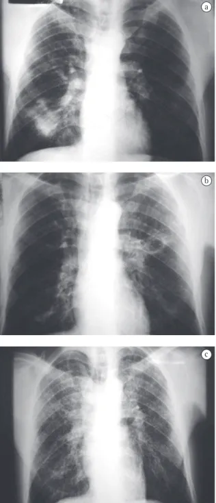
One case in particular seems to be illustrative of a true combination of PCM and TB. Eight years ago, the patient was definitively diagnosed with PCM through positive sputum smear testing for P. brasiliensis and was treated with trimethoprim-sulfamethoxazole for two years. The treatment was then suspended due to clinical improvement and negative serology. This patient again presented respiratory complaints five years after the end of the anti-PCM treatment, at which point he was diagnosed with TB (through positive sputum smear microscopy for acid-fast bacilli) and treated with Regimen I (rifampicin, isoniazid, and pyrazinamide). There was a good clinical response, and the patient was considered cured. Four months after the end of
the anti-TB treatment, the patient again complained of fever, weight loss, mild productive cough, and dyspnea, Recurrence of PCM was confirmed (Figure 5), and the treatment with trimethoprim-sulfamethoxazole was restarted.
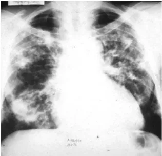
Figure 2 - Chest X-ray showing bilateral lung opacities predominantly in the middle lobes and with cavitation in the left upper lobe. Case of paracoccidioidomycosis concomitant with tuberculosis (acid-fast bacilli positive).
Figure 3 - Chest X-ray showing bilateral lung opacities predominantly in the upper lobes. Case of paracoccidioidomycosis previously treated for tuberculosis with no improvement (acid-fast bacilli negative).
Discussion
In the state of São Paulo, PCM and TB are common diseases. The incidence of TB is 45 cases per 100,000 inhabitants. However, neither the incidence nor the prevalence of PCM have been well-defined, since it is a disease for which reporting is not compulsory. Nevertheless, the prevalence of PCM is estimated to be 1 to 3 cases per 100,000 inhabitants
in endemic areas.(6-8)
In patients presenting a drop in cellular immu-nity, PCM and TB both occur in a more severe form, and such patients can present the two diseases concomitantly. Deficient production of certain
cytokines, such as IFN-γ, IL-12, and IL-23, as well as
of their receptors, predisposes patients to TB and to
PCM.(5) The pattern of cytokine production by CD4+ T
lymphocytes can determine the severity of the clin-ical profile of these two diseases. The predominance
of IFN-γ (produced by CD4+ T lymphocytes known as
T helper 1 cells) is generally associated with a more favorable clinical evolution, whereas the
predomi-nance of IL-4, IL-5, and IL-10 (produced by CD4+ T
lymphocytes designated T helper 2 cells) results in a
more severe disease progression.(9,10)
The majority of individuals with PCM are male (15:1) and are farm laborers or former farm laborers between 30 and 50 years of age. Although such indi-viduals are often oligosymptomatic, with few physical examination findings, they can present pronounced alterations on simple chest X-rays and on
high-reso-lution computed tomography scans of the chest.(11-15)
Pulmonary alterations are typically bilateral, predominantly in the middle lobes and spinal cord regions.(16,17)
In contrast, TB affects males and females of all ages, the majority of whom are symptomatic and present abnormal physical examination findings, as well as radiological alterations, which can also be bilateral but are typically most pronounced in the superior and posterior segments.
Despite these differences, it is not uncommon for the differential diagnosis between the two diseases to be difficult. There have been various reports of the PCM/TB combination. In a study of 338 cases
of PCM,(12) 46 (13.65%) of the patients were found
to have also been diagnosed with TB. The patients were divided into 3 groups: a) 19 cases, 10 of which were inactive (latent TB), and 9 of which were active (symptomatic TB at the time of the PCM diagnosis);
Figure 5 - Sequential imaging of a given patient: a) 1997. Opacity in the left lung base: negativity for acid-fast bacilli and positivity for Paracoccidioides brasiliensis; b) 2004. Cavitary lesion in the left upper lobe: positivity for acid-fast bacilli; good response to anti-tuberculosis treatment; and c) Diffuse micronodular opacities (miliary pattern). Negativity for acid-fast bacilli and positivity for
P. brasiliensis with clinical and radiological improvement after treatment with trimethoprim-sulfamethoxazole.
a
b
b) 17 cases of patients with PCM who later devel-oped TB; and c) 10 cases in which the two diagnoses were made simultaneously.
In another study,(1) 422 cases of PCM were
studied. Of those, 23 (5.5%) were also diagnosed with TB. Another group of authors studied 147 patients with PCM and determined that 28 (19%)
had concomitant TB.(2)
In a study involving 159 patients with PCM,(16)
24 patients (15.09%) were found to have concomi-tant TB.
These results, however, do not differentiate between the cases in which there was bacteriolog-ical confirmation of TB and those with a history of previous treatment for TB without bacteriological confirmation.
Another study showed that, of 43 patients with PCM, 8 (18.6%) had a history of previous treat-ment for TB, but only 3 (6.99%) presented positive
sputum smear microscopy results.(18) Even within
the indigenous population in Brazil, 2 cases that were treated as TB were reported to have later been
definitively diagnosed as PCM.(19)
In the present study, we found that, of the 227 cases evaluated, 36 (15.8%) had been previ-ously treated for TB, but only 18 (7.9%) had had bacteriological and laboratory test confirmation had been obtained in only 18 (7.9%). Those 18 patients benefited from the specific anti-TB medication, being referred to our facility, some years later, with clinical complaints and radiological alterations that culminated in the diagnosis of PCM. The remaining 18 patients sought treatment at our facility because the treatment they had received provided no clinical improvement.
It should be borne in mind that, although the combination of PCM and TB has been documented in the literature, misdiagnosis is common due to the superimposition of and the similarity between their clinical and radiological presentations. In addition, most of the patients initially sought treatment at basic health clinics, where health care workers have more experience in dealing with TB, due to its high incidence, and are less likely to make a diagnosis of PCM. This has rarely happened at tertiary-care teaching hospitals, where these diseases are commonly encountered and diagnoses are typically confirmed prior to the initiation of treatment.
The PCM/TB combination is uncommon, and it can be difficult to make the differential
diag-nosis on the basis of clinical and radiological data. Incorrect treatment increases the chances of pulmo-nary sequelae, such as fibrosis, bronchiectasis, and chronic respiratory failure. Therefore, it is necessary to carry out exhaustive bacteriological investigation prior to instituting a specific therapeutic regimen for TB, as well as to increase the accuracy of and the emphasis on testing for fungi in sputum, especially at basic health clinics.
References
1. Paniago AMM, Aguiar JIA, Aguiar ES, Cunha RV, Pereira GROL, Londero AT, et al. Paracoccidioidomycosis - A clinical and epidemiological study of 422 cases observed in Mato Grosso do Sul. Rev Soc Bras Med Trop. 2003;36(4):455-9. 2. Leão RC, Mendes E. Paracoccidioidomycosis, neoplasia
and associated infections. Allergol Immunopathol (Madr). 1980;8(3):185-8.
3. Dannenberg AM. Pathophysiology: Basic aspects In: Schlossberg D. Tuberculosis and nontuberculous mycobacterial infections. 4th ed. Philadelphia: W.B. Saunders Co.; 1999. p. 17-47.
4. Musatti CC. Imunidade celular. In: Del Negro GD, Lacaz CS, Fiorillo AM. Paracoccidioidomicose - Blastomicose sul-americana. São Paulo: Sarvier; 1982. p. 119-26.
5. Moraes-Vasconcelos D, Grumach AS, Yamaguti A, Andrade ME, Fieschi C, Beaucoudrey L, et al. Paracoccidioides brasiliensis disseminated disease in a patient with inherited deficiency in the beta1 subunit of the interleukin (IL)-12/ Il-23 receptor. Clin Infec Dis. 2005;41(4):31-7.
6. Santos WA, Silva BM, Passos ED, Zandonade E, Falqueto A. Associação entre tabagismo e paracoccidioidomicose: um estudo de caso - controle no Estado do Espírito Santo, Brasil. Cad Saúde Pública. 2003;19(1):245-53.
7. Martinez R, Ferreira MS, Mendes RP, Telles Filho FQ. Blastomicose sul-americana (Paracoccidioidomicose): Etioepidemiologia e ecologia. In: Veronesi R, Fiocaccia R, editors. Tratado de Infectologia. São Paulo: Atheneu; 1996. p. 1081-3.
8. Ministério da Saúde. Fundação Nacional de Saúde. Centro de Referência Prof. Hélio Fraga/Sociedade Brasileira de Pneumologia e Tisiologia. Controle de tuberculose: Uma proposta de integração ensino-serviço. Funasa/CRPHF/SBPT, 2002.
9. Marques Mello L, Silva-Vergara ML, Rodrigues V Jr. Patients with active infection with Paracoccidioides brasiliensis present a Th2 immune response characterized by high interleukin-4 and interleukin-5 production. Hum Immunol. 2002;63(2):149-54.
10. Schluger NW. The pathogenesis of tuberculosis - the first one hundred (and twenty-three) years. Am J Resp Cell Mol Biol. 2005;32(4):251-56.
11. Tarantino AB, Gonçalves AJR, Capone D, Aidé MA. Micoses pulmonares. In: Tarantino AB, editor. Doenças Pulmonares. 4a ed. Rio de Janeiro: Guanabara Koogan; 1997. p. 457-87.
13. Londero AT. Paracoccidioidomicose: patogenia, formas clínicas, manifestações pulmonares e diagnóstico. J Pneumol. 1986;(12):41-60.
14. Londero AT, Ramos CD. Paracoccidioidomicose. Estudo clínico e micológico de 260 casos observados no interior do Estado do Rio Grande do Sul. J Pneumol. 1990;(16):129-32. 15. Gonçalves AJR, Figueiredo CP, Barbosa LSG, Aguilar J,
Almeida SL, Andrade EM, et al. Paracoccidioidomicose - Considerações sobre algumas formas incomuns. Arq Bras Med. 1984;58(5):301-8.
16. Vale ACF, Guimarães RR, Lopes DJ, Capone D. Aspectos radiológicos torácicos em Paracoccidioidomicose. Rev Inst Med Trop São Paulo. 1992;34(2):107-15.
17. Muniz MAS, Marchiori E, Magnago M, Moreira LBM, Almeida Junior JG. Paracoccidioidomicose pulmonar - aspectos na tomografia computadorizada de alta resolução. Radiol Bras. 2002;35(3):147-54.
18. Quagliato R Jr. Considerações a respeito da Paracoccidioidomicose e sua correlação com níveis séricos de alfa-1 antitripsina [thesis].Campinas: Universidade Estadual de Campinas; 1998.