69
LETTERS DOI: Xxxxxxxxxxxxxxxxxxxxx
Multiple subcortical strokes caused by
mucormycosis in a patient with lymphoma
Múltiplos infartos subcorticais causados por mucormicose em paciente com linfoma
Pedro Enrique Jiménez Caballero, Alfonso Miguel Falcón García, Juan Carlos Portilla Cuenca, Ignacio Casado Naranjo
Mucormycosis is caused by a saprophytic fungal infection by non-septate hyphae of the genus Mucor. Five classic forms are recognized: rhinocerebral, pulmonary, gastrointestinal, cutaneous, and disseminated. Rhinocerebral mucormyco-sis (RCM) frequently occurs in immunosuppressed patients, such as those with uncontrolled diabetes mellitus or haema-tological malignancy1.
CASE REPORT
A 56-year-old man was diagnosed with follicular non-Hodgkin’s lymphoma. Four months prior to admission, he pre-sented a right headache attributed to rhinosinusitis. Biopsy showed mucormycosis. here was no afectation intracranial in the brain magnetic resonance imaging (MRI), but the pa-tient became blind due to involvement of both central reti-nal arteries. He underwent debridement of the paranasal si-nuses and treatment with liposomal amphotericin B for two months and posaconazole continuously.
He was admitted by recurrent episodes of nonluent aphasia, right sensory disturbance, and motor weakness. In the last episode, the symptoms persisted and the patient had transcortical motor aphasia, right hemiparesis with domi-nance of the lower limb (manual muscle testing: upper limb 2/5, lower limb 1/5) and right unilateral sensory impairment, which was restricted at his perioral area and homolateral dis-tal hand and foot.
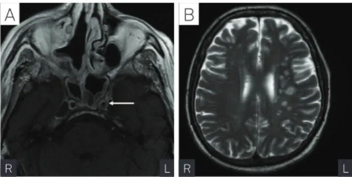
Hemogram showed lymphopenia (0,3x10⁹/L) and throm-bocytopenia (23x109/L). Blood clotting was normal. Plasma and urine ketone bodies were absent and other laboratory studies were normal. Echocardiogram, cardiac monitoring and Duplex of the supra-aortic trunk were normal. Transcranial duplex showed 70% stenosis at the M1 segment of left middle cerebral artery (MCA) with no other signiicant changes. MRI brain on T1 sequences with contrast (Fig A) showed an up-take at both cavernous sinus and internal carotid wall, which
Department of Neurology, San Pedro de Alcántara Hospital, Spain.
Correspondence: Pedro Enrique Jiménez Caballero; Calle Dionisio Acedo, 9 – Portal 7, 4-1ª, 10001 Cáceres - Spain; E-mail: pjimenez1010j@yahoo.es Conflict of interest: There is no conflict of interest to declare.
Received 19 June 2011; Received in final form 22 July 2011; Accepted 29 July 2011
determines a 70% arterial lumen stenosis in the left side. On T2 sequences (Fig B), multiple subcortical ischemic lesions in the left MCA territory could be seen.
he patient was diagnosed from multiple subcortical strokes in the territory of left MCA secondary to iniltration of its wall caused by mucormycosis. He was again treated with liposomal amphotericin B for two months, with clinical and radiological improvement. In the six-month follow-up, no new cerebrovascular disorders have appeared.
DISCUSSION
Mucor is attracted to blood vessels and invasion of their wall (particularly arterial) is the pathological trademark of the infection. The integrity of host defense mechanisms plays a key role. The presence of certain underlying dis-eases as lymphoma provides a favourable microenviron-ment for fungal growth. The disease usually spreads to the cavernous sinus, internal carotid artery and subsequent-ly to the brain2. Vascular manifestations of mucormycosis
Fig. Brain magnetic resonance image. (A) Axial T1-weighted image with contrast – uptake at both cavernous sinus and internal carotid wall (arrow) that determine an arterial lumen stenosis in the left side. (B) Axial T2-weighted image shows multiple subcortical ischemic lesions in the left middle cerebral artery territory.
R L R L
70 Arq Neuropsiquiatr 2012;70(1):69-70
References
1. Mathur SC, Friedman HD, Kende AI, Davis RL, Graziano SL. Cryptic mucor infection leading to massive cerebral infarction at initiation of antileukemic chemotherapy. Ann Hematol 1999;78:241-245.
2. Simmons JH, Zeitter PS, Fenton LZ, Abzug MJ, Fiallo-Scharer RV, Klingensmith GJ. Rhinocerebral mucormycosis complicated by internal carotid artery thrombosis in a pediatric patient with type 1 diabetes mellitus: a case report and review of the literature. Pediatr Diabetes 2005;6:234-238.
3. Takahashi S, Horiguchi T, Mikami S, Kitamura Y, Kawase T. Subcortical intracerebral hemorrhage caused by mucormycosis in a patient with
a history of bone-marrow transplantion. J Stroke Cerebrovasc Dis 2009;18:405-406.
4. Calli C, Savas R, Parildar M, Pekindil G, Alper H, Yunten N. Isolated pontine infarction due to rhinocerebral mucormycosis. Neuroradiology 1999;41:179-181.
5. Galetta SL, Wule AE, Goldberg HI, Nichols CW, Glaser JS. Rhinocerebral mucormycosis: management and survival after carotid occlusion. Ann Neurol 1990;28:103-107.
include pseudoaneurysms, partial thrombosis, narrowing and arteritic irregularities of intracranial arteries. Their pathologic basis is believed to be a combination of di-rect endothelial injury and growth of hyphae into the lu-men, with resultant distal infarcts and mycotic emboli. Vasculitis usually occurs in the supraclinoid portion of the internal carotid artery. Mycotic pseudoaneurysms, arterial dissection, or venous congestion could lead to intracranial
hemorrhages3. Rapid thickening and enhancement of the carotid artery wall on serial MRI establishes the nature of the arterial involvement by mucormycosis rather than atherosclerosis4.