Copyright © 2007 by Sociedade Brasileira de Pediatria
O
RIGINALA
RTICLEImpact of obesity on ventilatory function
Perran Boran,1 Gulnur Tokuc,2 Burcu Pisgin,3 Sedat Oktem,1 Zeliha Yegin,3
Ozlem Bostan3
Abstract
Objective:Although obesity was found to be associated with severe impairment of ventilation, most of the study
population has been morbidly obese adults. We aimed to explore the effects of mild obesity on ventilatory function in
the pediatric age group.
Methods:In a cross-sectional controlled study, 80 patients (M/F: 35/45), who were evaluated in our outpatient
clinic with the complaint of excess body weight, with no history of asthma or other atopic diseases were studied and
compared to a control group of 50 normal weight children controlled for age and sex. The mean age of patients was
9.7±2.5 years (7 to 15 years). Anthropometric measurements and spirometry were performed in all subjects. Forced
vital capacity (FVC) and forced expiratory volume in 1 second (FEV1) were used as measures of ventilatory function.
Results:There were no significant differences in FEV1%, FVC% and FEV1%/FVC% by study group (p > 0.05).
Only three patients had obstructive abnormalities documented on their pulmonary function tests (two had moderately
severe and one had mild obstructive abnormalities). No correlation was observed between pulmonary function
parameters and anthropometric measurements.
Conclusion:These data demonstrate that pulmonary function test parameters of the mildly obese children were
similar to those of the normal weight children. Anthropometric measurements had no significant effect on spirometric
measurements in children as they did on adults.
J Pediatr (Rio J). 2007;83(2):171-176:Obesity, ventilatory function, spirometry.
Introduction
Childhood obesity is associated with a range of adverse
consequences.1 Concerns about obese children include
metabolic and physical disorders other than psychosocial
stress. Abnormalities of the respiratory function have been
observed in many studies.2-4
Several mechanisms have been proposed on the possible
effects of obesity on pulmonary function. The most common
abnormalities reported are reduced expiratory reserve
volume and functional residual capacity due to reduced chest
wall and lung compliance and increased respiratory
resistance.5,6It is also believed that increased pulmonary
blood volume leads to congestion resulting in thickening of
the airway wall; thus reducing airway size.7
Although morbid obesity was found to be associated with
severe impairment of ventilation, studies on the effects of
mild obesity on ventilatory function are limited.3
1. MD. Pediatrician, 2nd Clinic of Pediatrics, Dr. Lutfi Kirdar Kartal Research and Training Hospital, Istanbul, Turkey. 2. MD. Associate professor, 2nd Clinic of Pediatrics, Dr. Lutfi Kirdar Kartal Research and Training Hospital, Istanbul, Turkey. 3. MD. Resident, 2nd Clinic of Pediatrics, Dr. Lutfi Kirdar Kartal Research and Training Hospital, Istanbul, Turkey.
Manuscript received Jul 31 2006, accepted for publication Dec 13 2006.
Suggested citation:Boran P, Tokuc G, Pisgin B, Oktem S, Yegin Z, Bostan O. Impact of obesity on ventilatory function. J Pediatr (Rio J). 2007;83(2):171-176.
doi 10.2223/JPED.
It was hypothesized that obesity can have adverse effects
on ventilatory function even in mildly obese children. The
purpose of this article is to explore the effects of simple
obesity on ventilatory function.
Methods
We carried out a cross-sectional controlled trial in 100
patients who were admitted to Dr. Lutfi Kirdar Kartal Training
and Research Hospital, pediatric outpatient clinic, with the
complaint of excess body weight, and compared to a control
group of normal weight children of similar ages, between
November 2004 and May 2005. The control group was
composed of healthy children who have attended our
outpatient clinic for their routine checkups, vaccines,
nutritional assessments and who have normal physical
examination.
Based on information from previous studies, a sample
size of 50 children per group was calculated to be adequate to
detect a difference.
In this study an obese subject was defined as one whose
BMI was above the 95th percentile according to sex- and
age-specific BMI reference range using the new charts
provided by the Centers for Disease Control and Prevention.8
Those with obesity secondary to an organic condition (one
patient had Hashimoto’s thyroiditis); having a chronic
cardiorespiratory or neuromuscular problem or history of
asthma and other atopic diseases were excluded from the
study. The remaining 80 patients (M/F: 35/45; mean age
9.7±2.5 years) with obesity were enrolled into the study.
They were compared with 50 healthy children with normal
weight (M/F: 20/30; mean age 9.2±2.08).
A questionnaire was administered by the investigators to
determine risk factors including daily TV viewing time, eating
habits, daily physical activities, and family history of obesity.
Parents were asked about the child's snoring, difficulty
breathing, observed apnea, cyanosis, struggling to breathe,
shaking the child to "make him or her breathe," watching the
child sleep, afraid of apnea, the frequency and loudness of
snoring, and daytime symptoms such as excessive daytime
sleepiness to determine obstructive sleep apnea (OSA)
symptoms.
An informed consent was obtained from the subjects and
their parents.
Anthropometric measurements and spirometry were
performed in all subjects. Height was measured to the
nearest 1 cm against a wall chart, and weight was measured
to the nearest 0.1 kg using an electronic digital scale. BMI was
calculated as weight (kg) divided by the square of height in
meters (kg/m²). Waist circumference was measured as the
minimum abdominal circumference between the xiphoid
process and the umbilicus. Hip circumference was measured
as the maximum circumference over the buttocks. The
waist-to-hip ratio (WHR) was calculated as the ratio between
these two circumferences. The height and weight of patients’
parents were also measured by the same physician and BMI
was calculated.
Spirometry (Spiromed- microplus M503, MAN5105,
spirometer) was performed in all subjects. The best of at least
three technically acceptable values for forced expiratory
volume in 1 second (FEV1) and forced vital capacity (FVC)
were selected. Forced vital capacity and FEV1 were used as
measures of ventilatory function. The pulmonary function
test results were expressed as percentages of predicted
normal values.9For the purpose of this study, the threshold of
abnormality was identified as less than 80% of the predicted
value. Obstructive airway disease was identified as a
decrease in the FEV1/FVC ratio to less than 80%. The various
pulmonary deficits were classified as “mild” (> 70%),
“moderate” (< 70% and > 60%), “moderately severe”
(< 60% and > 50%), and “severe” (< 50%). The reversibility
test was applied to subjects whose FEV1/FVC ratio decreased
to less than 80%. Salbutamol sulfate was used for the test
and it was inhaled twice and FEV1 was measured 15 minutes
later. If the difference in FEV1 before and after bronchodilator
inhalation was greater than or equal to 15%, the test was
accepted as positive.
The data were analyzed by SPSS software 10.0.
Numerical variables were evaluated by one-sample
Kolmogorov Smirnov test to assess whether they followed a
normal distribution. Since the parameters were not normally
distributed, a nonparametric Mann-Whitney U test was used.
Medians, 1st and 3rd quartiles were computed to describe
respiratory function parameters. A p value less than 0.05 was
considered statistically significant. The chi-square test was
used to compare sex and age distribution in both groups.
Regression models were built and the covariates were
tested by the enter and forward approach. Several risk
factors such as weight, body mass index, relative weight and
waist-to-hip ratio were included as independent variables.
Results
Eighty children with exogenous obesity aged between 7 to
15 years (mean age 9.7±2.5 years) were enrolled and
compared with 50 healthy normal weight children (mean age
9.2±2.08 years). The male/ female ratio of the obese and
normal weight children were 35/45 and 20/30, respectively.
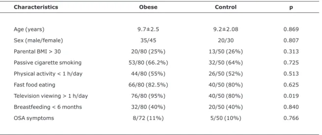
Demographic characteristics and obesity-associated risk
The children in the two groups were comparable for a
number of baseline characteristics, including age, sex, and
child feeding practices, parental obesity, passive cigarette
smoking, OSA symptoms and performance of physical
activity, except for television viewing. Significantly more
obese subjects are reported to watch television more than
one hour a day (p < 0.05).
Anthropometric measurements of the obese and control
groups are given in Table 2.
The mean values of weight, relative weight, BMI, and
WHR were significantly higher in the obese group, as
expected (p < 0.01). There were no significant differences in
age and sex by study group (p = 0.888).
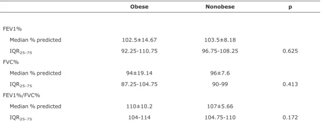
Mildly obese subjects compared to nonobese subjects did
not differ in any of the lung function measurements (Table 3).
Three patients in the obese group had obstructive
abnormalities documented on their pulmonary function tests
(two had moderately severe and one had mild obstructive
abnormality). The reversibility test was positive in these
three patients. These three patients had no asthma
symptoms such as dyspnea, wheezing, chronic cough or
previous history of atopy.
According to regression analyses, anthropometric
measurements had no significant effect on FEV1% (p = 0.3),
FVC% (p = 0.545), and FEV1/FVC% (p = 0.869).
Discussion
Many studies have demonstrated an association between
obesity and ventilatory abnormalities in adults.10-12
However, investigations into this issue in childhood are
limited2-4,13-15and studies conducted to date have yielded
conflicting results. Many have focused on extreme levels of
obesity, or have used a small sample size.
Chaussain et al., in their study of 39 obese children,
reported that lung compliance and resistance reflected as
vital capacity and residual volume were similar to those of the
control group.15 Bosisio et al., in their study of 23 obese
children, also found lung volumes to be within the normal
range.4Consistent with these studies, our results revealed
that FEV1 %, FVC % and FEV1%/FVC% were similar to those
of the control group.
Similar studies in children confirm reduced functional
residual capacity and static lung volumes. Mallory et al. found
that 3 out of 17 obese patients had restrictive and 8 out of 17
had obstructive changes in pulmonary function3Inselman et
al.2and Li et al.14found reductions in diffusing lung capacity
to be common among the obese children they studied. They
suggested that reductions in diffusing lung capacity seen in
children may reflect structural changes in the interstitium of
the lung, resulting in decreased alveolar surface area.
One possible explanation for these conflicting results may
be that most of the studies have focused on extreme levels of
Table 1- Baseline characteristics of children enrolled in the obese and control groups
Characteristics Obese Control p
Age (years) 9.7±2.5 9.2±2.08 0.869
Sex (male/female) 35/45 20/30 0.807
Parental BMI > 30 20/80 (25%) 13/50 (26%) 0.313
Passive cigarette smoking 53/80 (66.2%) 32/50 (64%) 0.725
Physical activity < 1 h/day 44/80 (55%) 26/50 (52%) 0.513
Fast food eating 66/80 (82.5%) 40/50 (80%) 0.625
Television viewing > 1 h/day 76/80 (95%) 40/50 (80%) 0.019
Breastfeeding < 6 months 32/80 (40%) 20/50 (40%) 0.840
OSA symptoms 8/72 (11%) 5/50 (10%) 0.766
obesity, or have used small sample sizes without a control
group. It is also possible that conventional respiratory
function tests are only mildly affected except in extreme
cases and that individuals with different levels of obesity will
exhibit a different response. Ray et al. emphasized that total
lung capacity and vital capacity may be reduced only in
extreme obesity.16
Although abnormalities of the respiratory function are a
common finding in adult obesity, we can not draw any
conclusions from adult studies since physiological function
and body fat deposition are different from those of children
and also there are so many confounding factors such as
smoking status, and an abnormal pulmonary function test
value can be considered to be caused by intrinsic lung disease
or factors other than obesity.
Previous studies suggested that patterns of fat deposition
are important in determining the consequences of obesity
and that high WHR is inversely related to spirometry and
Table 2- Anthropometric measurements of obese patients and controls
Weight (kg) Height (cm) BMI (kg/m2) Relative weight (%)
Waist/Hip circumference
Obese 51.2±15.7 140.4±15.6 25.5±2.1 139.3±15.3 0.89±0.1
Nonobese 29.6±10.2 139.2±16.2 17.1±1.9 95.5±12.2 0.81±0.2
p < 0.01 0.76 < 0.01 < 0.01 < 0.01
BMI = body mass index. Values expressed as means ± SD.
Table 3- Pulmonary function tests of obese and normal weight children
Obese Nonobese p
FEV1%
Median % predicted 102.5±14.67 103.5±8.18
IQR25-75 92.25-110.75 96.75-108.25 0.625
FVC%
Median % predicted 94±19.14 96±7.6
IQR25-75 87.25-104.75 90-99 0.413
FEV1%/FVC%
Median % predicted 110±10.2 107±5.66
IQR25-75 104-114 104.75-110 0.172
FEV1% = percentage predicted forced expiratory volume in 1 second; FVC% = percentage predicted forced vital capacity; IQR = interquartile range.
static lung volume.17 In our study, anthropometric
measurements are not correlated with spirometric
measurements in children as they are in adults. Although the
waist-to-hip ratio was significantly higher in the obese group,
it may not be sufficiently high to exert any effect on
pulmonary function.
It is also possible that anthropometric measurements
have failed to determine fat distribution accurately.
Conventional anthropometric measurements have been
criticized as being unreliable and insufficiently sensitive to
assess intraabdominal fat.18 A more valid and precise
measure of body fat distribution, such as measurements
obtained with modern investigation methods such as CT, MRI
or DEXA (dual energy X ray absorptiometry) would be
preferred, but we did not want to expose patients to radiation.
Furthermore, the deposition of visceral fat is very
age-dependent; in one study, visceral fat was shown to
increase in men from 12.4% of body surface at age younger
than 40 years to 18% after age 65.19 This increase was
independent of obesity. By contrast, the figure was 5.4% for
adolescents and adiposity for male and female children is
predominantly subcutaneous which may not constitute a
great health risk.18
There have been reports in the literature suggesting an
association between asthma and obesity.20,21Although three
patients had reversible obstructive abnormality documented
on their pulmonary function tests, they had no respiratory or
atopic symptoms previously and since we have not
performed any provocation tests, further studies are needed
to determine whether obesity causes or enhances bronchial
hyperreactivity.
Our study has certain limitations. First, it was a
cross-sectional study and, since measurements of the obese
subjects were taken at a single point in time, they may not
have accurately reflected the clinical status. Second,
radiological assessment would have been helpful in this
study, since it is capable of determining fat distribution more
accurately than anthropometric indices.
Conclusion
In conclusion, baseline pulmonary function test
parameters were not different between mildly obese and
normal weight children. Anthropometric parameters had no
significant effect on pulmonary function. Longitudinal studies
including physiological tests are needed to explore the effects
of different levels of obesity on pulmonary function in
children.
Acknowledgements
The authors are grateful to Haydar Sur for the statistical
analyses.
References
1. Deane S, Thomson A.Obesity and the pulmonologist. Arch Dis Child. 2006;91:188-91.
2. Inselma LS, Milanese A, Deurloo A. Effects of obesity on pulmonary function in children. Pediatr Pulmonol. 1993;16:130-7.
3. Mallory GB Jr., Fiser DH, Jackson R.Sleep associated breathing disorders in morbidly obese children and adolescents. J Pediatr. 1989;115:892-7.
4. Bosisio E, Sergi M, di Natale B, Chiumello G.Ventilatory volume flow rates, transfer factor and its components (membrane component, capillary volume) in obese adults and children. Respiration. 1984;45:321-6.
5. Pankow W, Podszus T, Gutheil T, Penzel T, Peter J H, Von Wichert P.Expiratory flow limitation and intrinsic positive end-expiratory pressure in obesity. J Appl Physiol. 1998;85:1236-43.
6. Zerah F, Harf A, Perlemuter L, Lorino H, Lorino AM, Atlan G. Ef-fects of obesity on respiratory resistance. Chest. 1993;103:1470-6.
7. Hogg JC, Pare PD, Moreno R.The effect of submucosal edema on airways resistance. Am Rev Respir Dis. 1987;135:S54-6.
8. National Center for Health Statistics. Hyattsville: pediatric growth charts provided by the CDC. http://www.cdc.gov/ growthcharts/2000. Access: 11/12/2005.
9. Standardization of spirometry, 1994 update.American Thoracic Society. Am J Respir Crit Care Med. 1995;152:1107-36.
10. De Lorenzo A, Maiolo C, Mohamed EI, Andreoli A, Petrone-De-Luca P, Rossi P. Body composition analysis and changes in airways function in obese adults after hypocaloric diet. Chest. 2001;119:1409-15.
11. Sahebjami H, Gartside PS.Pulmonary function in obese subjects with a normal FEV1/FVC ratio. Chest. 1996;110:1425-9.
12. Ferretti A, Giampiccolo P, Cavalli A, Milic-Emili J, Tantucci C. Expiratory flow limitation and orthopnea in massively obese subjects. Chest. 2001;119:1401-8.
13. Lazarus R, Colditz G, Berkey CS, Speizer FE.Effects of body fat on ventilatory function in children and adolescents: cross-sectional findings from a random population sample of school children. Pediatr Pulmonol. 1997;24:187-94.
14. Li A M, Chan D, Wong E, Yin J, Nelson E AS, Fok T F.The effects of obesity on pulmonary function. Arch Dis Child 2003; 88:361-363.
15. Chaussain M, Gamain B, La Torre AM, Vaida P, de Lattre J. Respiratory function at rest in obese children. Bull Eur Phsiopathol Respir. 1977;13:599-609.
16. Ray CS, Sue DY, Bray G, Hansen JE, Wasserman K.Effects of obesity on respiratory function. Am Rev Respir Dis. 1983;128:501-6.
18. Brambilla P, Manzoni P, Sironi S, Simone P, Del Maschio A, di Natale B, et al.Peripheral and abdominal obesity in childhood obesity. Int J Obes Relat Metab Disord. 1994;18:795-800.
19. Seidell JC, Oosterlee A, Deurenberg P, Hautvast JG, Ruijs JH. Abdominal fat depots measured with computed tomography: ef-fects of degree of obesity, sex and age. Eur J Clin Nutr. 1988;42:805-15.
20. Schachter LM, Salome CM, Peat JK, Woolcock AJ. Obesity is a risk for asthma and wheeze but not airway hyperresponsiveness. Thorax. 2001;56:4-8.
21. Bibi H, Shoseyov D, Feigenbaum D, Genis M, Friger M, Peled R, et al.The relationship between asthma and obesity in children: is it real or a case of over diagnosis?J Asthma. 2004;41:403-10.
Correspondence: Perran Boran