Copyright © 2007 by Sociedade Brasileira de Pediatria
R
EVIEWA
RTICLEPharmacologic support of infants
and children in septic shock
José Irazuzta,1 Kevin J. Sullivan,2 Pedro Celiny R. Garcia,3 Jefferson Pedro Piva3
Abstract
Objectives:Septic shock (SS) is a frequent cause for admission to the pediatric intensive care unit, requiring prompt recognition and intervention to improve outcome. Our aim is to review the relevant literature related to the
diagnosis and management of SS and present a sequential management for its treatment.
Sources:Non-systematic review of medical literature using the MEDLINE database. Articles were selected according to their relevance to the objective and according to the authors’ opinions.
Summary of the findings: The outcome of sepsis and SS is dependent on the early recognition and implementation of time-sensitive goal-directed therapies. These include rapid aggressive fluid resuscitation followed
by a well-designed pharmacotherapy. The goals of the resuscitation are the restoration of microcirculation and
improved organ tissue perfusion. Clinical and laboratory markers are needed to assess the adequacy of the
treatments. Altered pharmacokinetic and pharmacodynamic responses dictate that vasoactive agents should be
adjusted to achieve the predetermined goals. In initial resuscitation with isotonic solutions (> 60 mL/kg), either
crystalloid (normal saline) or colloid infusion could be used. Despite adequate fluid resuscitation, if: (a) wide pulse
pressure, low blood pressure, or bounding pulses (high cardiac output, low systemic vascular resistance – SVR) are
present, norepinephrine should be considered; (b) prolonged capillary refill, weak pulses, narrow pulse pressure,
normotensive (low cardiac output, high SVR), dopamine, epinephrine or dobutamine should be considered.
Adjunctive therapy with stress dose of corticosteroid is indicated in selected populations.
Conclusions: Septic shock hemodynamics is a changing process that requires frequent assessment and therapeutic adjustments.
J Pediatr (Rio J). 2007;83(2 Suppl):S36-45: Septic shock, sepsis, pediatric intensive care, fluid resuscitation, hemodynamic support, corticosteroids.
Introduction
A significant improvement in the outcome of sepsis and
septic shock (SS) over the last few years has been in large
part due to the utilization of aggressive fluid resuscitation and
to the implementation of time-sensitive goal-directed
therapies.1-4Early diagnosis of SS is paramount to initiate
therapy. SS presents as a constellation of signs of infection,
hemodynamic dysfunction and organ failure. The most
common symptoms are hypothermia or hyperthermia,
tachycardia, altered mental status, diminished (cold shock)
or bounding peripheral pulses (warm shock), prolonged
(> 3 seconds, cold shock) or brisk capillary refill (warm
shock), mottled or cool extremities, and diminished urine
output (< 1 mL/kg/h). A wide pulse pressure (diastolic blood
1. MD. Division of Pediatric Critical Care Medicine, Health Science Center, University of Florida, Jacksonville, FL, USA.
2. MD. Division of Pediatric Critical Care Medicine, Health Science Center, University of Florida, Jacksonville, FL, USA. Department of Pediatric Anesthesia and Critical Care Medicine, Nemours Children’s Clinic, Jacksonville, FL, USA.
3. MD, PHD. Department of Pediatrics, Hospital São Lucas and Medical School, Pontifícia Universidade Católica do Rio Grande do Sul (PUCRS), Porto Alegre, RS, Brazil.
Suggested citation:Irazuzta J, Sullivan KJ, Garcia PC, Piva JP. Pharmacologic support of infants and children in septic shock. J Pediatr (Rio J). 2007;83(2 Suppl):S36-45.
doi 10.2223/JPED.1623
pressure that is less than one-half the systolic pressure) is
sometimes observed; hypotension, not always present, is a
late sign of SS.
The lack of rapid restoration of adequate microcirculation
triggers a cascade of inflammation and disseminated
microthrombosis for which, in pediatrics, no effective
treatment is available at present. It is not possible to evaluate
the completeness of resuscitation by a single parameter; a
comprehensive evaluation of clinical or biochemical
measurements is needed.5 Inadequate early resuscitation
results in multiple organ system failure and in death days to
weeks after the initial presentation. A report showed that
every hour that went by without restoration of appropriate
circulation was associated with a two-fold increase in
mortality.6
During SS, the tissue oxygen supply is inadequate to
meet metabolic demands, which are significantly increased in
critical organs. Additionally, there is a maldistribution of
cardiac output with increased blood flow to skeletal muscles
at the expense of a relative hypoperfusion of the splanchnic
circulation. Thus, the therapeutic goals are to restore
effective intravascular blood volume, support the needs of an
increased cardiac output and oxygen delivery while
redirecting blood flow to essential organs and preventing
microthrombosis.
Septic shock is further classified as fluid-sensitive septic
shock, fluid-refractory septic shock (fail to improve with
adequate volume resuscitation), catecholamine-resistant SS
(fail to improve with fluids and catecholamines), and
refractory septic shock (fail to improve with fluids,
catecholamines and vasodilators). SS is a dynamic process;
so vasoactive medications and their infusion dose may have
to be changed and adjusted over time to maintain adequate
organ perfusion and microcirculation. Vasoactive agents
have varying effects on systemic vascular resistance (SVR)
(i.e., vasodilators or vasopressors) and pulmonary vascular
resistance, contractility (i.e., inotropy) and heart rate
(chronotropes). Age of the patient and changes in the
perfusion of liver and kidney affect the pharmacokinetics of
vasoactive medications (available drug in serum). The
pharmacodynamic response is affected by inflammation,
nitric oxide production and down-regulation of receptors.
Thus, the recommended infusion doses are approximations
and should be adjusted to achieve predetermined goals of
organ perfusion and microcirculation.
Initial assessment and management
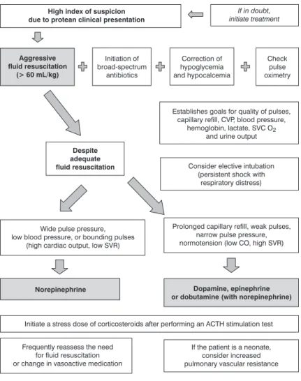
Initial resuscitation of infants and children centers on the
administration of isotonic solutions in quantities of 20 mL/kg
over 10 minutes repetitively while monitoring the patient’s
clinical response to treatment (Figure 1). There are no data
that confirm the superiority of either crystalloid (normal
saline) or colloid in children. All patients with SS suffer from
some degree of relative hypovolemia secondary to systemic
vasodilation, capillary leak, increased insensible loss, and
diminished oral intake, and may require up to 200 mL/kg of
intravenous fluids to adequately restore circulating volume.
This fluid resuscitation does not lead to an increased
incidence of acute respiratory distress syndrome (ARDS or
non-hydrostatic pulmonary edema).7It is often necessary to
begin treatment with vasoactive/inotropic medications
concomitantly with the initiation of fluid resuscitation in
patients who present with unstable hemodynamics (i.e.: low
heart rate, low cardiac output). During and after initial fluid
resuscitation, clinical and laboratory parameters regarding
the patient’s response to treatment should be evaluated.
Clinical evidence of positive response includes increased
strength of peripheral pulses, warmth of extremities,
decreased pulse rate, narrowing and normalization of blood
pressure, improvements in mental status and in urine output.
Unfortunately, clinical response to fluid resuscitation is a
relatively insensitive indicator of the completeness of
restoration of microvascular blood flow. The oxygen
saturation of the superior vena cava (SVC O2) (an indirect
measurement of cardiac output and oxygen utilization) and
serum lactate (the product of anaerobic metabolism) are
markers to assess microcirculation.5 Verification of
increasing and acceptable measurements of SVC O2(> 70%)
is recommended to demonstrate adequacy of systemic
oxygen delivery relative to demand.8This finding is especially
reassuring when serum lactate levels are declining. Elevated
central venous oxygen saturations in the setting of increasing
serum lactate may indicate the presence of cellular metabolic
failure and inability to extract and consume oxygen.
Patients with inadequate resolution of shock in response
to fluid resuscitation (fluid-refractory SS) require
optimization of oxygen-carrying capacity and systemic
oxygen delivery. Invasive monitoring of central venous
pressure (CVP) is instituted to ensure that satisfactory right
ventricular preload is present (CVP = 10-12), and
oxygen-carrying capacity is optimized by transfusion of
packed red blood cells to correct anemia (hemoglobin
concentration > 10 g/dL). Recent reports put into question
the ability of CVP to reflect adequacy of the preload favoring
other measurements of cardiac output in response to a fluid
challenge.9If despite these measures the patient continues
to have incomplete response to resuscitation, it is necessary
to institute pharmacologic therapy to support circulation.
Hemodynamics in pediatric SS
Adults and children have different adaptive responses
that must be considered when selecting vasoactive agents.
Among adult patients, the most common hemodynamic
output. SVR is diminished due to decreased vascular
responsiveness to catecholamines, alterations in
α
-adrenergic receptor signal transduction, and theelaboration of inducible nitric oxide synthase. After volume
loading, cardiac output increases despite diminished ejection
fraction, as a result of compensatory responses that include
ventricular dilatation and increased heart rate.10 Indeed,
significant myocardial depression may be present among
adult patients with SS. Pediatric patients demonstrate
diverse hemodynamic profiles during fluid-refractory SS:
58% have low cardiac index responsive to inotropic
medication ± vasodilators, 20% exhibit high cardiac index
and low SVR responsive to vasopressor therapy, and 22%
present both vascular and cardiac dysfunctions,
necessitat-ing the use of vasopressors and inotropic support.7 The
heterogeneity and changing pattern of the hemodynamic
presentation, during the initial hours, dictate that an
incorrect cardiotonic/vasoactive regimen should be
suspected when there is unresponsiveness to fluid therapy
and to vasoactive agents.
The relative ability of infants and children to augment
cardiac output through increased heart rate is limited by their
pre-existing elevated heart rate, which precludes
proportionate increases in heart rate without compromising
diastolic filling time (Table 1). Additionally, the increased
connective tissue content of the infant’s heart and diminished
content of actin and myosin limits the potential for acute
ventricular dilatation.11
Almost invariably there is relative hypovolemia often
associated with a maldistribution of cardiac output. In the
presence of respiratory distress, an elective tracheal
intubation followed by mechanical ventilation will contribute
to redistributing blood flow from respiratory muscles toward
other vital organs. However, it is imperative to have an
adequate fluid resuscitation before the intubation as the
change from spontaneous breathing to positive pressure
ventilation will decrease the effective preload to the heart.
When sedatives and analgesics are used, a vasodilator effect
could be observed, affecting tissue perfusion independent of
ACTH = adrenocorticotropic hormone; CVP = central venous pressure; SVC O2= oxygen satu-ration of the superior vena cava; SVR = systemic vascular resistance.
the presence of hypotension. In such situation, early
vasoactive /inotropic support should be considered.
The maldistribution of blood flow with hypoperfusion of
the splanchnic circulation, even when global cardiac output is
normal or increased, represents a special challenge. One of
the beneficial effects of potent vasopressors in sepsis is to
redirect blood flow away from the skeletal muscles to the
splanchnic circulation.
Pharmacologic agents for the support of pediatric SS
Pharmacologic support must be individualized as
different hemodynamic abnormalities exist in pediatric
patients, and the primary hemodynamic abnormalities
present in a given patient may change with time and
progression of the patient’s disease.
The pharmacologic agents may be classified as inotropic
medications, vasopressors, and vasodilators. Inotropic
medications increase cardiac output by increasing
myocardial contractility and/or heart rate. Vasopressors
elevate SVR by increasing the tone of arterial circulation, and
vasodilators decrease arterial resistance, resulting in
decreased afterload and increased cardiac output without
affecting contractility.
In many cases, a single drug may have combined effects
that result in the alteration of contractility and SVR, or may
have dose-dependent differential effects on contractility and
SVR. Additionally, there is wide inter-individual variability
with respect to the pharmacodynamics of these medications,
resulting in different effects in different individuals at the
same infusion rate. Lastly, the medications have direct effects
on cardiovascular system, indirect effects mediated through
the patient’s autonomic nervous system, or mixed effects by
both mechanisms. In this section, we review the
pharmacodynamics of many of the medications commonly
used to support patients in SS.
The medications traditionally used to support circulation
in patients with sepsis and shock include vasopressors
(dopamine, norepinephrine, and vasopressin) and inotropes
(epinephrine, dobutamine, and milrinone). Newer
medica-tions, which include fenoldopam and levosimendan, may find
application in the management of SS. Finally, in infants,
special consideration and management are sometimes
required in the management of pulmonary artery
hypertension and calcium homeostasis.
Vasopressors
Vasopressor therapy is required in patients with
diminished SVR. Patients in SS with diminished systemic
resistance and elevated cardiac output will often have warm
extremities, bounding peripheral pulses, widened pulse
pressure, and normal or low blood pressure. In the presence
of diminished cardiac output, peripheral perfusion is often
compromised, and the blood pressure is often low.
Vasopressor therapy is initiated to restore perfusion to vital
organs; however, in the presence of diminished myocardial
contractility, it may further compromise cardiac output.
Therefore, appropriate monitoring is indicated. While the
medications listed in this section have vasopressor activity,
dopamine and norepinephrine also have some inotropic
activity and may increase heart rate and contractility as well.
Table 1-Threshold heart rates and perfusion pressure MAP-CVP or MAP-IAP for age (modified from The Harriet Lane Handbook12)
Heart rate (bpm) MAP-CVP (cm H2O)
Threshold rates
Term newborn 120-180 bpm 55
Up to 1 year 120-180 bpm 60
Up to 2 years 120-160 bpm 65
Up to 7 years 100-140 bpm 65
Up to 15 year 90-140 bpm 65
Dopamine
Dopamine is a precursor of norepinephrine in the adrenal
medulla and a neurotransmitter in the central nervous
system. Dopamine produces systemic hemodynamic effects
that are dose-dependent; however, some effects are
indirectly mediated by the release of norepinephrine from
sympathetic vesicles. In adults, with infusion rates of less
than 3 µg/kg/min, it induces the activation of primarily
dopaminergic (DA) receptors. At doses of 3-5 µg/kg/min,
dopamine activates DA (80-100%) and β-adrenergic receptors (5-20%), and at doses of 5-10 µg/kg/min, it
activates predominantlyβ-receptors with a small amount of
α
-adrenergic receptor activation. At doses above 10µg/kg/min
α
-adrenergic receptor activation predominates.Dopamine has been traditionally used as a first line
medication for the support of circulation, and has been used
in many types of critical illnesses as a non-specific support for
splanchnic and renal blood flow. Recent evidence puts these
practices into question.
Dopamine dose-related effects are unpredictable across
the pediatric population; many clinicians prefer to titrate
medications that independently and specifically address the
abnormalities of cardiac output and systemic resistance.
Examples of this hemodynamic strategy would be the
administration of dobutamine and a nitrovasodilator to the
patient with low cardiac output and elevated systemic
resistance, or the administration of dobutamine and
norepinephrine to the patient with diminished cardiac output
and low SVR. Such a strategy circumvents the inherent
difficulty in using medications that are agonists to a broad
range of cardiovascular receptors in patients with variable
and changing hemodynamic abnormalities.
Dopamine has also been administered in the hope of
augmenting splanchnic and renal blood flow, and preventing
progression of acute renal failure. There is no evidence to
support this practice, and several large trials and
meta-analyses indicate that this practice is of no benefit.13In
addition to this, emerging evidence indicates that dopamine
has several deleterious side effects that may negatively
impact morbidity and mortality, including decreased oxygen
consumption and gastric mucosal pHi in the gut despite
increased splanchnic blood flow,14 impairment of gastric
motility,15 blunting of hypoxic respiratory drive in
mechanically ventilated patients,16impairment in ventilation
/perfusion matching with worsening of hypoxemia,17
impairment of anterior pituitary hormone secretion and
cell-mediated immunity, and aggravation of impaired thyroid
function seen in critical illness.18Finally, the use of dopamine
and other indirect acting inotropes/vasopressors in preterm
infants and infants less than 6 months of age may be less
effective because of the immaturity of
norepinephrine-containing synaptic vesicles in the sympathetic nervous
system.19This constellation of side effects, lack of efficacy,
and imprecise targeting of hemodynamic variables have led
some clinicians to prefer norepinephrine to dopamine as their
initial vasopressor of choice. Other clinicians prefer dopamine
as their first choice based on many years of successful clinical
experience in the setting of hypotension and/or low cardiac
output. It is recommended to initiate dopamine at
5 µg/kg/min and titrate not to exceed 10 µg/kg/min.
Norepinephrine
Norepinephrine is a direct agent and is naturally produced
in the adrenal gland. It is a potent vasopressor that redirects
blood flow away from the skeletal muscle to the splanchnic
circulation even in the presence of decreased cardiac output.
The majority of adult patients with SS present diminished
SVR to some degree, which results in maldistribution of
cardiac output and organ hypoperfusion. Close to 20% of
children with volume-refractory SS have low SVR.7 In
children with SS receiving sedatives or analgesics, the
incidence of low SVR may be higher, and norepinephrine
infusion could be the drug of choice.
Norepinephrine has been used extensively to elevate SVR
in septic adults and children. Because of the fear of excessive
vasoconstriction, norepinephrine has historically been
avoided. The available evidence, however, does not support
this bias.20 In doses beginning as low as 0.02 µg/kg/min,
norepinephrine has been titrated upward to increase SVR,
elevate diastolic blood pressure, and decrease pulse
pressure. Several reports have described the ability of
norepinephrine to restore hemodynamic stability in
adequately volume-resuscitated patients refractory to
therapy with dopamine. When compared with dopamine,
resuscitation with norepinephrine is associated with more
rapid resolution of lactic acidosis,21and animal data suggest
that norepinephrine use exerts a protective effect on renal
blood flow in SS.22Human studies have also demonstrated an
improvement in urine output,21and no deleterious effects on
splanchnic perfusion in SS.23One study on adult patients has
even recognized a survival advantage among adult SS
patients treated with norepinephrine when compared with
other vasopressors.24
The use of norepinephrine avoids concerns over
age-specific insensitivity to dopamine. However, safe and
effective use of norepinephrine is predicated upon several
presumptions. First, patients have been effectively
volume-resuscitated, as this is the first and most important
treatment for SS. Through the provision of adequate volume
resuscitation, excessive doses of norepinephrine can be
avoided and then minimize complications secondary to
excessive vasoconstriction. Second, through clinical,
laboratory, and/or invasive monitoring techniques, we are
Excessive norepinephrine administration in a patient who is
inadequately volume-resuscitated may mislead the clinician
into believing that a patient is hemodynamically stable when,
in fact, vital organ perfusion is compromised. In patients with
impaired contractility, the additional afterload imposed by
norepinephrine may substantially compromise cardiac
output. In some patients with both impaired or marginal
cardiac output and decreased systemic resistance, it may be
necessary to support myocardial contractility through the
addition of an agent such as dobutamine.
Vasopressin
Vasopressin (antidiuretic hormone) is synthesized in the
hypothalamus. Under normal conditions, blood levels are
kept constant at concentrations largely regulated by serum
osmolarity. Vasopressin is rapidly metabolized by the liver
and kidney with a half-life of 10-30 minutes.25Vasopressin is
also released in response to decreases in blood pressure with
serum levels increasing more than ten-fold to improve blood
pressure via vasoconstriction.26 At low concentrations,
catecholamines exert stimulatory effects upon vasopressin
release via central
α
-1 receptors, but at higher levels, theymay inhibit vasopressin release by stimulating
α
-2 and βreceptors.27 Hypoxia, acidosis, endotoxin, and cytokines
stimulate vasopressin release; nitric oxide inhibits its
secretion.27
The actions of vasopressin are mediated by G-protein
coupled receptors that are classified as V1, V2, V3, and
oxytocin receptors (OTR). V1 receptors are located in
vascular smooth muscle cells in the systemic, splanchnic,
renal, and coronary circulations. Activation of V1receptors
results in increased intracellular calcium concentrations,
smooth muscle contraction, and vasoconstriction.27 V 2 receptors mediate the antidiuretic actions of vasopressin in
the nephron, and V3 receptors play a role in secondary
messaging in the anterior pituitary gland. OTR are located in
the myometrium and mammary myoepithelial cells, where
they mediate smooth muscle contraction, and are present on
the surface of endothelial cells, where activation leads to
increased endothelial cell calcium concentrations, activation
of nitric oxide synthase, and elaboration of nitric oxide
resulting in vasodilation.27
Patients with severe sepsis are very sensitive to
exogenous administration of vasopressin.28In acute SS, an
early ten-fold increase in vasopressin levels is noted.
However, after more prolonged shock, vasopressin levels fall
toward normal, resulting in relative vasopressin
deficiency.28,29The cause of diminished vasopressin levels in
sepsis may be related to impaired osmoregulation or
impaired baroregulation, or to the inhibitory effects of
increased nitric oxide on vasopressin release, both of which
are conditions associated with severe sepsis.27
Exogenous administration of vasopressin produces blood
pressure elevation in patients with SS, whereas it produces
no pressor response in healthy patients. The mechanism for
this exaggerated sensitivity to vasopressin is not clear.
In patients with catecholamine-refractory SS and
elevated cardiac output with low systemic resistance,
vasopressin is initiated in low doses and titrated to the desired
clinical effect. In patients with persistent vasodilation
unresponsive to catecholamines, vasopressin may be
effective in restoring SVR and blood pressure.
In a personal note, we have used vasopressin in SS
refractory to norepinephrine (> 1 µg/kg/min) despite
adequate fluid resuscitation and corticosteroids. Vasopressin
was titrated until a positive response was observed, and
subsequently, norepinephrine infusion was amenable to be
reduced. Hyponatremia is a common side effect that is
generally observed after 24 h of drug infusion.
Vasopressin is a potent vasoconstrictor and may
precipitate coronary, mesenteric, and cutaneous ischemia in
high doses. There is evidence to suggest that vasopressin
may produce neutral or beneficial effects on renal blood flow
and urine output.30It is prudent to monitor cardiac output
when initiating and titrating therapy with potent
vasoconstrictors in patients with marginal cardiac output and
diminished myocardial contractility. Addition of inotropic
support in patients with decreased cardiac output may be
required.
Inotropes
Inotropic medications are used to improve cardiac output
in patients with diminished myocardial contractility. They are
often administered concomitantly with vasopressors in
patients with diminished SVR or with a vasodilator in patients
with elevated systemic resistance. Milrinone and dobutamine
possess some vasodilatory properties and decrease afterload
while improving the contractile state of the myocardium.
Epinephrine, depending upon the dose administered, may
produce decrease in systemic resistance at low doses, or may
elevate systemic resistance at higher doses, while increasing
myocardial contractility. Dobutamine and epinephrine can
increase myocardial oxygen consumption and may produce
varying degrees of dysrhythmias and myocardial ischemia.
Epinephrine
Epinephrine is a direct agent that is naturally produced in
the adrenal gland and the principal stress hormone with
widespread metabolic and hemodynamic effects.
Epineph-rine is a naturally occurring catecholamine that possesses
potent inotropic and chronotropic effects. Epinephrine
infusions may be initiated at doses of 0.02 µg/kg/min and
titrated upward (up to 1.0 µg/kg/min) to achieve the desired
a central venous catheter, but can be administered through
an intraosseous needle or peripheral intravenous catheter
until central access is achieved. Subcutaneous infiltration of
epinephrine may result in soft tissue necrosis, which may be
antagonized by subcutaneous infiltration with phentolamine.
Epinephrine is a reasonable selection for the treatment of
patients with low cardiac output and poor peripheral
perfusion as it increases heart rate and myocardial
contractility.31 Depending on the dose administered,
epinephrine may exert variable effects on SVR. At low doses
(generally considered to be < 0.3 µg/kg/min) epinephrine
exerts greaterβ-2 adrenergic receptor activation, resulting in vasodilation in skeletal muscle and cutaneous vascular beds,
shunting the blood flow away from the splanchnic
circulation.32 At higher doses,
α
-1 adrenergic receptoractivation becomes more prominent and may increase SVR
and heart rate. For patients with markedly elevated systemic
resistance, epinephrine may be administered simultaneously
with a vasodilator.
Adult patients have been noted to exhibit decreased
intestinal mucosal pH in response to epinephrine infusion, but
it is not known whether gut injury occurs in septic children
receiving epinephrine infusions.32 Epinephrine increases
gluconeogenesis and glycogenolysis resulting in elevated
serum glucose concentrations. As a side effect of
gluconeogenesis stimulation, the skeletal muscle releases
more lactic acid to be transported to the liver for glucose
synthesis (the Cori cycle). As such, patients receiving
epinephrine may demonstrate elevated lactic acid
concentrations independent of any changes in organ
perfusion. Thus, serum lactate concentrations must be
followed closely while initiating epinephrine therapy as they
may not strictly reflect oxygen supply-demand balance.33
Dobutamine
Dobutamine is a synthetic agonist with a complex
stimulation ofβ-1,β-2,
α
-1 andα
-2 adrenergic receptors directly or through a metabolite. Dobutamine increasesmyocardial contractility and heart rate. Dobutamine lowers
systemic resistance in part by reflex withdrawal of
sympathetic tone. This hypotensive effect is pronounced and
seems to be more often observed in adult patients than in
small children. Dobutamine is considered for patients with
diminished cardiac output when accompanied with elevated
SVR.34 It is administered by continuous infusion of
3-20 µg/kg/min. In the setting of diminished contractility and
output and diminished systemic resistance, dobutamine has
been administered along with norepinephrine in order to
normalize both indices of hemodynamic function.35 Of
significant interest, dobutamine at 5 µg/kg/min seems to
increase splanchnic blood flow by a direct effect on the
microvasculature, independent of increasing cardiac
output.36A significant drop in blood pressure, unacceptable
tachycardia, increased myocardial oxygen consumption, and
atrial and ventricular dysrhythmias are undesirable potential
side effects.
Milrinone
Milrinone is a phosphodiesterase type III (PDE III)
inhibitor that produces its hemodynamic effects by inhibiting
the degradation of cyclic AMP in smooth muscle cells and
cardiac myocytes. PDE III inhibitors therefore work
synergistically with catecholamines, which produce their
hemodynamic effects by increasing the production of cyclic
AMP. Milrinone is useful in the treatment of patients with
diminished myocardial contractility and output and elevated
SVR as it mediates increased contractility and output, and
decreases systemic resistance.37 Additionally, milrinone
mediates its effects through mechanisms independent of
adrenergic receptors and is not affected by the
down-regulation and desensitization of these adrenergic
receptors.
As a vasodilator, milrinone administration may result in
decreased systemic blood pressure, and it may be necessary
to administer volume infusion in order to correct or prevent
hypotension. Milrinone has a long half-life of 2-6 hours, and
as such, it may take several hours to reach steady state
serum concentrations. In order to achieve rapid serum levels,
some clinicians administer a loading bolus of 50 µg/kg over
10-30 minutes. This must be done with caution in patients
with sepsis and shock as this may precipitate hypotension,
requiring volume infusion and/or vasopressor infusion. Some
clinicians divide the loading dose into a series of “mini-loads”
which are given over a more prolonged period of time to
minimize hypotension and test the patient’s ability to tolerate
the loading dose. The infusion dose of milrinone is
0.25-0.75 µg/kg/min. Because of its long half-life, it is
advisable to stop milrinone infusion if serious side effects
such as dysrhythmia, hypotension, or excessive vasodilation
occur. Additionally, because milrinone is predominantly
excreted in the urine, dosage may need to be adjusted in
response to deteriorating renal function in order to avoid
toxicity.
Vasodilators
Vasodilator medications are occasionally required in the
treatment of septic pediatric patients with markedly elevated
systemic resistance and normal or decreased cardiac output.
Vasodilators decrease SVR and improve cardiac output by
decreasing ventricular afterload. Some authors suggest the
use of nitroprusside as first line vasodilator due to its short
half life, because in case of hypotension, it could be rapidly
reversed after the infusion is interrupted. Nitroprusside is
infused at an initial rate of 0.5 µg/kg/min to a maximum dose
to sodium nitroprusside, which includes sodium thiocyanate
accumulation in the setting of renal failure, and cyanide
toxicity with prolonged high-dose infusions. Other clinicians
utilize milrinone as a vasodilator in situations of: a) refractory
SS with profound myocardial dysfunction or high SVR; and b)
pulmonary complications and suspectedly high pulmonary
vascular resistance – acute respiratory distress syndrome
(ARDS) or refractory hypoxemia.
Other drugs
Rescue from refractory shock has been described using
two newer medications that share PDE activity.
Levosimendan increases calcium sensitivity of the contractile
apparatus, and exerts some type III PDE inhibitor activity.
Enoximone is also a type III PDE inhibitor with more
selectivity for the preservation of cyclic AMP produced byβ-1 receptor activation in myocardial cells, and hence it improves
cardiac performance with less risk of undesired hypotension.
Fenoldopam is a selective postsynaptic dopamine (D1)
agonist utilized to prevent renal failure in shock. Fenoldopam
decreases peripheral SVR with increased renal and
splanchnic blood flow. It is six times as potent as dopamine in
producing renal vasodilation. Fenoldopam is infused at a dose
between of 0.1-1 µg/kg-/min.38
Thyroid dysfunction should be considered in the presence
of individuals with abnormal chromosome 21, central
nervous system diseases and pan-hypopituitary states. If T4
and T3 serum hormones are low, oral thyroxin (or
intravenous liothyronine) should be administered. Some
authors have described improvement in myocardial function
after thyroid hormone supplementation in SS.
Corticosteroids
Stress dose of corticosteroids may have an indication in a
selected patient population. Children are more likely to have
absolute adrenal insufficiency defined by a basal cortisol <
7 µg/dL and/or an increment after adrenocorticotropic
hormone (ACTH) stimulation < 18 µg/dL. Patients at risk of
inadequate cortisol production include those with purpura
fulminans, children who have previously received steroid
therapies for chronic illness, and patients with pituitary or
adrenal abnormalities.39Currently, ACTH stimulation test is
recommended to be performed with 1 µg of intravenous
corticotrophin instead of the high dose of 250 µg, which can
mask adrenal insufficiency.40
The unresponsiveness to vasoactive agents observed
during catecholamine-resistant SS is sometimes reversed by
the administration of hydrocortisone.41The hydrocortisone
dose should be titrated to the resolution of shock:
2-30 mg/kg/day divided every 6 h or 1-2 mg/kg/h as a
continuous infusion. Corticosteroids should be weaned off
after the resolution of SS, but maintained for a minimum of 5
to 7 days.
A syndrome of relative adrenal insufficiency (or
dysfunction) has been described (baseline cortisol
> 18 µg/dL with cortisol increment after ACTH stimulation
< 9 µg/dL). The administration of prolonged hydrocortisone
and fludrocortisone (6 mg/kg/d cortisol equivalent x 7 days)
is recommended for adults with relative adrenal insufficiency.
This practice is customary in some pediatric centers, but
there are not enough data to recommend steroid therapy for
adrenal dysfunction in the pediatric population.42
Glycemic control
Several studies have associated hyperglycemia with
higher mortality among critically ill patients. Hyperglycemia
is attributed to the peripheral resistance to insulin and
increased gluconeogenesis. Peripheral resistance to insulin is
proportional to the length and severity of the diseases and
closely associated with the outcome.
A strict glycemic control, between 80-110 mg/dL,
decreases mortality and morbidity in adult surgical
patients.43Retrospective studies involving children seem to
demonstrate similar results. However, it is challenging to
maintain this strict control in pediatrics without hypoglycemic
events; so a more liberal approach is often practiced. Glucose
levels and the length of hyperglycemia are independently
associated with mortality.44 A higher mortality rate was
associated with glucose above 178 mg/dL (odds ratio = 2.6)
in one pediatric study.45
Pulmonary hypertension
Although inhaled nitric oxide therapy is the treatment of
choice for uncomplicated persistent pulmonary hypertension
of the newborn (PPHN), metabolic alkalinization remains an
important initial resuscitative strategy during shock in
neonates. PPHN in the setting of SS can be reversed when
acidosis is corrected. For centers with access to inhaled nitric
oxide, this is the only selective pulmonary vasodilator
reported to be effective in the reversal of PPHN.
Extracorporeal membrane oxygenation (ECMO) remains the
therapy of choice for patients with refractory PPHN and
sepsis.1
ECMO is a viable therapy for refractory shock in neonates
and children. It is important to remember that neonates could
be exceedingly sensitive to hypocalcemia, hypoglycemia or
the lack of thyroid hormone.1
Conclusions
The outcome of sepsis and SS is in part dependent on the
implementation of time-sensitive goal-directed therapies.
aggressive fluid resuscitation, followed by a well-designed
pharmacotherapy. The goals of resuscitation are geared
toward the restoration of microcirculation and improvement
of organ tissue perfusion. Clinical and laboratory markers are
needed to assess the adequacy of treatment. Altered
pharmacokinetic and pharmacodynamic responses dictate
that vasoactive agents should be adjusted to achieve the
predetermined goals. SS hemodynamics is a changing
process that requires frequent assessment and therapy
adjustments. Adjunctive therapy with corticosteroids is
indicated in selected cases. Treatment during the initial hours
affects the outcome weeks later.
References
1. Carcillo JA, Fields AI. Clinical practice parameters for hemodynamic support of pediatric and neonatal patients in septic shock. Crit Care Med. 2002;30:1365-78.
2. Carcillo JA, Davis AL, Zaritsky A.Role of early fluid resuscitation in pediatric septic shock. JAMA. 1991;266:1242-5.
3. Lin SM, Huang CD, Lin HC, Liu CY, Wang CH, Kuo HP.A modified goal-directed protocol improves clinical outcomes in intensive care unit patients with septic shock: a randomized controlled trial. Shock. 2006;26:551-7.
4. Trzeciak S, Dellinger RP, Abate NL, Cowan RM, Stauss M, Kilgan-non JH, et al.Translating research to clinical practice: a 1-year experience with implementing early goal-directed therapy for septic shock in the emergency department. Chest. 2006;129:225-32.
5. Varpula M, Tallgren M, Saukkonen K, Voipio-Pulkki LM, Pettila V.
Hemodynamic variables related to outcome in septic shock. Int Care Med. 2005;31:1066-71.
6. Han YY, Carcillo JA, Dragotta MA, Bills DM, Watson RS, Wester-man ME, et al.Early reversal of pediatric-neonatal septic shock by community physicians is associated with improved outcome. Pediatrics. 2003;112:793-9.
7. Ceneviva G, Paschall JA, Maffei F, Carcillo JA.Hemodynamic sup-port in fluid-refractory pediatric septic shock. Pediatrics. 1998;102:e19.
8. Rivers E, Nguyen B, Havstad S, Ressler J, Muzzin A, Knoblich B, et al.Early goal-directed therapy in the treatment of severe sepsis and septic shock. N Engl J Med. 2001;345:1368-77.
9. Michard F, Alaya S, Zarka V, Bahloul M, Richard C, Telboul JL.
Global end-diastolic volume as an indicator of cardiac preload in patients with septic shock. Chest. 2003;124:1900-8.
10. Parker MM, McCarthy KE, Ognibene FP, Parrillo JE. Right ventricular dysfunction and dilatation, similar to left ventricular changes, characterize the cardiac depression of septic shock in humans. Chest. 1990;97:126-31.
11. Feltes T, Pignatelli R, Kleinert S, Mariscalco M.Quantitated left ventricular systolic mechanics in children with septic shock utilizing noninvasive wall stress analysis. Crit Care Med. 1994;22:1647-58.
12. National Heart, Lung, and Blood Institute. The Harriet Lane Handbook, 13th ed.Report of the Second Task Force on Blood Pressure Control in Children--1987. Pediatrics. 1987;79:1-25.
13. Kellum JA, Decker J.Use of dopamine in acute renal failure: a meta-analysis. Crit Care Med. 2001;29:1526-31.
14. Jakob SM, Ruokonen E, Takala J. Effects of dopamine on systemic and regional blood flow and metabolism in septic and cardiac surgery patients. Shock. 2002;18:8-13.
15. Dive A, Foret F, Jamart J, Bulpa P, Installe E.Effect of dopamine on gastrointestinal motility during critical illness. Int Care Med. 2000;26:901-7.
16. van de Borne P, Oren R, Somers V.Dopamine depresses minute ventilation in patients with heart failure. Circulation. 1998;98:126-31.
17. Shoemaker WC, Appel PL, Kram HB, Duarte D, Harrier HD, Ocampo HA.Comparison of hemodynamic and oxygen transport effects of dopamine and dobutamine in critically ill surgical patients. Chest. 1989;96:120-6.
18. Van den Berghe G, de Zegher F.Anterior pituitary function during critical illness and dopamine treatment. Crit Care Med. 1996;24:1580-90.
19. Padbury JF, Agata Y, Baylen BG, Ludlow JK, Polk DH, Habib DM, et al. Pharmacokinetics of dopamine in critically ill newborn infants. J Pediatr. 1990;117:472-6.
20. Sakr Y, Reinhart K, Vincent JL, Sprung CL, Moreno R, Ranieri VM, et al. Does dopamine administration in shock influence outcome? Results of the Sepsis Occurrence in Acutely Ill Patients (SOAP) Study. Crit Care Med. 2006;34:589-97.
21. Martin C, Papazian L, Perrin G, Saux P, Gouin F.Norepinephrine or dopamine for the treatment of hyperdynamic septic shock?
Chest. 1993;103:1826-31.
22. Bellomo R, Kellum JA, Wisniewski SR, Pinsky MR. Effects of norepinephrine on the renal vasculature in normal and endotoxemic dogs. Am J Respir Crit Care Med. 1999;159:1186-92.
23. LeDoux D, Astiz ME, Carpati CM, Rackow EC.Effects of perfusion pressure on tissue perfusion in septic shock. Crit Care Med. 2000;28:2729-32.
24. Martin C, Viviand X, Leone M, Thirion X.Effect of norepinephrine on the outcome of septic shock. Crit Care Med. 2000;28:2758-65.
25. Holmes CL, Patel BM, Russell JA, Walley KR. Physiology of vasopressin relevant to management of septic shock. Chest. 2001;120:989-1002.
26. Mutlu G, Factor P.Role of vasopressin in the management of septic shock. Intensive Care Med. 2004;30:1276-91.
27. Barrett LK, Singer M, Clapp LH.Vasopressin: mechanisms of ac-tion on the vasculature in health and in septic shockCrit Care Med. 2007;35:33-40.
28. Landry DW, Levin HR, Gallant EM, Ashton RC Jr., Seo S, D'Alessandro D, et al.Vasopressin deficiency contributes to the vasodilation of septic shock. Circulation. 1997;95:1122-5.
29. Sharshar T, Blanchard A, Paillard M, Raphael JC, Gajdos P, An-nane D.Circulating vasopressin levels in septic shock. Crit Care Med. 2003;31:1752-8.
30. Luckner G, Dunser MW, Jochberger S, Mayr VD, Wenzel V, Ulmer H, et al.Arginine vasopressin in 316 patients with advanced vasodilatory shock. Crit Care Med. 2005;33:2659-66.
32. De Backer D, Creteur J, Silva E, Vincent JL.Effects of dopamine, norepinephrine, and epinephrine on the splanchnic circulation in septic shock: which is best?Crit Care Med. 2003;31:1659-67.
33. Beale RJ, Hollenberg SM, Vincent JL, Parrillo JE.Vasopressor and inotropic support in septic shock: an evidence-based review. Crit Care Med. 2004;32:S455-65.
34. Ruffolo RR Jr.The pharmacology of dobutamine. Am J Med Sci. 1987;294:244-8.
35. Zhou SX, Qiu HB, Huang YZ, Yang Y, Zheng RQ.Effects of norepinephrine, epinephrine, and norepinephrine-dobutamine on systemic and gastric mucosal oxygenation in septic shock. Acta Pharmacol Sin. 2002;23:654-8.
36. De Backer D, Creteur J, Dubois MJ, Sakr Y, Koch M, Verdant C, et al.Effects of dopamine, norepinephrine, and epinephrine on the splanchnic circulation in septic shock: which is best?Crit Care Med. 2006;34:403-8.
37. Lindsay CA, Barton P, Lawless S, Kitchen L, Zorka A, Garcia J, et al. Pharmacokinetics and pharmacodynamics of milrinone lactate in pediatric patients with septic shock. J Pediatr. 1998;132:329-34.
38. Brienza N, Malcangi V, Dalfino L, Trerotoli P, Guagliardi, Bortone D, et al. A comparison between fenoldopam and low-dose dopamine in early renal dysfunction of critically ill patients. Crit Care Med. 2006;34:707-14.
39. Joosten KF, de Kleijn ED, Westerterp M, de Hooq M, Eijck FC, Hop WC, et al.Endocrine and metabolic responses in children with meningococcal sepsis: striking differences between survivors and nonsurvivors. J Clin Endocrinol Metab. 2000;85:3746-53.
40. Sarthi M, Lodha R, Vivekanandhan S, Arora NK.Adrenal status in children with septic shock using low-dose stimulation test. Ped Crit Care Med. 2007;8:23-8.
41. Casartelli CH, Garcia PC, Piva JP, Branco RG.[Adrenal insuf-ficiency in children with septic shock]. J Pediatr (Rio J). 2003;79 Sup 2:S169-76.
42. Markovitz BP, Goodman DM, Watson RS, Bertoch D, Zimmerman J.A retrospective cohort study of prognostic factors associated with outcome in pediatric severe sepsis: what is the role of steroids?Pediatr Crit Care Med. 2005;6:270-4.
43. van den Berghe G, Wouters P, Weekers F, Verwaest C, Bruyninckx F, Schetz M, et al.Intensive insulin therapy in the critically ill patients. N Engl J Med. 2001;345:1359-67.
44. Srinivasan V, Spinella PC, Drott HR, Roth CL, Helfaer MA, Nadkarni V.Association of timing, duration, and intensity of hyperglycemia with intensive care unit mortality in critically ill children. Pediatr Crit Care Med. 2004;5:329-36.
45. Branco RG, Garcia PC, Piva JP, Casartelli CH, Seibel V, Tasker RC.
Glucose level and risk of mortality in pediatric septic shock. Pediatr Crit Care Med. 2005;6:470-2.
Correspondence: Jose Irazuzta
Division of Pediatric Critical Care Medicine Wolfson Children’s Hospital, Third Floor 800 Prudential Drive
32207 – Jacksonville, FL – USA Tel.: +1 (904) 202.8758