Difference between
Toxoplasma gondii
and
Neospora
caninum
Tao Lei, Hui Wang, Jing Liu, Huizhu Nan, Qun Liu*
Key Laboratory of Animal Epidemiology and Zoonosis, Ministry of Agriculture, and National Animal Protozoa Laboratory, College of Veterinary Medicine, China Agricultural University, Beijing, China
Abstract
Toxoplasma gondii(T. gondii) andNeospora caninum(N. caninum) are both obligate intracellular protozoan parasites and share many common morphological and biological features. Despite these similarities the two parasites differ dramatically in virulence in mice, but the factors involved in virulence differences between the two parasites remain unknown. A secreted serine-threonine kinase called rhoptry protein 18 (ROP18) was identified to play a crucial role on virulence differences among different T. gondii clonal lineages. Intriguingly, we found that ROP18 in Nc1 strain of N. caninum (NcROP18) is a pseudogene due to several interrupting stop codons in the sequence in our previous studies. We assume that the difference of ROP18 leads to virulence difference betweenT. gondiiandN. caninum.We constructed a transgenicN. caninumNc1 stain by transfecting the TgROP18 from theT. gondiiRH strain. Phenotype and virulence assays showed that the expression of TgROP18 inN. caninumdid not affect the motility and cell invasion, but resulted in a significant increase in intracellular parasite proliferation and virulence in mice. Immunity-Related GTPase (IRG) phosphorylation assay showed that the transgenic parasite Nc1-TgROP18 was able to phosphorylate IRGs asT. gondiidid. The present study indicated that the ROP18 plays a crucial role in virulence of the closely related parasitesT. gondiiandN. caninumand it is indeed a key factor responsible for the virulence difference betweenT. gondiiandN. caninum.
Citation:Lei T, Wang H, Liu J, Nan H, Liu Q (2014) ROP18 Is a Key Factor Responsible for Virulence Difference betweenToxoplasma gondiiandNeospora caninum. PLoS ONE 9(6): e99744. doi:10.1371/journal.pone.0099744
Editor:Ira J Blader, University at Buffalo, United States of America
ReceivedFebruary 10, 2014;AcceptedMay 17, 2014;PublishedJune 13, 2014
Copyright:ß2014 Lei et al. This is an open-access article distributed under the terms of the Creative Commons Attribution License, which permits unrestricted use, distribution, and reproduction in any medium, provided the original author and source are credited.
Funding:This study was supported by Natural Science Foundation of Beijing (6131001), the Natural Science Foundation of China (31302075), the National Special Research Programs for Non-Profit Trades (Agriculture) (200903036) and the earmarked fund for Modern Agro-industry Technology Research System. The funders had no role in study design, data collection and analysis, decision to publish, or preparation of the manuscript.
Competing Interests:The authors have declared that no competing interests exist. * E-mail: [email protected]
Introduction
Toxoplasma gondii(T. gondii) andNeospora caninum(N. caninum) are closely related protozoan parasites of the phylum Apicomplexa [1]. They are both obligate intracellular parasites that cause a wide range of diseases in different host species. They also share many common morphological and biological features, such as develop-ing in intermediate hosts, reproducdevelop-ing asexually, or to move between intermediate and definitive hosts, reproducing sexually [2]. Because of the similarities between the two parasites, N. caninum was initially misidentified asToxoplasma[3–4].
Despite these similarities the two parasites differ dramatically in virulence in experimental animals. The RH wild-type strain ofT. gondiicauses lethal infection in all strains of laboratory mice with one tachyzoite (LD100<1) [5–6], whereas the Nc1 wild-type strain ofN. caninumis much less virulent with a median lethal dose (LD50) 107tachyzoites or higher (unpublished data). A secreted serine-threonine kinase called rhoptry protein 18 ofT. gondii(TgROP18), which can bind to and phosphorylate immunity-related GTPases (IRGs) [7], was identified as the key virulence factor ofT. gondii [5]. In previous studies, we found that ROP18 in Nc1 strain ofN. caninum (NcROP18) is a pseudogene due to several interrupting stop codons in the sequence, which was confirmed by the findings of Reid et al [4].
We suspected that ROP18 might be responsible for the virulence difference betweenT. gondiiandN. caninum. In order to test this hypothesis, we constructed the Nc1 strain ofN. caninum stably expressing the TgROP18 gene using pyrimethamine-resistant DHFR-TS and green fluorescent protein (GFP) genes as double-selection markers, and evaluated the phenotypes and virulence of the transgenic parasite.
Materials and Methods
Ethics statement
All experiments with animals in this study were performed in strict accordance with the recommendations in the Guide for the Care and Use of Laboratory Animals of the Ministry of Science and Technology of China. All experimental procedures were approved by the Institutional Animal Care and Use Committee of China Agricultural University (The certificate of Beijing Labora-tory Animal employee, ID: 18049). All efforts were made to minimize animal suffering.
Parasite culture and preparation
Medicine, Japan), which is a transgenic parasite by transfecting Nc1 wild-type strain with the pDMG plasmid and expressing green fluorescent protein (GFP), were propagated as tachyzoites by serial passages in human foreskin fibroblast (HFF) cell as previously described [8]. Briefly parasites were cultured in the Dulbecco’s Modification of Eagle’s Medium (DMEM) (pH 7.4) supplemented with L-glutamine, 10% heat-inactivated fetal bovine serum (FBS), penicillin (100 U/mL) and streptomycin (100 ug/ mL) at 37uC and 5% CO2. Parasites were harvested and isolated by washing in cold phosphate-buffered saline (PBS), centrifuga-tion, resuspension in cold PBS, syringing three times through a 27-gauge needle, filtering through a 5.0mm pore filter (Millipore, USA), washing twice with PBS, and finally centrifugation at 2,000 rpm for 10 min [9].
Construction of the transfer vector pDMG-TgROP18 The genomic DNA was extracted fromT. gondiiRH tachyzoites with phenol-chloroform and precipitated with ethanol [10]. A synonymous mutation at the EcoRV site of the TgROP18 gene (GenBank: JX045330) except the stop codon was obtained by PCR amplification of two overlapping sections (1,243 bp and 465 bp respectively) using the following primer pairs: F1+R2 and F2+R1. Primer sequences were: F1 59 -cgGATATCATGTTTTC-GGTACAGCGG-39, R1 59 -ccgATGCATTTCTGTGTGGAG-ATGTTC-39, F2 59 -GTTCAAGCTCAGGGAATT-GTGCA-TACGGACATTAAACCGGCGAATT-39, R2 59 -AATTCGCC- GGTTTAATGTCCGT-ATGCACAATTCCCTGAGCTTGA-AC-39. Primers F1 and R1 were introduced to the EcoRV and NsiI sites (underlined), respectively. After purification, the PCR product was double digested with EcoRVandNsiI(NEB, USA). Then the recovered fragment was inserted into the pDMG vector (kindly provided by Professor Xuenan Xuan, Obihiro University of Agriculture and Veterinary Medicine, Japan), which is a transfer vector for constructing recombinantT. gondiiexpressing foreign genes [11–12]. The resulting plasmid was designated as pDMG-TgROP18. The TgROP18 gene was fused with the reporter gene GFP, and the fused TgROP18-GFP gene is under the control ofT. gondiiGRA1 promoter. The pDMG-TgROP18 was used as a transfer vector to transfectN. caninum.
Transfection and selection ofN. caninumstably expressing TgROP18
Transfection ofN. caninum was carried out by electropora-tion as described previously [12–13]. Freshly lysed-out N. caninum Nc1 tachyzoites were washed and resuspended at 2– 56107/ml with 50mg of pDMG-TgROP18 in a cytomix buffer (120 mM KCl, 0.15 mM CaCl2,10 mM K2HPO4-KH2PO4, 25 mM Hepes, 2 mM EDTA, 5 mM MgCl2, pH 7.6) supple-mented with 2 mM ATP and 5 mM glutathione. The parasites were transferred to a 0.2 cm gap cuvette and electroporated with 2 kV at 25mFd and 50 V with the Gene Pulser Xcell electroporation system (BioRad, USA). After electroporation, the parasites were allowed to recover for 15 min at room temperature before inoculation to HFF cells grown in 25 cm2 T-flasks. Recombinant parasites were selected on HFF cells in the presence of pyrimethamine at a concentration of 1mM. After 10 generations of selection, the pyrimethamine-resistant and fluorescent parasites were isolated by flow cytometry (Beckerman MoFlo XDP, USA), and the isolated recombinant parasite stably expressing TgROP18 was desig-nated as Nc1-TgROP18.
Production of recombinant TgROP18 and Irgb6 protein and of Anti-TgROP18 and Anti-Irgb6 serum
The expression of TgROP18 and Irgb6 protein as the (His)6-tag fusion protein in Escherichia coli(E. coli) and production of Anti-TgROP18 and Anti-Irgb6 serum in BALB/c mice and rabbits were carried out as described previously [14]. The open reading frame of TgROP18 without the stop codon was amplified by PCR using primers introduced to theBamHIandXhoIsites (underlined): 59-cgGGATCCATGTTTTCGGTACAGCGG-39 and 59 -ccgC-TCGAGTTCTGTGTGGAGATGTTC-39. Total RNA was ex-tracted from Ana-1 cells (kindly provided by Dr Xiangmei Zhou, China Agricultural University), which is a murine macrophage cell line, with the Trizol reagent (Invitrogen, USA), and cDNA was synthesized by using EasyScript First-Strand cDNA Synthesis SuperMix kit (TransGen, China). The cDNA was used as the template for Irgb6 cloning. The open reading frame of Irgb6 without the stop codon was amplified by PCR using primers introduced to the BamHI and XhoI sites (underlined): 59 -cgGGATCCATGGCTTGGGCCTCCAGCTTTGA-39 and 59 -ccgCTCGAGA-GCTTCCCAGTACTCGGGGGGCT-39. The PCR products were inserted into the expression vector pET-28a (Novagen, Germany) and then incorporated intoE. colifor protein expression. The recombinant proteins fused to a (His)6-tag were expressed and purified using HisTrap FF purification columns (Novagen, Germany) as described by the manufacturer. Female specific-pathogen-free (SPF) BALB/c mice aged 6 weeks old and New Zealand white rabbits were purchased from the Laboratory Animal Center of Academy of Military Medical Sciences (Beijing, China). For the first injection, mice were immunized subcutane-ously with 100mg of purified recombinant TgROP18 or Irgb6 protein in an equal volume of Freund’s complete adjuvant (Sigma, USA), while rabbits were immunized subcutaneously with 1 mg of purified recombinant TgROP18 or Irgb6 protein in an equal volume of Freund’s complete adjuvant. The second and third injections were carried out in 2 and 4 weeks post-primary injection with 50mg (mice) or 500mg (rabbits) of the antigen in Freund’s incomplete adjuvant (Sigma, USA). TgROP18 and Anti-Irgb6 serums were collected 2 weeks after the last immunization.
Determination of TgROP18 expression in Nc1-TgROP18 Immunofluorescence assay (IFA). The recombinant N. caninum stably expressing TgROP18 isolated by flow cytometry was firstly confirmed by IFA as previously described [15]. Briefly, the parasites were fixed with 4% (v/v) paraformaldehyde (PFA) and permeabilized with 0.2% Triton-100 in PBS for 20 min, and then blocked with PBS supplemented with 3% BSA for 30 min at room temperature. The samples were incubated for 60 min with mouse Anti-TgROP18 serum diluted at 1:100 in 1% BSA-PBS as the primary antibodies, washed and incubated for 60 min with Texas Red-conjugated Anti-mouse IgG (Proteintech, USA) as the secondary antibody. Finally, the parasites were observed under a laser confocal scanning microscope (Leica TCS SP5 II, Germany). PCR. The genomic DNA was extracted from theN. caninum Nc1 strain, Nc1-GFP strain, transgenic Nc1-TgROP18 strain, and T. gondiiRH strain. PCR detection was performed using specific primers for TgROP18 (primer sequences: 59 -ATGTTTTGGTA-CAGCGG-39 and 59-TTCTGTGTGGAGATGTTC-39) and GFP (primer sequences: 59-ATGCATAAAGGAGA-AGAAC-39
and 59-TTATTTGTATAGTTCATCCAT-39).
as the endogenous reference gene. qRT-PCR was performed using specific primers for TgROP18 (primer sequences: 59 -TGA-GAAGGCGGAT-TCTGGATG-39and 59 -CCTTAACAGCCA-ACTCTTCATTGGTCT-39) and 18sRNA (primer sequences: 59 -ATTAGATACAGAACCAACCCAC-39and 59 -TGAATGATC-CGTCGCAGAC-39).
Western blot. Total lysates were prepared from purified Nc1, Nc1-GFP, Nc1-TgROP18 and T. gondii RH tachyzoites and subjected to sodium dodecyl sulfate-polyacrylamide gel electro-phoresis as described previously [15]. Following electroelectro-phoresis, separated proteins were transferred onto polyvinylidene fluoride (PVDF) membranes (Millipore, USA). The PVDF membrane was blocked with 5% (w/v) skim milk diluted in PBS for 60 min at room temperature before incubation with mouse Anti-TgROP18 serum (dilution 1:500) and rabbit Anti-GFP polyclonal antibody (dilution 1:1,000; Proteintech, USA) on a separate membrane. After washing in PBST, the PVDF membrane was incubated with goat Anti-mouse IgG horseradish peroxidase (HRP)-labelled secondary antibody (dilution 1:10,000; Proteintech, USA) or goat Anti-rabbit IgG HRP-labelled secondary antibody (dilution 1:10,000; Proteintech, USA). Labelled proteins were visualized with ECL chemiluminescence reagents (CoWin Biotech, China).
Plaque assay
The plaque assay was performed as described previously [16]. Monolayers of HFF grown in 6-well plates were infected with tachyzoites and incubated for 7 days at 37uC. The cells were fixed with 4% PFA for 20 min, and then stained with Crystal violet for 10 min and washed with water. Plaques were visualized under the microscope (46objective). The Nc1 wide-type strain incubated for 30 min at 50uC was used as the negative control.
Transmigration assay
Freshly egressed parasites were added to the upper compart-ment of the Transwell system (Corning, USA) containing 200mL ringer buffer (155 mM NaCl, 3 mM KCl, 2 mM CaCl2, 1 mM MgCl2, 3 mM NaH2PO4, 10 mM Hepes, 10 mM Glucose, pH 7.4). The lower compartment below the membrane (3mm pore) contained 500mL of the same solution. The parasites were incubated at 37uC in a CO2incubator and samples for counting from the lower compartment were taken at the indicated times.
Gliding motility assay
The gliding motility assay was carried out as described previously [17–18]. Eight-well glass chamber slides were coated overnight at 4uC with 50% fetal bovine serum in PBS (pH7.4) and wash slides with PBS before use. Freshly lysed parasites were filtered, pelleted, and resuspended in HHE (Hank’s Balanced Salt, 10 mM HEPES, 1 mM EGTA) and allowed to glide on previously FBS coated slides at 37uC for 15 min. Parasites were fixed with 4% PFA and IFA using the Anti-TgSAG1 antibody (RH strain) or Anti-NcSRS2 antibody (Nc1, Nc1-GFP and Nc1-TgROP18 strains) was performed to visualize the trails.
Cell invasion assay
Cell invasion assays were performed as described previously [19]. Freshly egressed parasites were inoculated on HFF cells seeded on 24-well plates for 30 min. The extracellular parasites were removed by washing three times with PBS. The cells were further incubated for 24 h before fixation for IFA (Nc1 and RH strains) or visualization under a fluorescence microscope (Nc1-GFP and Nc1-TgROP18 strains). The number of vacuoles representing successful invasion events was counted. The Nc1
strain incubated for 30 min at 50uC before use as the negative control. Three independent experiments were performed.
Intracellular growth assay
For intracellular growth assays, freshly lysed parasites were inoculated on cells seeded on 24-well plates for 30 min. The extracellular parasites were removed by washing three times with PBS and the resulting intracellular parasites allowed to grow for 24 h before fixation for IFA (Nc1 and RH strains) or visualization under the fluorescence microscope (Nc1-GFP and Nc1-TgROP18 strains). The rate of intracellular growth was monitored by counting the number of parasites per vacuole. The parasites of at least 100 vacuoles were counted for each condition, and the results are representative of three independent experiments.
Virulence assay in mice
Female BALB/c mice of 6 weeks old were purchased from the Laboratory Animal Center of Academy of Military Medical Sciences (Beijing, China). They were housed under specific pathogen-free conditions for 7 days before manipulation. Food and water were freely available throughout the experiments. The tachyzoites of Nc1, Nc1-GFP, Nc1-TgROP18 and RH strains grown in HFF cells monolayers were purified from freshly lysed HFF cells. The tachyzoites were injected intraperitoneally (IP) mice at 10, 103 or 106 per animal. All animals were monitored three times a day for clinical signs and mortality for 30 days post injection. The mice were humanely euthanized when they were unable to reach food or water for more than 24 h and lost 20% normal body weight. All mice were monitored three times a day (every 8 hours). The mice were humanely euthanized by cervical dislocation after anesthetization. The mice were anesthetized by subcutaneous injection of Atropine (0.02 mg/kg) before euthana-sia.
IRG phosphorylation assay
IRG phosphorylation assay was performed by IFA on Ana-1 cells. Briefly, IFNc-stimulated Ana-1 cells were seeded on coverslips and infected with freshly harvested parasites. At 12 h after challenge, coverslips were fixed for IFA. The following immunoreagents were used: mouse Anti-TgROP18 serum (dilu-tion 1:100), Anti-Irga6 phosphopeptide Ab T102-555 (1:1000) (kindly provided by Professor J.C. Howard, University of Cologne, Germany), fluorescein isothiocyanate (FITC)-conjugated goat Anti-mouse antibody (dilution 1:100; Proteintech, USA) and Texas Red-conjugated goat Anti-rabbit antibody (dilution 1:100; Proteintech, USA). Finally, the parasites were observed under a laser confocal scanning microscope.
Statistical analysis
Statistical calculations were performed as described previously [20]. P values were calculated in Excel using the Student’s t-test assuming equal variance, unpaired samples, and using 2-tailed distribution, where P#0.005 was considered significant, and where P#0.001 was considered extremely significant. Means and standard deviations (SD) were also calculated in Excel.
Results
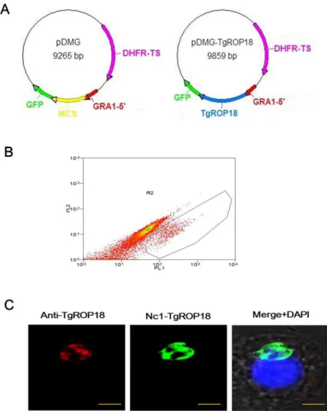
electroporation, and the drug-resistant recombinant parasites with bright green fluorescence were observed under the fluorescence microscope 24 h after transfection. After 10 generations of selection by pyrimethamine, a recombinant N. caninum stably expressing TgROP18 was isolated by flow cytometry (Fig. 1B) and designated as Nc1-TgROP18.
Identification of Nc1-TgROP18
The expression of TgROP18 in the isolated parasites was confirmed by IFA, PCR, qRT-PCR, and western blot. When analyzed by IFA, TgROP18 was found at the apical end of Nc1-TgROP18 tachyzoites, colocalizing exactly with GFP, which was similar to that observed for T. gondii and is consistent with localization in rhoptries (Fig. 1C). Electrophoresis of the PCR products showed that both the GFP and TgROP18 target genes (717 bp and 1,662 bp, respectively) were amplified from the genomic DNA of the Nc1-TgROP18 strain, while only the GFP target gene was amplified from the genomic DNA of the Nc1-GFP strain, the TgROP18 target gene from the genomic DNA of RH
strain, and neither gene from the genomic DNA of the Nc1 wild-type strain (Fig. 2A). As shown in Fig. 2B, transgene of TgROP18 in the Nc1-TgROP18 strain was expressed just a little below the RH wide-type strain (not statistically significant) on the mRNA level. Western blot analysis confirmed the expression of both GFP and TgROP18. The TgROP18 and GFP fusion protein (,87 kDa) expressed by the Nc1-TgROP18 strain was recognized by both Anti-TgROP18 and Anti-GFP polyclonal antibodies, while only the TgROP18 protein (,60 kDa) was detected with polyclonal antibodies against TgROP18 in RH strain, and only the GFP protein (,27 kDa) was detected with polyclonal antibodies against GFP in the Nc1-GFP strain (Fig. 2C). Collectively, these results demonstrated that the transgenic parasite Nc1-TgROP18 was successfully constructed.
Phenotype assays of Nc1-TgROP18
Plaque formation measures parasite survival and replication in cell culture, and reflects parasite motility on the surface of the host cell layer, invasion, intracellular growth and egress [21]. Any
Figure 1. Construction of Nc1-TgROP18.(A) Plasmid map of transfer vectors pDMG and pDMG-TgROP18, respectively. DHFR, dihydrofolate reductase-thymidylate synthase; MCS, multiple cloning site; GFP, green fluorescent protein. (B) The recombinant N. caninumstably expressing TgROP18 was isolated by flow cytometry. (C) IFA Localization of TgROP18 in Nc1-TgROP18. The recombinantN. caninumstably expressing TgROP18 was confirmed by IFA using mouse anti-rTgROP18 serum as the primary antibody. Scale bar, 5mm.
change of these phenotypes of the transgenic Nc1-TgROP18 can be determined by the plaque assay. Seven days after inoculation of HFF cells, the transgenic Nc1-TgROP18 strain formed plaques of similar size to theT. gondiiRH strain, but much bigger than those of theN. caninumNc1 and Nc1-GFP strains (Fig. 3), indicating that at least one of the steps in the lytic cycles of the Nc1-TgROP18 strain was increased compared to the Nc1 wild-type strain and the expression of TgROP18 in Nc1 strain was most likely responsible for the change.
More detailed analyses were undertaken to determine which step has changed. Transmigration assays using the Transwell system showed that the motility of the Nc1-TgROP18 strain was comparable with that of the Nc1 and Nc1-GFP strain, but slightly below the RH strain (not statistically significant) (Fig. 4A), suggesting transfection of Nc1 with TgROP18 did not affect its motilityin vitro, which was further confirmed by the finding of the gliding motility assay (Fig. 4B). Similar results were obtained when cell invasion assays were performed, in which the transgenic Nc1-Figure 2. Identification of Nc1-TgROP18.(A) Electrophoresis of the PCR products. Both the GFP and TgROP18 target genes (717 bp and 1,662 bp, respectively) were amplified from the genomic DNA of the Nc1-TgROP18 strain. (B) qRT-PCR comparison of TgROP18 expression in RH strain and Nc1-TgROP18 strain. Data are mean6SD (error bars) of three independent experiments. (C) and (D) Expression of TgROP18 in the cloned parasites was confirmed by Western blot analysis. The TgROP18 and GFP fusion protein (,87 kDa) was recognized by both GFP and
TgROP18 strain showed no alteration of invasion compared with the other three strains (Fig. 4C). Then intracellular replication assays were carried out to establish whether stably expressing TgROP18 in Nc1 strain improved the ability of the parasite to proliferate in host cells. Parasites that invaded host cells 24 h after inoculation were analyzed for intracellular growth by counting the number of parasites per vacuole. Because of the lack of synchronization of host cell invasion, the intracellular vacuoles contained 2, 4, 8, or 16 parasites. As shown in Fig. 4C, the distribution of 4 parasites per vacuole did not significantly differ between the four strains. In contrast, the number of the vacuoles containing 2 parasites of the transgenic Nc1-TgROP18 strain was significantly lower than that for the Nc1 wide-type strain and the Nc1-GFP strain, while the rate of the vacuoles containing 8 parasites of the transgenic Nc1-TgROP18 strain was significantly higher than that for the Nc1 wide-type strain and the Nc1-GFP strain. Moreover, a small percentage of vacuoles containing 16 parasites, which was absent in the Nc1 and Nc1-GFP strains, were observed in the transgenic Nc1-TgROP18 strain. There was no statistically significant difference in intracellular replication between the Nc1-TgROP18 strain and the T. gondii RH strain. These findings indicated that TgROP18 expression inN. caninum led to a significant increase in parasite proliferation, and suggested that ROP18 was probably responsible for the intracellular proliferation ofT. gondiiandN. caninum.
Virulence assay in mice
Since the results above demonstrated that expression of TgROP18 in N. caninum led to increased intracellular parasite proliferation, the mouse infection assay was then performed to assess the contribution of TgROP18 to virulence of the transgenic parasite. Mice were inoculated IP with the transgenic Nc1-TgROP18 strain, Nc1 wide-type strain, Nc1-GFP strain and RH wide-type strain at 10, 103or 106tachyzoites per mouse. All mice injected with tachyzoites of the Nc1-TgROP18 strain died within 5,16 days post infection, and the mortality kinetics was similar to that observed in mice inoculated the RH strain. In contrast, there was no death of the mice infected with Nc1 wide-type and
Nc1-GFP tachyzoites at any dose level in 30 days after infection (Fig. 5). The data showed that transfection of Nc1 with TgROP18 enhanced dramatically the virulence of Nc1 compared to the parental Nc1 wide-type, suggesting that TgROP18 was responsi-ble for parasite proliferation.
IRG phosphorylation assay
To determine whether transgenic parasite Nc1-TgROP18 is able to phosphorylate IRGs we examined the recruitment of IRGs to the parasitophorous vacuole (PV) based on the protein Irga6, which is treated as one of the most important indicator of the host IRGs family and implicated in resistance toT. gondii. We observed that only Nc1-TgROP18 was able to phosphorylate Irga6 (Fig. 6A, B), indicating the transgenic parasite can inactivate the IRGs due to the transgenic expression of TgROP18.
Discussion
T. gondiiandN. caninumare closely related protozoan parasites, but the two parasites differ greatly in virulence. The factors involved in virulence differences between the two parasites have not been investigated. In this study, our findings identified ROP18 as a key factor responsible for virulence difference between T. gondiiandN. caninumfor the first time.
T. gondii is an obligate intracellular parasite belonging to the phylum of Apicomplexa, which includes a great number of important human and animal pathogens such asCryptosporidium, Eimeria, Neospora,Plasmodium, andTheileria[21–22]. Because of its importance as an opportunistic pathogen and experimental advantages on genetic manipulation in the laboratory, T. gondii has emerged as a major model for the study of intracellular parasitism [23]. N. caninum is also an obligate intracellular protozoan parasite [24], which is known to have morphological and biological characteristics highly similar toT. gondii. Because of the similarities between the two parasites,N. caninumwas initially misidentified as T. gondii for many years [3–4]. The use of N. caninumas a heterologous system for the expression of genes from T. gondiihas been explored, and it may prove to be useful for the Figure 3. Plaque assay.The indicated strains grew on HFF cells for 7 days before fixation and staining with Crystal violet. The Nc1 strain incubated for 30 min at 50uC was used as the negative control. Three independent experiments were performed and results of one representative experiment are shown here. Scale bar, 200mm.
identification of parasite factors that are involved in the phenotypic differences between these two closely-related parasites [25–26], which was confirmed by transgenic expression of TgROP18 from T. gondii RH strain in N. caninumNc1 strain in the present study.
Most strains ofT. gondiibelong to one of three distinct clonal lineages, referred to as type I, type II and type III [27–28]. Despite having less than 2% genetic diversity, the threeToxoplasmalineages differ significantly in virulence among other differences. Type I strains are the most virulent and cause mortality at doses as low as one parasite (LD100<1) [29], whereas type II and type III have median lethal doses in mice ranging from 102 to 105 (LD5< 102,105) [30–31]. Forward genetic analysis accompanied by quantitative trait locus (QTL) mapping revealed that TgROP18, a highly polymorphic serine-threonine kinase, which secretes from a specialized apical organelle named rhoptries during host cell invasion [32–33], and is subsequently delivered to the cytosolic
side of the PV, plays a crucial role on virulence differences among T. gondii clonal lineages. TgROP18 differs dramatically in expression among the three clonal lineages ofT. gondii, and more exactly type III express 1,000-fold lower levels compared to type I and type II [5–6]. Transgenic expression of TgROP18 from RH strain, a type I lineage, in the avirulent type III CTG strain causes a four log increase in virulence in the mouse model [5]. Consistent with this, overexpression of TgROP18 in type I strain results in a significant increase in intracellular parasite proliferation rate, which is closely related to virulence [22]. In contrast, clean deletion of TgROP18 in the virulent type I strain leads to virulence attenuation [34–35].
N. caninumis much less virulent in mice even compared to the avirulent type III strain ofT. gondii, but factors responsible for the virulence difference between the two closely-related parasites remain unknown. TgROP16, which is a tyrosine kinase that directly phosphorylates the host signal transducer and activator of Figure 4. Phenotype assays of Nc1-TgROP18.(A) Transmigration assay in the Transwell system. Data are represented as mean6SD (error bars) of three independent experiments. (B) Gliding motility assay. The trails were stained with the anti-TgSAG1 antibody (RH strain) or anti-NcSRS2 antibody (Nc1, Nc1-GFP and Nc1-TgROP18 strains). The arrow indicates a trail. Scale bar, 5mm. (C) Cell invasion assay showed no difference in cell invasion between the transgenic Nc1-TgROP18 strain and the untransfected Nc1 orT. gondiiRH strain. Data are mean6SD (error bars) of three independent experiments. (D) Intracellular replication assay of the transgenic Nc1-TgROP18 compared to the Nc1 strain, Nc1-GFP strain and the RH strain. Asterisks indicate statistically significant results (*P#0.005; **P#0.001), as determined with the Student’s t test. Data are mean6SD (error bars) of three independent experiments.
Figure 5. Virulence assay in mice.Mice (n = 5) were injected with 10, 103or 106tachyzoites and were monitored for 30 days. All mice injected with the Nc1-TgROP18 strain andT. gondiiRH strain died within 16 days post infection, compared with no death of mice infected with Nc1 wide-type or Nc1-GFP tachyzoites. Three independent experiments were performed and one representative is shown here.
doi:10.1371/journal.pone.0099744.g005
Figure 6. IRG phosphorylation assay.(A) and (B) Localization of phosphorylated Irga6 in IFNc-stimulated Ana-1 cells infected with Nc1 wide-type strain or transgenic Nc1-TgROP18 strain, respectively. TgROP18 localized with mouse Anti-TgROP18 serum (FITC, green), Irga6 localized with rabbit Anti-Irga6 phosphopeptide Ab T102-555 (Texas Red, red), and nuclei stained with DAPI (blue). Scale bar, 2.5mm.
transcription 3 (STAT3) and STAT6 [36–37], was identified as another key virulence factor of T. gondii, but it may be not responsible for virulence difference between T. gondii and N. caninum, due to the fact that ROP16 ofN. caninum(NcROP18) is also able to phosphorylate STAT family of proteins (unpublished data). Intriguingly, we found that NcROP18 is a pseudogene due to several interrupting stop codons in the sequence by accident in our previous studies, which was confirmed by the findings of Reid AJ [4]. Therefore, we suspected that the difference of the ROP18 gene leads to virulence difference betweenT. gondiiandN. caninum. Thus a transgenic parasite was constructed by transfecting the TgROP18 of RH strain intoN. caninumNc1 stain. The phenotypes and virulence assays show that transgenic expression of TgROP18 inN. caninumdoes not affect its motility and invasion, but leads to a dramatical increase in intracellular parasite proliferation rate and virulence in mice.
T. gondii is capable of infecting essentially any warm-blooded animals by actively invading nucleated host cells and forming PV [38], which provides a protective niche for the parasite to avoid immune clearance and promote intracellular proliferation. A family of immunity-related GTPases (IRGs) plays an important role in resistance to T. gondii and a lot of other intracellular pathogens, including Chlamydia, Mycobacteria, Leishmania, Listeria, andSalmonella[39–40]. ForT. gondii, recruitment of IRGs on the PVM leads to its disruption, thereby resulting in parasite degradation [41–42]. The serine-threonine kinase TgROP18 of virulent type I strain is able to phosphorylate IRGs on key threonine residues in switch region I of the GTPase domain, which results in block IRG recruitment and protects the parasite from attack by the mouse immune system [43–44].N. caninumas well as type II and type III strains ofT. gondiiare unable to inactivate the IRGs because of active ROP deficient, which was confirmed by our observations in the present study [45–46]. Recent studies have revealed that a pseudokinase named TgROP5 was required for the catalytic activity of the active TgROP18 in type I strain, and both the two proteins were necessary to avoid IRG recruitment on
PVM ofT. gondii[20]. Transfection of the virulent ROP18 allele into avirulent type III strain enhanced parasite growth and caused a four log increase in virulence in the mouse model [5], indicating that TgROP5 performs similar function in both virulent type I strain and avirulent type III strain of T. gondii. Therefore, we speculated ROP5 of N. caninum (NcROP5) could regulate the activity of TgROP18 based on the results of the present study that the transgenic parasite Nc1-TgROP18 was able to phosphorylate IRGs as T. gondii did. Another possibility is that a novel and undiscovered N. caninum rhoptry protein plays the same role as TgROP5 [4].
In conclusion, the present study confirmed that TgROP18 plays a crucial role on virulence ofT. gondiifrom a different angle by expression ofT. gondiigenes inN.caninum. More significantly is that this is the first study to investigate what is responsible for the virulence difference between T. gondii and N. caninum, and our findings finally identify ROP18 is indeed the key factor of virulence difference between the two closely related protozoan parasites.
Acknowledgments
We are grateful to Professor JC Howard and Dr T Steinfeldt (University of Cologne, Germany) for kindly providing the anti Irga6 phosphopeptide Ab T102-555. We thank Professor Xuenan Xuan (Obihiro University of Agriculture and Veterinary Medicine, Japan) for kindly providing the pDMG plasmid and the Nc1-GFP strain. We thank Dr Xiangmei Zhou (China Agricultural University) for kindly providing the Ana-1 cells. We also thank Dr Jin Zhu (Therapeutic Goods Administration, Australia) and Professor Xiaojiang Chen (University of Southern California, USA) for their assistance in the preparation of the manuscript.
Author Contributions
Conceived and designed the experiments: TL JL QL. Performed the experiments: TL HZN. Analyzed the data: TL HW JL QL. Contributed reagents/materials/analysis tools: QL. Wrote the paper: TL QL.
References
1. Dubey JP, Carpenter JL, Speer CA, Topper MJ, Uggla A (1988) Newly recognized fatal protozoan disease of dogs. J Am Vet Med Assoc 192: 1269– 1285.
2. Dubey JP, Barr BC, Barta JR, Bjerkas I, Bjorkman C, et al. (2002) Redescription ofNeospora caninumand its differentiation from related coccidia. Int J Parasitol 32: 929–946.
3. Kaufmann H, Yamage M, Roditi I, Dobbelaere D, Dubey JP, et al. (1996) Discrimination ofNeospora caninumfromToxoplasma gondiiand other apicom-plexan parasites by hybridization and PCR. Molecular and Cellular Probes 10: 289–297.
4. Reid AJ, Vermont SJ, Cotton JA, Harris D, Hill-Cawthorne GA, et al. (2012) Comparative genomics of the apicomplexan parasites Toxoplasma gondii and Neospora caninum: Coccidia differing in host range and transmission strategy. PLoS pathogens 8: e1002567.
5. Taylor S, Barragan A, Su C, Fux B, Fentress SJ, et al. (2006) A secreted serinethreonine kinase determines virulence in the eukaryotic pathogen Toxoplasma gondii. Science 314: 1776–1780.
6. Saeij JPJ, Boyle JP, Coller S, Taylor S, Sibly LD et al. (2006) Polymorphic secreted kinases are key virulence factors in toxoplasmosis. Science 314: 1780– 1783.
7. Fentress SJ, Behnke MS, Dunay IR, Moashayekhi M, Rommereim LM, et al. (2010) Phosphorylation of immunity-related GTPases by a parasite secretory kinase promotes macrophage survival and virulence. Cell Host Microbe 16: 484–495.
8. Gaskell EA, Smith JE, Pinney JW, Westhead DR, McConkey GA (2009) A unique dual activity amino acid hydroxylase inToxoplasma gondii. PLoS one 4: e4801.
9. Zhang HS, Muller KA, Compaore Eung-goo L, Liao M, Zhang GH, et al. (2007) Apical membrane antigen 1 is a cross-reactive antigen betweenNeospora caninumandToxoplasma gondii, and the anti-NcAMA1 antibody inhibits host cell invasion by both parasites. Mol Biochem Parasitol 151: 205–212.
10. Wang L, Chen H, Liu D, Huo X, Gao J, et al. (2013). Genotypes and mouse virulence ofToxoplasma gondiiisolates from animals and humans in China. PLoS one 8: e53483.
11. Nishikawa Y, Xuan X, Makala L, Vielemeyer O, Joiner KA, et al. (2003) Characterization ofToxoplasma gondiiengineered to express mouse interferon-gamma. Int J Parasitol 2003, 33: 1525–1535.
12. Zhang GH, Huang XH, Boldbaatar D, Battura B, Battsetsega B, et al. (2010) Construction ofNeospora caninumstably expressing TgSAG1 and evaluation of its protective effects againstToxoplasma gondiiinfection in mice. Vaccine 28: 7243– 7247.
13. Sibley LD, Messina M, Niesman IR (1994) Stable DNA transformation in the obligate intracellular parasite Toxoplasma gondii by complementation of tryptophan auxotrophy. Proc Natl Acad Sci USA 91: 5508–5512.
14. Cui X, Lei T, Yang DY, Hao P, Li B, et al. (2012)Toxoplasma gondiiimmune mapped protein-1 (TgIMP1) is a novel vaccine candidate against toxoplasmosis. Vaccine 30: 2282–2287.
15. Cui X, Lei T, Yang DY, Hao P, Liu Q (2012) Identification and characterization of a novelNeospora caninumimmune mapped protein 1. Parasitology 139: 998– 1004.
16. Santos JM, Ferguson DJP, Blackman MJ, Soldati-Favre D (2011) Intramem-brane cleavage of AMA1 triggersToxoplasmato switch from an invasive to a replicative mode. Science 331: 473–477.
17. Daher W, Plattner F, Carlier MF, Soldati-Favre D (2010). Concerted action of two formins in gliding motility and host cell invasion byToxoplasma gondii. PLoS pathogens 6: e1001132.
18. Sheiner L, Santos JM, Klages N, Parussini F, Jemmely N, et al. (2010). Toxoplasma gondiitransmembrane microneme proteins and their modular design. Molecular microbiology 77(4): 912–929.
19. Meissner M, Schluter D, Soldati-Favre D (2002) Role ofToxoplasma gondiimyosin A in powering parasite gliding and host cell invasion. Science 298: 837–840. 20. Behnke MS, Fentress SJ, Mashayekhi M, Li LX, Taylor GA, et al. (2012) The
21. Plattner F, Yarovinsky F, Romero S, Didry D, Carlier MF, et al. (2008). Toxoplasmaprofilin is essential for host cell invasion and TLR11-dependent induction of an interleukin-12 response. Cell Host Microbe 3: 77–87. 22. El Hajj H, Lebrun M, Arold ST, Vial H, Labesse G, et al. (2007) ROP18 is a
rhoptry kinase controlling the intracellular proliferation ofToxoplasma gondii. PLoS pathogens 3: e14.
23. Kim K, Weiss LM (2004) Toxoplasma gondii: the model apicomplexan. Int J Parasitol 34: 423–432.
24. Howe DK, Sibley LD (1999) Comparison of the major antigens ofNeospora caninumandToxoplasma gondii. Int J Parasitol 29:1489–1496.
25. Howe DK, Mercier C, Messina M, Sibley LD (1997) Expression ofToxoplasma gondiigenes in the closely-related apicomplexan parasiteNeospora caninum. Mol Biochem Parasitol 86: 29–36.
26. Beckers CJ, Wakefield T, Joiner KA (1997) The expression of Toxoplasma proteins inNeospora caninumand the identification of a gene encoding a novel rhoptry protein. Mol Biochem Parasitol 89: 209–223.
27. Grigg ME, Bonnefoy S, Hehl AB, Suzuki Y, Boothroyd JC (2001) Success and virulence in Toxoplasma as the result of sexual recombination between two distinct ancestries. Science 294: 161–165.
28. Su C, Evans D, Cole RH, Kissinger JC, Ajioka JW, et al. (2003) Recent expansion ofToxoplasmathrough enhanced oral transmission. Science 299: 414– 416.
29. Sibley LD, Boothroyd JC (1992) Virulent strains ofToxoplasma gondiicomprise a single clonal lineage. Nature 359: 82–85.
30. Saeij JP, Boyle JP, Boothroyd JC (2005) Differences among the three major strains ofToxoplasma gondiiand their specific interactions with the infected host. Trends Parasitol 21: 476–481.
31. Howe DK, Summers BC, Sibley LD (1996) Acute virulence in mice is associated with markers on chromosome VIII inToxoplasma gondii. Infect Immun 64: 5193– 5198.
32. Haukansson S, Charron AJ, Sibley LD (2001)Toxoplasmaevacuoles: a two-step process of secretion and fusion forms the parasitophorous vacuole. EMBO J 20: 3132–3144.
33. Bradley PJ, Ward C, Cheng SJ, Alexander DL, Coller S, et al. (2005) Proteomic analysis of rhoptry organelles reveals many novel constituents for hostparasite interactions inT. gondii. J Biol Chem 280: 34245–34258.
34. Fentress SJ, Sibly LD (2010) The secreted kinase ROP18 defendsToxoplasma’s border. Bioessays 33: 693–700.
35. Reese ML, Zeiner GM, Saeij JP, Boothroyd JC, Boyle JP (2011) Polymorphic family of injected pseudokinases is paramount inToxoplasmavirulence. Proc Natl Acad Sci USA 108: 9625–9630.
36. Yamamoto M, Standley DM, Takashima S, Saiga H, Okuyama M, et al. (2009) A single polymorphic amino acid onToxoplasma gondiikinase ROP16 determines the direct and strain-specific activation of Stat3. J Exp Med 206: 2747–2760. 37. Ong YC, Reese ML, Boothroyd JC (2010) Toxoplasma rhoptry protein 16
(ROP16) subverts host function by direct tyrosine phosphorylation of STAT6. J Biol Chem 285:28731–28740
38. Sibley LD (2004) Intracellular parasite invasion strategies. Science 304: 248– 253.
39. Shenoy AR, Kim BH, Choi HP, Matsuzawa T, Tiwari S, et al. (2008) Emerging themes in IFN-gamma-induced macrophage immunity by the p47 and p65 GTPase families. Immunobiol 212: 771–784.
40. Taylor GA, Feng CG, Sher A (2007) Control of IFN-gamma-mediated host resistance to intracellular pathogens by immunity-related GTPases (p47 GTPases). Microb Infect 9: 1644–1651.
41. Ling YM, Shaw MH, Ayala C, Coppens I, Taylor GA, et al. (2006) Vacuolar and plasma membrane stripping and autophagic elimination ofToxoplasma gondii in primed effector macrophages. J Exp Med 203: 2063–2071.
42. Martens S, Parvanova I, Zerrahn J, Griffiths G, Schell G, et al. (2005) Disruption of Toxoplasma gondii parasitophorous vacuoles by the mouse p47-resistance GTPases. PLoS pathogens 1: e24.
43. Fentress SJ, Behnke MS, Dunay IR, Moashayekhi M, Rommereim LM, et al. (2010) Phosphorylation of immunity-related GTPases by a parasite secretory kinase promotes macrophage survival and virulence. Cell Host Microbe 16: 484–495.
44. Steinfeldt T, Konen-Waisman S, Tong L, Pawlowski N, Lamkemeyer T, et al. (2010) Phosphorylation of mouse immunity-related GTPase (IRG) resistance proteins is an evasion strategy for virulent Toxoplasma gondii. PLoS Biol 8: e1000576
45. Zhao Y, Ferguson DJ, Wilson DC, Howard JC, Sibley LD, et al. (2009) Virulent Toxoplasma gondii evade immunity-related GTPas-mediated parasite vacuole disruption within primed macrophages. J Immunol 182: 3775–3781. 46. Khaminets A, Hunn JP, Konen-Waisman S, Zhao YO, Preukschat D, et al.