Morphological Study of Isolated Ovarian Preantral
Follicles Using Fibrin Gel Plus Platelet Lysate after
Subcutaneous Transplantation
Ali Reza Rajabzadeh, M.Sc.1, 2, Hossein Eimani, Ph.D.1, 3*, Homa Mohseni
Koochesfahani, Ph.D.2, Abdol-Hossein Shahvardi, Ph.D.1,
Rouhollah Fathi, Ph.D.1
1. Department of Embryology at Reproductive Biomedicine Research Center, Royan Institute for Reproductive Biomedicine, ACECR, Tehran, Iran
2. Department of Cell and Molecular Biology, Faculty of Biological Sciences, Kharazmi University, Tehran, Iran 3. Department of Anatomy, Faculty of Medicine, Baqiyatallah (a.s.) University of Medical Sciences,
Tehran, Iran
*Corresponding Address: P.O.Box: 16635-148, Department of Embryology at Reproductive Biomedicine Research Center, Royan Institute for Reproductive Biomedicine, ACECR, Tehran, Iran
Email: eimanih@royaninstitute.org
Received: 22/Dec/2012, Accepted: 10/Mar/2014
Abstract
Objective: Ovarian and follicle transplantation may preserve fertility in young cancer
sur-vivors. In this study, we have transplanted preantral follicles using ibrin gel as a carrier and ibrin gel supplemented with platelet lysate (PL) as a rich source of angiogenic and growth factors. The purpose of this study was to evaluate the role of ibrin gel and PL in
follicle transplantation.
Materials and Methods: In this experimental study, ovaries were taken from
14-day-old Naval Medical Research Institute (NMRI) mice. Preantral follicles were dissected from the ovaries and encapsulated into fibrin gel supplemented with 5, 10, 15 or 20% PL, then transplanted back into the same donor mice. Fibrin gels supplemented with PL that contained preantral follicles were placed in a subcutaneous pocket in the back of the neck of the recipient, donor mouse (the same mouse that follicles were collected). After 14 days the grafts were processed and embedded in paraffin blocks,
then serially sectioned for histological evaluation. We counted the follicles and
clas-sified them according to stage (preantral or antral). Data were presented as mean ± standard error of mean (SEM) and analysed by analysis of variance (ANOVA) and the
Kruskal-Wallistest.
Results: The mean percentage of recovered follicles encapsulated and transplanted
in each group were 33.30 ± 2.47 (fibrin gel), 31.96 ± 1.90 (fibrin gel+5% PL), 34.02 ± 2.44 (fibrin gel+10% PL), 48.31 ± 2.06 (fibrin gel+15% PL) and 17.60 ± 2.79 (fibrin gel+20% PL). There was a signiicant increase in the recovery rate of grafted folli
-cles with fibrin gel+15% PL (48.31%; p<0.001). The percentage of preantral folli-cles showed no significant difference in all groups (p<0.05). The percentage of antral fol
-licles showed a significant decrease in fol-licles grafted with fibrin gel+20% PL when compared to the other groups (11.77%; p<0.005) but no significant difference was observed in the other groups.
Conclusion: The use of PL in follicle transplantation can improve ovarian follicular survival rate.
Keywords:Transplantation, Follicle, Ovary
Cell Journal(Yakht eh), Vol 17, No 1, Spring 2015, Pages: 145- 152
Citation: Rajabzadeh AR, Eimani H, Mohseni Koochesfahani H, Shahvardi AH, Fathi R. Morphological study of isolated
Introduction
Recent improvements in cancer treatments have significantly increased survival rates. Unfortu -nately treatments such as chemotherapy and ra -diotherapy can cause severe damage to the ovaries and mature ovarian failure. Ovarian cryopreserva -tion prior to chemotherapy/radiotherapy and auto-transplantation post-treatment can restore fertility to cancer patients. Options for fertility preserva -tion in patients at risk for premature ovarian failure (POF) include embryo cryopreservation, oocyte cryopreservation and ovarian tissue cryopreserva -tion followed by subsequent transplanta-tion (1).
The major impediment to transplantation is that the graft is completely dependent on the establish -ment of neovascularization, thus a significant ratio of follicles may be lost because of ischemia and hypoxia. Ischemia in the transplant from delayed revascularization is one of the transplantation con -cerns. In the absence of direct vascular anasto -mosis, the success and survival of ovarian grafts is dependent upon vascularization. In the mouse, revascularization begins during the first 48 hours after transplantation (2). Hypoxia-induced delayed revascularization in the early stages of ovarian transplantation causes the loss of large numbers of immature follicles. On the other hand, numer -ous growth factors such as platelet-derived growth factors (PDGF) (3), vascular endothelial growth factor (VEGF), insulin-like growth factor (IGF) and basic fibroblast growth factor (bFGF) (4, 5) in -volved in neovascularization and follicular growth can be used to improve transplantation. Minimiz -ing the initiation duration of revascularization can improve graft function and follicular survival rate, and reduce premature follicle depletion.
As an alternative strategy, cryopreservation and transplantation of isolated ovarian follicles has a number of potential advantages over ovarian tis-sue transplantation. First, ovarian tistis-sue has a more complex structure with different cell types making the choice of a suitable cryopreservation protocol challenging. Cryopreserved-thawed follicles can be suspended in a plasma clot (6, 7) or collagen gel (8) permitting auto-transplantation via a mini -mally invasive approach. Isolated ovarian follicle transplantation can negate the risk of cancer recur -rence that is of concern in auto-transplantation of cryopreserved-thawed ovarian tissue in cancer pa-tients in remission (9).
Extracted activated platelets [platelet lysate (PL)] are a rich source of angiogenic and growth factors (10, 11) which may have an important role in improving transplant survival. Fibrin gel due to its availability, biodegradation and biocompatibil -ity has been used as a delivery vehicle and suitable preservative for transplantation of pancreatic islets (12), muscle-derived stem cells (13) and keratino -cytes (14). In this study, as an alternative method, we have autologously transplanted isolated prean -tral follicles by using fibrin gel supplemented with PL and analyzed the results.
Materials and Methods
Experimental design
In this experimental study, female Naval Medi -cal Research Institute (NMRI) mice (Pasteur In -stitute, Tehran, Iran) were housed and bred at the Laboratory Animal Breeding Center of Royan Institute (Tehran, Iran). Animals were kept at a temperature of 19-22˚C and 50% humidity un˚ -der light-controlled conditions (12 hours light: 12 hours dark) and provided sterile food and water ad libitum. The experiments were approved by the lo -cal Ethics Committee at Royan Institute.
One of the ovaries (right ovary) was taken from 14-day-old female NMRI mice after they were anesthetized with ketamine/xylazine (Sigma, Ger-many). A total of 1919 preantral follicles were dissected from the ovaries and divided into five groups: I. encapsulated into fibrin gel (group 1; 24.31 follicles per mouse), II. fibrin gel+5% PL (group 2; 21.52 follicles per mouse), III. fibrin gel+10% PL (group 3; 22.41 follicles per mouse), IV. fibrin gel+15% PL (group 4; 22.55 follicles per mouse) and V. fibrin gel+20% PL (group 5; 22.17 follicles per mouse). The follicles were then trans -planted back into the same donor mice. Grafted encapsulated follicles were harvested after 14 days and histologically evaluated for survival and de -velopment to the next stages.
Platelet preparation
PC was stored at -70˚C until use. Then, samples that contained approximately 1×10⁹ platelets/ml were selected and frozen at -70˚C to obtain PL that contained platelet-released growth factors. Frozen PC was rapidly thawed at 37˚C and centrifuged at 3000 g for 30 minutes at 4˚C to remove the platelet bodies.
Collection of preantral follicles
Mice were anesthetized by ketamine and xy -lazine. After disinfecting the dorsal skin, a small incision was made and the ovaries removed. Pre -antral follicles were mechanically collected from ovaries as described previously (15). Briely, for mechanical isolation, preantral follicles were dis -sected using a 29 gauge needle attached to 1ml syringe in alpha-minimum essential medium (α-MEM, Gibco-Invitrogen, Genmany)+10% fe -tal bovine serum (FBS, Sigma, Germany) at 37˚C. Round-shaped preantral follicles with 2-3 layers of granulosa cells and the spherical oocyte centrally located within the follicle were considered to be morphologically normal and used in the experi -ments.
Follicle encapsulation and transplantation
Fibrinogen and thrombin solutions were pre -pared from Cell Science Research Center of Roy -an Institute (Tehr-an, Ir-an).
For group 1, preantral follicles were suspend -ed in 10 μl of thrombin solution and mix-ed with 5 μl of reconstituted fibrinogen solution to form the fibrin gel. For group 2, preantral follicles were suspended in a mixture of 9.5 µl of thrombin and 0.75 µl of PL followed by the addition of 4.75 µl of fibrinogen solution (fibrin gel+5% PL). For group 3, preantral follicles were suspended in a mixture of 9 µl of thrombin plus 1.5 µl of PL and mixed with 4.5 µl of fibrinogen solution (fibrin gel+10% PL). Group 4 preantral follicles were suspended in a mixture of 8.5 µl of thrombin and 2.5 µl of PL, then mixed with 4.25 µl of fibrinogen solution (fi -brin gel+15% PL). For group 5, preantral follicles were suspended in a mixture of 8 µl of thrombin and 3 µl of PL, after which 4 µl of fibrinogen solu -tion was added (fibrin gel+20% PL). In all groups, after 3 to 5 minutes, the fibrin gel/fibrin gel sup-plemented with PL that contained preantral folli -cles were placed in a subcutaneous pocket in the posterior area of the mouse’s neck. Each mouse
re-ceived the follicles that were collected from it. The incision was sutured with polyethylene suture 6 (Ethicon, USA) and mice were allowed to recover. After waking up, the mice were transferred to the animal house and kept for 14 days under standard conditions.
Histological assessments
After two weeks, the mice were anesthetized and an incision was made in the posterior sections of their necks. Grafted follicles associated with their encompassing tissue were excised and immedi -ately placed in Bouin’s solution. For all groups the fixed follicles were dehydrated and embedded in paraffin wax (Merck, Germany) serially sectioned in 6 μm sections and stained with hematoxylin–eo -sin (H&E) for histological evaluations. Each five sections were observed and analyzed by light mi -croscope (Nikon, Japan) after which the numbers of morphologically normal follicles were counted on the basis of Gougeon’s study (16). Eosinophilia of the ooplasm, contraction and clumping of the chromatin material, and wrinkling of the nuclear membranes of the oocytes were regarded as signs of atresia (16). The follicles were classified ac -cording to stage as preantral or antral follicles. Preantral follicles were confirmed by the presence of two or more layers of cuboidal granulosa cells without antrum. Antral follicles were identified as having an antral cavity (17).
Data were presented as mean ± standard error of mean (SEM) and analysed by analysis of variance (ANOVA) and the Kruskal-Wallis test. A prob -ability of p<0.05 was considered statistically sig -nificant. Statistical package for the social sciences (SPSS) software (version 16; SPSS Inc., Chicago, IL, USA) was used for data analysis.
Results
31.96 ± 1.90 (fibrin gel+5% PL), 34.02 ± 2.44 (fi -brin gel+10%PL), 48.31 ± 2.06 (fi-brin gel+15% PL) and 17.60 ± 2.79 (fibrin gel+20% PL) per grafted fibrin gel for each group (Table 1, Fig.1A). A comparison between the five groups showed a significant increase in the recovery rate of graft -ed follicles encapsulat-ed with fibrin gel+15% PL (48.31%; p<0.001) and a significant decrease in grafted follicles with fibrin gel+20% PL compared
to the other groups (17.60%, p<0.001, Fig.1A). The percentage of preantral follicles showed no significant differences in all groups (p<0.05). However the percentage of antral follicles sig -nificantly decreased in the grafted follicles with fibrin gel+20% PL compared to the other groups (11.77%, p<0.005) but no significant difference was observed in the other groups (Fig.1B).
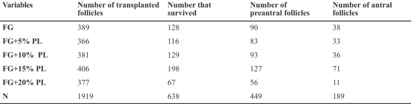
Table 1: Number of transplanted follicles and survived follicles, preantral/antral follicles 14 day post transplantaion
Number of antral follicles
Number of preantral follicles Number that
survived Number of transplanted
follicles Variables
38 90
128 389
FG
33 83
116 366
FG+5% PL
36 93
129 381
FG+10% PL
71 127
198 406
FG+15% PL
11 56
67 377
FG+20% PL
189 449
638 1919
N
FG; Fibrin gel, PL; Platelet lysate and N; Number.
Fig.1: Histological evaluaion and comparison of follicular recovery rate. Number of follicles transplanted to the under skin of the posterior
neck and number that survived. A. Preantral and antral rate and B. Between grated isolated preantral follicles with ibrin gel and ibrin gel supplemented with platelet lysate (PL) at various doses, transplanted for 14 days under the skin in the posterior area of the neck. *; Signiicntly difrent from other groups in antral rate (p<0.05) and Signiicant diference between non-idenical leter (p<0.01).
Fig.2: Histologic secions of murine isolated follicle grats auto-transplanted under the skin in the posterior neck for 14 days. Hematoxylin
and eosin (H&E) secion of murine skin issue (around the follicles), preantral follicles [A, B (arrow head), E (arrowhead), I] and antral (B, D, E, H) follicles in the grat. Original magniicaion: A-I: ×100; D: ×200. (Black arrows: hair follicles, white arrows: follicular blood vascu-latures).
FG: Fibrin gel and PL: Platelet lysate.
Morphologically, follicles at all stages looked well preserved (Fig.2). Preantral follicles appeared as rounded structuresthat contained intact oocytes with large nuclei, and spherical or slightly ovoid in shape. The oocyte was surrounded by more than two layers of granulosa cells (Fig.2A, B, E, I). Healthy-looking
Discussion
Loss of follicular pool is caused mostly by is -chemia and not providing a rapid the blood supply. Isolated follicles due to their lack of stroma may connect faster into newly formed vasculatures. Therefore, in this study isolated follicles have been used for auto-transplantation. For this purpose, the fibrin gel used in this study fixed the follicles at the transplantation site due to its sticky nature and connected the host tissue and graft to each other. This might enhance revascularization via facilita -tion of cell infiltra-tion from the host tissue and par -acrine interactions.
Although the role of angiogenic cytokines such as VEGF, PDGF and FGF in survival, migra -tion and prolifera-tion of endothelial cells is well known, several observations have shown that the extracellular matrix (ECM) is equally important (18). To promote migration, cytokine function is entirely dependent on the endothelial cell binding to the ECM which is important during the growth of new blood vessels from existing vessels. Bio -materials are effective in other systems to increase angiogenesis (19). In this study fibrin gel, as the biodegradable scaffold, has been used to maintain the follicles at the graft site.
PL is a concentration of human platelet growth factors in a small volume of plasma obtained by lysing the platelet bodies through temperature-shock. Therefore, PL contains all the fundamen -tal growth factors secreted by platelets to initiate tissue regeneration and angiogenesis, including PDGFs, bFGF, VEGF, IGF-1 and transforming growth factor-b (TGF-b) (10, 20). Allare potent angiogenic factors and endothelial cell mito -gens.
In this study, we observed that isolated fol -licles survived and grew in non-capsulated sites such as subcutaneous spaces. The survival rate in group 4 (fibrin+15% PL) compared with group 1 (fibrin gel) was associated with a sig -nificant increase. This increased survival was probably due to the presence of a certain dose (15%) of PL and appeared to be the result of a decrease in ischemia conditions due to the pres -ence of angiogenic and growth factors which led to the provision of a more rapid blood sup -ply to the graft. However, growth and matura -tion (antral to the preantral) were not signifi
-cantly different between the two groups. Hence, it appeared thatangiogenic and growth factors in PL could only have increased angiogenesis and sprouting new blood vessels (Fig.3).
This study aimed to evaluate whether trans-plantation of isolated ovarian follicles encap -sulated in fibrin supplemented with PL might have biological properties appropriate for use in fertility preservation. Our results were con-sistent with a study conducted by Dolmans et al. (7) which reported that loss of grafted fol -licles (approximately 50%) was related to is -chemia and delayed revascularization. It seems, although revascularization in transplanted fol -licles encapsulated in fibrin gel supplemented with PL was facilitated, the lack of stromal cells around the grafted follicles caused no observed significant difference in survival rate to the same another studies (7, 21).
After transplantation the follicles encapsulated in fibrin gel supplemented with PL (15%) had a higher survival rate than follicles encapsulated in PL-free fibrin gel (48 vs. 32%). This indicated that PL which contained angiogenic and growth factors caused more rapid revascularization than group 1 which did not use PL. We showed that different doses of PL added to the fibrin had different effects on follicular survival rate. Survival rates increased significantly at the 15% dose compared to the 5, 10 and 20% doses.
Fig.3: Morphological evaluaion of neovascularizaion in follicle grats ater 14 days. White and black arrows demonstrate grated follicles
and blood vasculature around the grats, respecively. A. Grated follicles encapsulated in ibrin gel without platelet lysate (PL) and B. Grated follicles encapsulated in ibrin gel supplemented with 15% PL.
Conclusion
Due to the widespread biological effects of this substance whether or not the follicles grafted have developmental potential and also identification of possible side effects of this product more research should be done. According to the results the use of PL in ovarian follicle transplantation could im -prove survival of the follicles.
Acknowledgments
This research was financially supported by a
grant-in-aid for scientific research from Royan In -stitute, Tehran, Iran. There is no conlict of interest in this article.
References
1. Kim SS, Battaglia DE, Soules MR. The future of human ovarian cryopreservation and transplantation: fertility and
beyond. Fertil Steril. 2001; 75(6): 1049-1056.
2. Dissen GA, Lara HE, Fahrenbach WH, Costa ME, Ojeda SR. Immature rat ovaries become revascularizedrapidly
after autotransplantation and show a gonadotropin- de-pendent increase in angiogenicfactor gene expression.
Endocrinology. 1994; 134(3): 1146-1154.
3. Raica M, Cimpean AM. Platelet-derived growth fac
-A
tor (PDGF)/PDGF receptors (PDGFR) axis as target for antitumor and antiangiogenic therapy. Pharmaceuticals. 2010; 3(3): 572-599.
4. Bruno JB, Matos MHT, Chaves RN, Celestino JJH, Sarai
-va MVA, Lima-Verde IB, et al. Angiogenic factors and ovarian follicle development. Anim Reprod. 2009; 6(2): 371-379.
5. Webb R, Garnsworthy PC, Gong JG, Armstrong DG. Control of follicular growth: local interactions and nutri
-tional influences. J Anim Sci. 2004; 82 E-Suppl: E63-74.
6. Carroll J, Gosden RG. Transplantation of frozen-thawed mouse primordial follicles. Hum Reprod. 1993; 8(8): 1163-1167.
7. Dolmans MM, Martinez-Madrid B, Gadisseux E, Guiot Y, Yuan WY, Torre A, et al. Short-term transplantation of iso -lated human ovarian follicles and cortical tissue into nude
mice. Reproduction. 2007; 134(2): 253-262.
8. Carroll J, Whittingham DG, Wood MJ, Telfer E, Gosden RG. Extra-ovarian production of mature viable mouse oo
-cytes from frozen primary follicles. J Reprod Fertil. 1990; 90(1): 321-327.
9. Ajala T, RaiJ, Larsen-Disney P, Howell R. Fertility pres
-ervation for cancer patients: a review. Obstet Gynecol Int. 2010; 2010: 160386.
10. Doucet C, Ernou I, Zhang Y, Llense JR, Begot L, Holy X, et al. Platelet lysates promote mesenchymal stem cell expansion: a safety substitute for animal serum in cell-based therapy applications. J Cell Physiol. 2005; 205(2): 228-236.
11. Sanchez AR, Sheridan PJ, Kupp LI. Is platelet-rich plasma
the perfect en-hancement factor? A current review. Int J Oral Maxillofac Implants. 2003; 18(1): 93-103.
12. Andrades P, Asiedu C, Rodriguez C, Goodwin KJ, McCarn J, Thomas JM. Subcutaneous pancreatic islet transplanta
-tion using ibrin glue as a carrier. Transplant Proc. 2006; 39(1): 191-192.
13. Xu Y, Song YF, Lin ZX. Transplantation of muscle-derived
stem cells plus biodegradable ibrin glue restores the ure -thral sphincter in a pudendal nerve-transected rat model.
Braz J Med Biol Res. 2010; 43(11): 1076-1083.
14. Grant I, Warwick K, Marshall J, Green C, Martin R. The co-application of sprayed cultured autologous
keratino-cytes and autologous ibrin sealant in a porcine wound model. Br J Plast Surg. 2002; 55(3): 219-227.
15. Cortvrindt R, Smitz J, Van Steirteghem AC. In-vitro matu
-ration, fertilization and embryo development of immature oocytes from early preantral follicles from prepuberal mice in a simpliied culture system. Hum Reprod. 1996; 11(12): 2656-2666.
16. Gougeon A. Dynamics of follicular growth in the human: a model from preliminary results. Hum Reprod. 1986; 1(2): 81-87.
17. Li F, Tao Y, Zhang Y, Li Y, Fang F, Liu Y, et al. Follicle
growth and oocyte development after ovary
transplanta-tion into back muscle of immune-intact adult castrated male mice. Reproduction. 2010; 140(3): 465-76. 18. Davis GE, Senger DR. Endothelial extracellular matrix:
biosynthesis, remodeling, and functions during vascular morphogenesis and neovessel stabilization. Circ Res. 2005; 97(11): 1093-1107.
19. Donnez J, Squiflet J, Dolmans MM. Frozen-thawed ovar
-ian tissue retransplants. Semin Reprod Med. 2009; 27(6): 472-478.
20. Eppley BL, Woodell JE, Higgins J. Platelet quantiication
and growth factor analysis from platelet-rich plasma:
im-plications for wound healing. Plast Reconstr Surg. 2004; 114(6): 1502-1508.
21. Aerts JM, Martinez-Madrid B, Leroy JL, Van Aelst S, Bols PE. Xenotransplantation by injection of a suspension of
isolated preantral ovarian follicles and stroma cells under