J. Evid. Based Med. Healthc., pISSN- 2349-2562, eISSN- 2349-2570/ Vol. 3/Issue 22/Mar. 17, 2016 Page 986
A COMPARATIVE STUDY BETWEEN HYDROFIBER DRESSING & POVIDONE DRESSING IN
DIABETIC FOOT ULCERS
Rithin Suvarna1, Komaragiri Viswanadh2, Hanumanthappa M. B3, N. Devidas Shetty4
1Professor, Department of General Surgery, A. J. Institute of Medical Sciences, Mangaluru. 2Resident, Department of General Surgery, A. J. Institute of Medical Sciences, Mangaluru. 3Professor, Department of General Surgery, A. J. Institute of Medical Sciences, Mangaluru. 4Professor, Department of General Surgery, A. J. Institute of Medical Sciences, Mangaluru.
ABSTRACT
BACKGROUND
Diabetic Foot Ulcer (DFU) is the most common complication of Diabetes Mellitus (DM). It occurs in 15% of all patients with DM. Major increase in mortality among diabetic patients, observed over the past 20 years is considered to be due to the development of macro and micro vascular complications, including failure of the wound healing process. Non-healing chronic diabetic ulcers are often treated with extracellular matrix replacement therapy.so far, it is a common trend in diabetic foot care domain to use advanced moist wound therapy. At present, there are various categories of moist dressings available such as povidone dressings, adhesive backing film, silicone coated foam, hydrogels, hydrocolloids etc.
AIMS & OBJECTIVES
1) To study efficacy of hydrofiber dressings and wound healing in diabetic foot ulcers.
2) To compare the safety, final outcome and patient compliance in hydrofiber and povidone dressing.
MATERIALS & METHODS
This prospective, observational study is carried in our department from September 2014 to September 2015. All the patients with age of 30 years and above were admitted in AJIMS satisfying the inclusion and exclusion criteria were taken into the study. The patients were followed until the ulcer is treated.
STATISTICAL ANALYSIS
Data are presented as Mean and Standard Deviation (SD). Fischer’s exact test and Chi-Square Test were used as appropriate. GraphPad Prism Version 6.0h was used to analyze data and to prepare graphs. A ‘P’ value below the level of 0.05 was taken as statistically significant.
RESULTS
Among the patients who were studied, hydrofiber dressings helped in the better & faster healing of DFU.
CONCLUSION
In comparison with povidone gauze dressings to hydrofiber dressings in diabetic foot ulcers, the latter is more efficacious in controlling the infection, minimal usage of antibiotics, faster healing rate and patient compliance during dressings.
KEYWORDS
Hydrofiber dressing, Povidone dressing, Diabetic Foot Ulcer.
HOW TO CITE THIS ARTICLE: Suvarna R, Viswanadh K, Hanumanthappa MB et al. A comparative study between hydrofiber dressing & povidone dressing in diabetic foot ulcers. J. Evid. Based Med. Healthc. 2016; 3(22), 986-991.
DOI: 10.18410/jebmh/2016/226
INTRODUCTION: Diabetic Foot Infection (DFI) is a leading cause for hospital admission in India which is due to multiple precursors mainly neuropathy, peripheral vascular disease and impaired wound healing.1,2 It has been reported
that about 25% diabetics develop severe foot problems at some point in their lifetime and often end with amputation.3
The risk of lower limb amputation is 15-46 times higher in diabetics than that in non-diabetics.4 The impaired
circulation in diabetic foot limits the access of phagocytes favouring the development of infection. DFI are often polymicrobial like Escherichia coli, Proteus species (spp),
Pseudomonas spp., Staphylococcus aureus and
Enterococcus spp. are the most frequently involved micro-organisms.5,6,7 Non-healing or chronic wounds are a
significant health care problem today, the quest for better wound healing agents is perhaps one of the oldest challenges for medical practice. One such agent is hydrofiber dressing.
Submission 17-02-2016, Peer Review 02-03-2016, Acceptance 09-03-2016, Published 16-03-2016. Corresponding Author:
Dr. Komaragiri Viswanadh, Flat No. 501, Petmarc Apartments,
3rd Kottara Cross, Mangaluru – 575004, Karnataka.
J. Evid. Based Med. Healthc., pISSN- 2349-2562, eISSN- 2349-2570/ Vol. 3/Issue 22/Mar. 17, 2016 Page 987
Antiseptics, such as iodine-based preparations, are commonly used on wounds, although there is no evidence to support a beneficial effect. Typically, they are applied to locally infected wounds, usually in combination with systemic antibiotics. Povidone-iodine has long been used as a skin antiseptic, but its antimicrobial effect on wounds is doubtful. Disadvantages of povidone dressing are it causes maceration of the surrounding skin, removal of healthy granulation tissue while changing the dressing.
In vitro studies with hydrofiber dressing have demonstrated microbicidal activity against wide range of pathogens upto a period of 14 days. Hydrofiber dressing absorbs and retains exudate, the harmful components such as bacteria contained within exudate, directly into its fibres. Ionic silver starts killing a broad spectrum of pathogens. Hydrofiber dressing in its gelled state conforms to the wound surface, thereby reducing dead space. Hydrofiber dressing in its gelled state does not damage tender, granulating wound tissue or healthy tissue surrounding exudating wounds during dressing changes and reduces pain & trauma during dressing removal. Vertical wicking locks exudate in hydrofiber dressing which may reduce the risk of maceration of peri-wound skin. Hydrofiber dressing absorbs wound fluid and creates a soft gel, which maintains a moist wound environment that aids autolytic debridement and supports
body’s healing process.
MATERIAL & METHODS: A Prospective, Randomised study was conducted in our department with the aim to evaluate the pattern of healing in diabetic foot ulcer patients with different dressings. We included the patients of type 1 & type 2 DM patients having foot lesions with their consent. Prior approval from Institutional Ethical Committee was obtained. The total number of diabetic foot ulcer patients enrolled in the prospective study was 100 cases (September 2014 to September 2015). We excluded the patients with foot lesions due to other causes like arterial diseases, traumatic foot ulcers, immune compromised patients & diabetic patients with malignancy and patients with ketoacidosis. Thirty patients were excluded for not satisfying the inclusion criteria. Each group contained 35 patients, which were either managed by hydrofiber dressings or by povidone dressings.
A detailed history and examination was recorded on a predesigned proforma with sketching of foot ulcers on transparent sheets with different colours. All the foot lesions
were classified as per Wagner’s classification and university
of Texas diabetic wound classification. Wound swab samples were obtained by rubbing the swab stick in the deepest accessible area of the lesion while ensuring aseptic techniques. The samples were sent for bacteriological assessment to determine the presence of infective
organisms. X-rays of the affected limb & colour Doppler examination were done during first visit. The detailed demographic profile & other details like complete blood count, fasting and post-prandial blood sugar levels, glycosylated haemoglobin, renal and liver function tests, urine routine, serum electrolytes, lipid profile pertaining to disease & treatment were recorded on a predesigned case record form.
The materials used are either Hydrofiber dressing with silver ions (H group) or with Povidone based gauze dressing (P group). The dressing techniques included as follows, all wounds were cleaned with sterile saline prior to assessment and dressing application. The sterile hydrofiber dressing with silver ions was left in place for upto 5 days. The method of use of the sterile, povidone gauze dressing was similar except for keeping the wound moist before use on dry wounds and to change the dressing daily.
Data are presented as Mean and Standard Deviation
(SD). Fischer’s exact test and Chi-Square Test were used as appropriate. GraphPad Prism Version 6.0h was used to
analyze data and to prepare graphs. A ‘P’ value below the
level of 0.05 was taken as statistically significant.
RESULTS: The mean age of presentation was 54.6±10.6 years (H group, n=35) and 56.6±12.6 years (P group, n=35) and the mean duration of diabetes was 8.20±10.06 years (H group) and 9.06±11.06 years (P group), (Table 1). The most common presenting complaint is a non-healing ulcer. The necessary investigations required in both the groups were
comparable (Table 2). As per Wagner’s classification, the
maximum lesions were grade 3-deep ulcer with abscess or osteomyelitis, however comparable distribution of ulcer grading between the groups. (Fig. 1)
After treating both the study groups with their respective dressings, hydrofiber dressing showed better healing of the foot ulcer with higher percentage of patients when compared to povidone dressing. (Fig. 2)
J. Evid. Based Med. Healthc., pISSN- 2349-2562, eISSN- 2349-2570/ Vol. 3/Issue 22/Mar. 17, 2016 Page 988
No. of Patients (Povidone)
No. of
Patients (Hydrofiber) Percent
Statistical Significance Age (years): 31-40 41-50 51-60 61-70 71-80 4 4 10 12 5 4 4 11 10 6 11% 11% 31% 31% 16% P= 0.98 Chi-Square Test Sex: Male Female 31 4 32 3 90% 10% P= 0.71 Fischer Exact Test
Types of DM:
Type 1 Type 2 5 30 6 29 16% 84% P=0.74 Chi-Square Test
Family h/o diabetes
Present Absent 15 20 14 21 41% 59% P=1.0 Fischer Exact Test
Table 1: Sociodemographic characteristics of patients (n=70)
Investigations Povidone dressing
Hydrofiber Dressing
Statistical Significance
X-ray foot 35 35 -
Colour Doppler 30 30
P=0.84 Chi-Square
Test
a) Normal study 23 21
b) Stenosed
vessels 7 9
Table 2: Investigations of patients
Organisms (Culture &sensitivity) Betadine Dressing Hydrofiber
Dressing Total
Statistical Significance
Staphylococcus sp. 14 7 21
E. coli 8 4 12
Proteus sp. 5 3 8 P=0.0008
Pseudomonas sp. 4 2 6 Chi-Square
Klebsiella sp. 3 1 4 Test
No organisms 1 18 19
Total 35 35 70
Table 3: Distribution of the isolates between the two groups
Level Povidone Dressing (n=35) Hydrofiber dressing (n=35) Statistical Significance
1 5 10 P=0.0015
2 12 21 Chi-Square
3 18 4 Test
Table 4: Usage of antibiotics
Characteristics Povidone Dressing (n=35) Hydrofiber Dressing (n=35)
Conservative antibiotics 35 35
Incision & drainage 06 08
Debridement and dressing 10 14
Amputation (total) a) Transmetatarsal b) Ray
c) Digital d) Below knee e) Above knee
Skin graft for chronic ulcer
13 02 03 05 02 01 06 05 0 02 03 0 0 08
Table 5: Management
Fig. 1: Pre-Treatment Grading of ulcer
J. Evid. Based Med. Healthc., pISSN- 2349-2562, eISSN- 2349-2570/ Vol. 3/Issue 22/Mar. 17, 2016 Page 989
Fig. 2: Post treatment ulcer grading
Group 1-Povidone dressing; Group 2-Hydrofiber
dressing; Fischer’s exact test, p<0.0001.
Fig. 3: Management of ulcer
Group 1-povidone dressing; Group 2-hydrofiber dressing.
Fig. 4: Final Outcome
Group 1-povidone dressing; Group H-hydrofiber dressing; chi-square; p=0.0007.
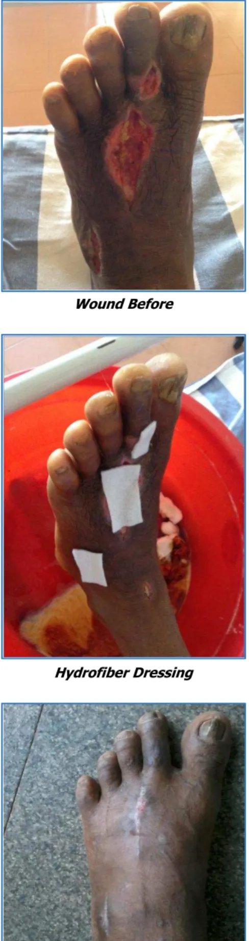
Sequential Photographs:
Wound Before
Hydrofiber Dressing
J. Evid. Based Med. Healthc., pISSN- 2349-2562, eISSN- 2349-2570/ Vol. 3/Issue 22/Mar. 17, 2016 Page 990
DISCUSSION: Foot ulcer infection is the most common and dangerous adversary in the diabetic foot. Therefore, it is also vital to identify symptoms of ulcer infection and address infection proactively. While the incidence of infection was somewhat lower than that reported elsewhere, infection was the most common adverse event, underscoring the strong need to address the microbial burden in the ulcer.
Important measures of such effects in future studies may include antibiotics prescribed, days to discontinue antibiotic usage, and the relative effects of topical and systemic antimicrobial agents.
In a recent study,8 it took 20 weeks for 31% of Diabetic
foot ulcers receiving good standard wound care to heal; the same percent healed during 8 weeks in the hydrofiber-dressing group.
Proportions healed or improved during 8 weeks (88% in hydrofiber group) compare favourably with 71-72% reported clinically improved in a 12-week trial managing
Wagner grade I/II DFU’s with iodine gauze based dressings.9
Smaller trials of various durations report similar results with other wound dressings.10-18
In larger trials, a collagen-oxidized regenerated cellulose dressing19 took 10 weeks for a similar percentage of wounds
to heal, dermal substitute 12 weeks20 and a human skin
equivalent approximately 5 weeks.21 The results are also
comparable to results reported for non-infected DFU’s managed with hyaluron based autologous dermal and epidermal grafts.22
Clinically, these findings support the role of a hydrofiber dressing with ionic silver as an effective option to include with appropriate off-loading, accommodative footwear and debridement in a non-ischaemic DFU.
The significant greater depth reduction and overall wound bed improvement at final clinical evaluation were noted in the group using the ionic silver hydrofiber technology.
The microbial sequestration properties, so observed differences may result from the dressing fibers, ionic silver in the hydrofiber dressing or nature of the wound-dressing interface.
These suggest that once the burdens of exudate and its microbial contents are addressed, ulcer depth reduces as granulation tissue fills the wound, further increasing the tissue barrier against invading organisms.
Of note, a recent 12-week prospective North American study of hydrofiber dressing with silver ions compared with saline moistened gauze showed a greater reduction in the ulcer depth in hydrofiber dressing group, a finding supporting the depth reduction results in the study.
The more positive effects of the ionic silver Hydrofiber
dressing on overall wound condition (more
improvement/less deterioration) for the patients initially prescribed antibiotics might be expected from use of a dressing which absorbs and holds wound exudate and microorganisms within its fibres where bacteria are killed by the ionic silver, providing an effective topical antimicrobial barrier for up to 14 days.23
One hypothesis generated by this research is that the capacity to absorb, hold and kill bacteria in infected wound fluid in the Hydrofiber silver dressing may work in synergy with systemic antibiotics, which may not always reach organisms on the wound surface.
Results of this study suggest that subjects prescribed systemic antibiotics for their DFUs (i.e. with clinically infected ulcers or those at significant risk of infection) may benefit particularly from the hydrofiber dressings with ionic silver.
This hypothesis of topical and systemic antimicrobial synergy merits additional research.
Overall, while healing speed, was comparable in the two groups, the silver ion hydrofiber-dressed subjects in this study experienced significantly greater depth reduction. There was a trend toward more overall wound improvement/ less deterioration with silver ion hydrofiber-dressed subjects than with iodine gauze- hydrofiber-dressed subjects, statistically significant in subjects prescribed antibiotics on enrolment for the study ulcer.
The study generates interesting hypothesis regarding potential synergy between topical silver and systemic antibiotics.
CONCLUSION: In comparison with povidone gauze dressings to hydrofiber dressings in diabetic foot ulcers, the latter is more efficacious in controlling the infection, minimal usage of antibiotics, faster healing rate and patient compliance during dressings.
ACKNOWLEDGEMENT: We would like to thank all the consultant surgeons of Department of General Surgery in the institute for allowing us to analyze their cases.
We would also like to thank Dr. Aravind for his support. The authors confirm that there are no known conflicts of interest, which are associated with this publication, and that there has been no financial support for this work that could have influenced its outcome.
REFERENCES:
1. American Diabetes Association. Consensus
development conference on diabetic foot wound care,
boston, massachusetts. Diabetes Care.
1999;22(8):1354-60.
2. Viswanathan V. Epidemiology of diabetic foot and management of foot problems in India. Int J Low Extrem Wounds 2010;9(3):122-26.
3. Viswanathan V, Jasmine JJ, Snehalatha C, et al. Prevalence of pathogens in diabetic foot infection in South Indian type 2 diabetic patients. J Assoc Physicians India 2002;50:1013-1016.
4. Armstrong DG, Lavery LA, Quebedeaux TL, et al. Surgical morbidity and the risk of amputation due to infected puncture wounds in diabetic versus nondiabetic adults. South Med J 1997;90(4):384-9. 5. Anandi C, Alaguraja D, Natarajanet V, et al.
J. Evid. Based Med. Healthc., pISSN- 2349-2562, eISSN- 2349-2570/ Vol. 3/Issue 22/Mar. 17, 2016 Page 991
6. Gadepalli R, Dhawan B, Sreenivas V, et al. A clinico-microbiological study of diabetic foot ulcers in an Indian tertiary care hospital. Diabetes Care 2006;29(8):1727-32.
7. Alavi SM, Khosravi AD, Sarami A, et al. Bacteriologic study of diabetic foot ulcer. Pak J Med Sci 2007;23(5):681-684.
8. Margolis DJ, Kantor J, Berlin JA. Healing of diabetic neuropathic foot ulcers receiving standard treatment. Diabetes Care 1999;22(5):692–695.
9. Apelqvist J, Ragnarson Tennvall G. Cavity foot ulcers in diabetic patients: a comparative study of cadexomer iodine ointment and standard treatment. An economic analysis alongside a clinical trial. Acta Derm Venereol 1996;76(3):231-235.
10.Ahroni JH, Boyko EJ, Pecoraro RE. Diabetic foot ulcer healing: extrinsic vs. intrinsic factors. Wounds 1993;5:245-255.
11.Motta GJ, Milne CT, Corbett LQ. Impact of antimicrobial gauze on bacterial colonies in wounds that require packing. Ostomy Wound Manage 2004;50(8):48–62. 12.Bale S, Baker N, Crook H, et al. Exploring the use of an
alginate dressing for diabetic foot ulcers. J Wound Care 2001;10(3):81-84.
13.Lohmann M, Thomsen JK, Edmonds ME, et al. Safety and performance of a new nonadhesive foam dressing for the treatment of diabetic foot ulcers. J Wound Care 2004;13(3):118-120.
14.Foster AVM, Greenhill MT, Edmonds ME. Comparing two dress- ings in the treatment of diabetic foot ulcers. J Wound Care 1994;3:224-228.
15.Piagessi A, Baccetti F, Rizzo L, et al. Sodium carboxyl-methyl-cellulose dressings in the management of deep ulcerations of diabetic foot. Diabet Med 2001;18(4):320-324.
16.Blackman JD, Senseng D, Quinn L, et al. Clinical evaluation of a semipermeable polymeric membrane dressing for the treat- ment of chronic diabetes foot ulcers. Diabetes Care 1994;17(4):322-325.
17.Vazquez JR, Short B, Findlow AH, et al. Outcomes of hyaluronan therapy in diabetic foot wounds. Diabetes Res Clin Pract 2003;59(2):123-127.
18.Mason JO, Keefe C, Hutchinson A, et al. A systematic review of foot ulcers in patients with type 2 diabetes mellitus. II: treatment. Diabet Med 1999;16(11):889-909.
19.Veves A, Sheehan P, Pham HT. A randomized, controlled trial of promogran (a collagen/oxidized regenerated cellulose dressing) vs. standard treatment in the management of diabetic foot ulcrs. Arch Surg 2002;137(7):822-827.
20. Marston WA, Hanft J, Norwood P, et al. The efficacy
and safety of dermagraft in improving the healing of chronic diabetic foot ulcers: results of a prospective randomized trial. Diabetes Care 2003;26(6):1701-1705.
21. Veves A, Falanga V, Armstrong DG, et al. Graftskin, a
human skin equivalent, is effective in the management of noninfected neuropathic diabetic foot ulcers: a prospective randomized multicenter clinical trial. Diabetes Care 2001;24(2):290-295.
22. Caravaggi C, Giglio RD, Pritelli C, et al.
HYAFF-11-based autologous dermal and epidermal grafts in the treatment of noninfected diabetic plantar and dorsal foot ulcers: a prospective, multicenter, controlled,
randomized clinical trial. Diabetes Care
2003;26(10):2853-2859.