Copyright © 2013 The Korean Society of Plastic and Reconstructive Surgeons
This is an Open Access article distributed under the terms of the Creative Commons Attribution Non-Commercial License (http://creativecommons.org/
licenses/by-nc/3.0/) which permits unrestricted non-commercial use, distribution, and reproduction in any medium, provided the original work is properly cited. www.e-aps.org
INTRODUCTION
A person with diabetes may experience one or more of its many complications, such as neuropathy, retinopathy, nephropathy, and arterial occlusive disease. One of the most important clini-cal manifestations of diabetes is diabetic foot ulcer, the most frequent cause of lower leg amputation [1]. A diabetic foot ulcer has a complicated etiology, and can easily progress to a chronic
ulcer. herefore, a multifactorial approach is needed [2]. Correction of peripheral vasculopathy, adequate debridement, and wound management including infection control are im-portant for treatment of a diabetic foot ulcer. If the diabetic foot ulcer is supericial, a simple wound dressing may be adequate. However, if the ulcer is suiciently deep, with a layer of subcu-taneous fat, along with exposed bone or ligament, a surgical procedure, such as skin grat or lap operation, is needed [3]. A
Treatment of Diabetic Foot Ulcer Using Matriderm
In Comparison with a Skin Grat
Hyojin Jeon, Junhyung Kim, Hyeonjung Yeo, Hoijoon Jeong, Daegu Son, Kihwan Han
Department of Plastic and Reconstructive Surgery, Keimyung University School of Medicine, Daegu, KoreaCorrespondence: Junhyung Kim Department of Plastic and Reconstructive Surgery, Keimyung University School of Medicine, 56 Dalseong-ro, Jung-gu, Daegu 700-712, Korea Tel: +82-53-250-7635 Fax: +82-53-255-0632 E-mail: med69@dsmc.or.kr Background For patients with neuropathy, vasculopathy, and impairment of wound
healing, treatment of a diabetic foot ulcer poses many challenges. A large number of dermal analogues have been invented in an effort to overcome these challenges. Matriderm, a dermal analogue, is made from bovine collagen and elastin. This study was conducted in order to evaluate the effectiveness of Matriderm for treatment of diabetic foot ulcers, in comparison with skin grafting.
Methods Sixty patients with diabetic foot ulcer were included in this prospective study. The average age of the patients, who had type II diabetes mellitus, was 58 years old. The patients were allocated to an experimental or control group with their consents. The patients were selected with their consent for inclusion in an experimental group and a control group. Patients in the experimental group received a Matriderm appliance and a split-thickness skin graft, while those in the control group received only a split-thickness skin graft.
Results A shorter hospitalization period (7.52 weeks) was observed in the experimental group than in the control group (9.22 weeks), and a shorter period of time (8.61 weeks) was required for complete healing, compared with the control group (12.94 weeks), with statistical signiicance (P < 0.05). A higher elasticity ratio of the affected side to the non-affected side was observed in the experimental group, compared with the control group (P < 0.01).
Conclusions Matriderm enables effective healing and improves elasticity in treatment of patients with diabetic foot ulcer.
Keywords Skin / Diabetes complications / Skin transplantation
Received: 19 Mar 2013 • Revised: 15 May 2013 • Accepted: 20 Jun 2013
pISSN: 2234-6163 • eISSN: 2234-6171 • http://dx.doi.org/10.5999/aps.2013.40.4.403 • Arch Plast Surg 2013;40:403-408
This article was presented at the 70th Congress of the Korean Society of Plastic and Reconstructive Surgeons on November 9-11, 2012 in Seoul, Korea.
No potential conlict of interest relevant to this article was reported.
skin graft is a relatively simple procedure, but has limitations, including a high possibility of loss of grat skin and low endur-ance against friction and pressure. On the other hand, a lap op-eration is efective and durable for deep ulcers but has a narrow patient spectrum and is associated with severe postoperative complications, including lap necrosis.
he purpose of this study was to investigate the efectiveness of Matriderm (Skin Health, Billerbeck, Germany), an artiicial dermis, by comparison with skin grat only in treatment of dia-betic foot ulcer [4,5].
METHODS
Patients
Ater receiving institutional review board approval, a prospec-tive study was conducted. From January 2011 to January 2012, 60 patients (male, 37; female, 23) who underwent treatment of diabetic foot ulcer in our department, with a mean age of 58 years (range, 34 to 73 years) and a postoperative follow-up period of 8.8 months (range, 6 to 12 months) were evaluated for enrollment in the study.
Proper wound bed preparation was performed before the study, including debridement (removing all non-viable tissues), controlling infection (based on the results of bacterial culture), and maintaining a balanced moist environment.
Inclusion criteria were type 2 diabetic patients with a chronic foot ulcer, lasting more than four weeks ater appropriate man-agement, measuring more than 1 cm2 in size, three sequential
negative wound culture tests, and being covered with healthy granulation tissue ater proper wound bed preparation. Exclu-sion criteria were a wound depth of Wagner grade 3, or a Uni-versity of Texas wound classiication greater than 3, or a wound
with vasculopathy of less than 0.9 on the ankle brachial index (ABI) [6,7].
After sufficient explanation of the operation, including the costs of Matriderm, the 60 patients were divided equally into a control group (n = 30), receiving a skin grat only, and an ex-perimental group (n = 30), receiving the Matriderm appliance combined with a skin grat, with the consent of the patients.
Surgical technique
In both groups, adequate debridement was performed until healthy tissue was exposed with pin point bleeding. After de-bridement, Matriderm was applied, soaked with normal saline, and a split-thickness skin graft was performed in one stage in the experimental group (Fig. 1). Only a split-thickness skin grat was performed in the control group (Fig. 2). Wet-to-dry com-pressive dressing was administered until the grat skin had taken in both groups. Ater the grat skin had taken, ointment (Con-netivina gel, Fidia Farmaceutici Spa., Abano Terme, Italy) and foam dressing (Mepilex, Mölnlycke Health Care, Gothenburg, Sweden) were applied daily.
Assessment
For comparison of two groups, four factors were measured. 1) The length of the hospital stay; 2) The length of complete wound epithelialization; 3) he number of completely healed patients at postoperative 12 weeks; 4) The elasticity ratio of the afected lesion and the contralateral region at the complete wound epithelialization point, using a skin diagnosis system (Aramo-TS, Aram Huvis Co. Ltd, Sungnam, Korea) [8].
To obtain objective results, the investigator responsible for evaluating the elasticity ratio was blinded. Complete healing was defined as complete wound epithelialization without any
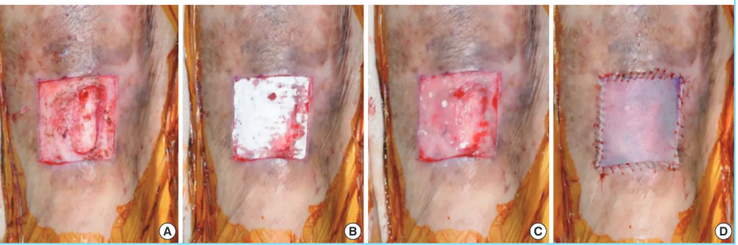
Fig. 1. Experimental group with Matriderm appliance
(A) Debridement was performed until pin point bleeding and healthy granulation were observed. (B) Matriderm was applied to the wound. (C) Complete hydration of Matriderm was performed. (D) A split-thickness skin graft was performed in one stage.
Postoperative evaluation Control group
Experimental
group P-value
Hospital stay (wk) 7.36 5.63 0.015
Complete healing period (wk) 8.61 12.94 0.010
Complete healing rate (%) 80.00 93.30 0.023
Elasticity ratio (affected/ non-affected side)
0.19 0.72 0.030
transudate or exudate from the wound [9]. SPSS ver. 12.0 (SPSS Inc., Chicago, IL, USA) was used for performance of the statistical analysis. he chi-squared test was used for data analy-sis. Signiicance was set at a value of P ≤ 0.05.
RESULTS
In total, 60 patients (100%) participated in the study without follow-up loss. A summary of the demographic details of the control and experimental groups, including age, HbA1c (%), wound size, wound depth, and ABI value, is shown in Table 1. No statistically signiicant diferences in the various characteris-tics were observed between the groups.
he length of the hospital stay was shorter in the experimental group, at 7.52 weeks, than in the control group (9.22 weeks, P < 0.05). In addition, the length of wound epithelialization was shorter in the experimental group, at 8.61 weeks, than in the control group (12.94 weeks, P < 0.05). The number of com-pletely healed patients at postoperative 12 weeks was larger in the experimental group, with 28 patients (93.3%), than in the control group. With 24 patients (80.0%, P < 0.05). he elasticity ratio of the afected lesion and contralateral region at the
com-plete wound epithelialization point was higher in the experi-mental group, at 0.72, than in the control group (0.19, P < 0.01). A summary of the outcomes of these four factors is shown in Table 2. There was no occurrence of complications, such as a rejection response to the artificial dermis, infection, bleeding problems, or loss of grat skin. Also, during the follow-up period, there was no recurrence in either group.
Case 1
A 69-year-old male with type 2 diabetes controlled by oral hypoglycemic agents for 11 years suffered from an unhealed diabetic foot ulcer on the tibial side of the right great toe. De-bridement was performed primarily, followed by application of Matriderm and a split-thickness skin graft. The patient stayed at the hospital for six weeks and wound epithelialization lasted eight weeks; the elasticity ratio was 0.81 (Fig. 3).
Case 2
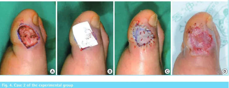
A 34-year-old female with type 2 diabetes controlled by oral hypoglycemic agents for three years sufered from an unhealed diabetic foot ulcer on the right foot dorsum after trauma. De-bridement was performed primarily, followed by application of Matriderm and a split-thickness skin grat. he patient stayed in the hospital for ive weeks, and wound epithelialization took six weeks; the elasticity ratio was 0.75 (Fig. 4).
Characteristic Control group
Experimental
group P-value
No. of patients 30 30
-Sex (male/female) 18/12 19/11 0.43
Age (yr) 59.00 58.70 0.53
HbA1C (%) 7.51 7.83 0.41
Initial size of ulcer (cm2) 26.30 29.20 0.78
Ankle brachial index 0.98 1.05 0.83
Wagner grade 1/2 4/26 5/25 0.37
University of Texas wound classiication depth 1A/2A
4/26 5/25 0.37
Table 1. Clinical characteristics of patients Fig. 2. Control group without Matriderm appliance
(A) Debridement was performed until pin point bleeding and healthy granulation were observed. Subcutaneous tissue was intact with healthy features. (B) Only a split-thickness skin graft was performed.
A B
DISCUSSION
Diabetic foot ulcer is not simply a problem of diabetes compli-cations; it also degrades the quality of life of patients, and can cause fatal complications, such as sepsis. According to the lat-est statistics for 2003, diabetic foot ulcers occurred in 1.2% of patients with diabetes foot disease, which is 47.9% of all cases of foot disease, 54.4% of all cases of foot amputation [1,4].
In diabetic patients, peripheral circulation disorders due to cal-ciication of the arteries and capillaries, thickening of the base-ment membrane, diabetic peripheral neuropathy, and hyper-glycemia due to lack of growth factors and cell metabolism are causes of impaired wound healing. herefore, surgical treatment
Fig. 4. Case 2 of the experimental group
A B C D
Fig. 3. Case 1 of the experimental group
A B C D
for diabetic foot ulcer with primary closure, skin grat, and lap application are important [2]. Primary closure is possible only for small wounds, and skin grating is easy to perform; however, the latter does not take well in deep wounds and has low du-rability. Flap application can overcome the limitations of skin grating; however, it is burdened with the risk of complications, such as lap necrosis, especially in the case of common diabetes with a poor vascular state.
In the past 10 years, dermis, collagen, and elastic fibers have emerged as important components of wound healing. Cuono et al. [10], a synthesized functional dermal tissue with elastic iber, has excellent resilience for prevention of scar formation. Matri-derm, which was used in our study, is a three-dimensional ma-A 69-year-old male had a diabetic foot ulcer on the tibial side of the right great toe. The patient stayed in the hospital for six weeks, and had healed completely at eight weeks postoperation. The elasticity ratio measured was 0.81. (A) After debridement, the periosteum was exposed. (B) Matriderm was applied to the periosteum. (C) A split-thickness skin graft was performed. (D) Image of completely healed state at eight weeks post- operation.
trix composed of collagen ibrils and elastin ibers for support of dermal regeneration in one stage. It induces dermis regeneration as a scafold, and decreases scar tissue formation. It also provides optimal dermal wound bed preparation as regenerated skin with extensive formation of rete ridges and capillary loops but with the absence of skin appendages and is ready for relatively thin-ner subsequent skin grating [11]. Collagen is obtained from the bovine dermis and contains dermal collagen types I, III, and V. Elastin is obtained from the bovine nuchal ligament by hydroly-sis. In addition, Matriderm has excellent hemostatic properties and thus reduces the risk of hematoma formation under grated skin. he non-use of chemical cross-linking of collagen results in biocompatibility [4,10]. In our study, the experimental group with Matriderm showed a shorter length of hospital stay and length of complete wound epithelialization and showed a higher elasticity ratio of the afected lesion and contralateral region at the complete wound epithelialization point. We concluded that Matriderm induced dermal regeneration and facilitated the tak-ing of the grat skin. Patients with chronic wounds like diabetic foot ulcers seem to hope for an especially quick return to their daily lives, which was possible in the experimental group.
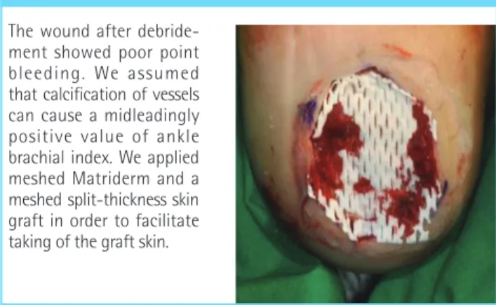
Although the patients had a good ABI score of 1.05, the wound showed poor pin-point bleeding ater debridement (Fig. 5). We assumed that calciication of the vessels can cause a misleadingly good ABI score. We applied a meshed Matriderm and meshed split-thickness skin grat in order to facilitate taking of the grat skin [12]. We expect that meshed Matriderm would also be efective on wounds with a bleeding tendency, such as those as-sociated with liver cirrhosis or chronic renal failure with dialysis. In our study, elasticity was the most important component for data analysis, considering the predictive factors for recurrence [13,14]. Although the surgeon and patients were aware of the treatment received, the investigator was blinded and evaluated the elasticity objectively using a skin diagnosis system
(Aramo-TS). The probe for the system can cause constant negative pressure, as much as 300 mm Hg. By this negative pressure, the sensor of the probe measured the changes in the skin in contact with the probe, and the value of the elasticity was then determined. To take into account the patient’s own skin charac-teristics, we compared both groups according to the elasticity ratio of the afected lesion and contralateral region [15]. As we continue to do follow-up with the patients, we expect lower recurrence in the experimental group, given its higher elasticity, than control group with lower elasticity.
In treatment of diabetic foot ulcer, the experimental group, which received the Matriderm appliance and a split-thickness skin grat, showed a shorter hospital stay, shorter epithelializa-tion period, and higher elasticity ratio than the control group, who received a split-thickness skin grat only. According to these results, we believe that Matriderm can be an efective treatment for diabetic foot ulcer.
REFERENCES
1. Chung CH, Kim DJ, Kim J, et al. Current status of diabetic foot in Korean patients using national health insurance data-base. J Korean Diabetes Assoc 2006;30:372-6.
2. Greenhalgh DG. Wound healing and diabetes mellitus. Clin Plast Surg 2003;30:37-45.
3. Kim SH, Kim JW, Kim JB, et al. Multifactorial factors of dia-betic foot on diabetes mellitus comparative clinical study. J Korean Soc Plast Reconstr Surg 2002;29:83-90.
4. Hant JR, Surprenant MS. Healing of chronic foot ulcers in diabetic patients treated with a human fibroblast-derived dermis. J Foot Ankle Surg 2002;41:291-9.
5. Wetzig T, Gebhardt C, Simon JC. New indications for arti-icial collagen-elastin matrices? Covering exposed tendons. Dermatology 2009;219:272-3.
6. Wagner FW Jr. he diabetic foot. Orthopedics 1987;10:163-72.
7. Lavery LA, Armstrong DG, Murdoch DP, et al. Validation of the Infectious Diseases Society of America’s diabetic foot infection classiication system. Clin Infect Dis 2007;44:562-5.
8. Hashmi F, Malone-Lee J. Measurement of skin elasticity on the foot. Skin Res Technol 2007;13:252-8.
9. Eum SJ, Han SK, Gu JH, et al. Treatment of diabetic ulcer us-ing autologous ibroblast-hyaluronic acid complex. J Korean Soc Plast Reconstr Surg 2009;36:548-54.
10. Cuono C, Langdon R, McGuire J. Use of cultured epider-mal autografts and derepider-mal allografts as skin replacement ater burn injury. Lancet 1986;1:1123-4.
Fig. 5. Meshed Matriderm appliance with poor vascular condition
11. Hamuy R, Kinoshita N, Yoshimoto H, et al. One-stage, simultaneous skin grafting with artificial dermis and basic ibroblast growth factor successfully improves elasticity with maturation of scar formation. Wound Repair Regen 2013; 21:141-54.
12. Han SK, You HJ. Wound coverage using advanced technol-ogy in Korea. J Korean Med Assoc 2011;54:594-603. 13. Young MJ, Adams JE, Anderson GF, et al. Medial arterial
calcification in the feet of diabetic patients and matched
non-diabetic control subjects. Diabetologia 1993;36:615-21.
14. Weiner RD, Hlad LM, McKenna DR. Recurrence of dia-betic pedal ulcerations following tendo-achilles lengthening. Diabet Foot Ankle 2011;2:6417.